Dynamic Changes in Antimicrobial Resistance in Fecal Escherichia coli from Neonatal Dairy Calves: An Individual Follow-Up Study - MDPI
←
→
Page content transcription
If your browser does not render page correctly, please read the page content below
animals
Article
Dynamic Changes in Antimicrobial Resistance in
Fecal Escherichia coli from Neonatal Dairy Calves:
An Individual Follow-Up Study
Sang-Ik Oh † , Seungmin Ha † , Jae-Hee Roh, Tai-Young Hur and Jae Gyu Yoo *
 National Institute of Animal Science, Rural Development Administration, 1500 Kongjwipatjwi-ro,
 Wanju 55365, Korea; ohsangik@korea.kr (S.-I.O.); justusha@korea.kr (S.H.); snowida@korea.kr (J.-H.R.);
 tyohur@korea.kr (T.-Y.H.)
 * Correspondence: vetjack@korea.kr; Tel.: +82-63-238-7220; Fax: +82-63-238-7235
 † These authors contributed equally to this work.

 Received: 5 August 2020; Accepted: 29 September 2020; Published: 1 October 2020

 Simple Summary: Antimicrobial resistance in food animals is a global public health concern. In dairy
 farms, young calves typically carry high levels of antimicrobial-resistant (AMR) Escherichia coli, and
 may act as a potential reservoir. Fecal E. coli were isolated and tested for susceptibilities to eight
 common antimicrobials from 19 newborn dairy calves using individual follow-up at daily and weekly
 intervals. Shedding of AMR E. coli first appeared at 2–3 days after birth. The majority of fecal E. coli
 from neonatal calves (≤28 days old) were resistant to streptomycin, sulfisoxazole, and tetracycline.
 A tetracyclines-associated resistance gene (tetB) was predominant in the fecal E. coli from neonatal
 calves, and was also detected in maternal colostrum samples from the mothers of the tested calves.
 These results suggest the potential of antimicrobial resistance genes being shared between neonatal
 calves and their mothers’ colostrum. Neonatal calves with a history of treatment with ceftiofur also
 shed AMR E. coli resistant against beta-lactams. Moreover, these results provide new insights for
 controlling the spread of antimicrobial resistance on dairy farms.

 Abstract: The prevalence of antimicrobial-resistant (AMR) Escherichia coli is typically higher in
 the feces of young dairy calves than in the feces of older cattle; however, the underlying factors
 contributing to this difference are poorly understood. In this study, AMR fecal E. coli from neonatal
 calves were characterized both at phenotypic and genotypic levels by individual follow-up sampling.
 Antimicrobial resistance profiles of E. coli isolates from the maternal colostrum were also determined.
 Most of the fecal AMR E. coli emerged in the calves at 2–3 days of age. The tetB was the most prevalent
 resistance gene detected among AMR fecal E. coli from
Animals 2020, 10, 1776 2 of 12
caused the emergence of multidrug-resistant bacteria, which could result in treatment failure [2].
Although the increased prevalence of resistant pathogenic bacteria is of prime concern, commensal
bacteria, especially non-pathogenic Escherichia coli, are also considered as a potential reservoir of
antimicrobial resistance [4]. Furthermore, the horizontal transfer of antimicrobial resistance genes
(ARGs) is possible between commensal bacteria and pathogenic strains [1,5].
In dairy cattle, the prevalence of antimicrobial-resistant (AMR) E. coli and ARGs is age-dependent,
with a higher prevalence detected in earlier stages of life, especially between 2–4 weeks old dairy
calves [5–7]. Several studies have evaluated the factors contributing to this high prevalence of AMR
E. coli in young dairy calves, and some studies have suggested the milk diet as an important risk
factor [1,6–10]. Supplementation of calf milk with antimicrobials, incidence of diarrhea, use of feed
additives with biocides and heavy metals, and vitamin supplements has also been suggested to
contribute to the high AMR prevalence in young calves [1,6,8,9]. Previous studies have proposed that
maternal colostrum, being the first feed, may be an important vehicle for AMR E. coli in neonatal
calves [1,11,12]. A recent study revealed that fecal E. coli from 2-day-old calves share appropriately
90% of their ARGs with the E. coli in their maternal colostrum, thereby suggesting that the colostrum
serves as a vector for ARGs transmitted to dairy calves [1]. In addition, Doyle et al. [13] suggested that
the presence of feces on the teat surface is the most abundant contributor to the raw milk microbiota,
indicating that AMR E. coli and their ARGs in the colostrum are environment-specific contaminants.
However, most of these previous studies focused on animals without considering the detailed prior
history of antibiotic use and clinical signs of disease [6,9,10]. Moreover, few studies have specifically
investigated AMR fecal E. coli in neonatal calves with individual follow-up of short term intervals from
the day of birth, which makes it difficult to specify when the AMR fecal E. coli emerges in newborn
calves [5]. As a result, the epidemiology and dynamic changes of AMR fecal E. coli in neonatal calves
are not fully understood.
To address these knowledge gaps, we evaluated changes in the antimicrobial resistance patterns
of fecal E. coli isolated from newborn dairy calves using individual follow-up fecal sampling from
the day of birth (once daily and weekly follow-up sampling). This detailed sampling approach with
corresponding information of the tested animals is expected to help discriminate the precise timing of
AMR fecal E. coli shedding from neonatal calves. Furthermore, we focused on investigating the role
of two risk factors (colostrum and antibiotic use) which are related to the emergence timing of AMR
fecal E. coli in neonatal calves. The study outcomes offer novel insights into the potentially influential
factors determining a high AMR prevalence in young dairy calves.
2. Materials and Methods
2.1. Animals
The study was performed at a large breeding dairy farm in Korea. A total of 29 newborn dairy
calves who were born between October 2019 and April 2020 and were available for analysis were
included in this study (Table 1). Newborn calves were separated from their dams within a few
hours after birth and housed in a separate hutch. All neonatal calves were fed their respective dams’
colostrum within the first 12 h after birth until 3 days post-birth. After 3 days, all calves were fed
whole milk until 28 days. The diet was supplemented with concentrated feed and Timothy hay as of
between day 7 and 28 onward, respectively. The health and welfare of the calves included in the study
were individually supervised by a designated veterinarian, who provided the detailed medical history
for the calves and their dams. All experiments were approved by the Institutional Animal Care and
Use Committee at the National Institute of Animal Science, Korea (approval number: NIAS 2019-369).
2.2. Sample Collection
The detailed scheme for sampling is shown in Figure 1. In experiment 1, all of the dairy calves
(n = 10) which were born during this experiment period were subjected to once-daily sampling 1Animals 2020, 10, x FOR PEER REVIEW 3 of 11
2.2. Sample
Animals 2020, Collection
10, 1776 3 of 12
The detailed scheme for sampling is shown in Figure 1. In experiment 1, all of the dairy calves
(n = 10)
to 7 days which
afterwere born
birth. duringthese
Among this experiment
10 calves, six period were colostrum
maternal subjected to once-daily
samples (15 sampling
mL each) 1from to 7
days after birth. Among these 10 calves, six maternal colostrum samples
the corresponding dam were collected prior to feeding. Colostrum was harvested aseptically after(15 mL each) from the
corresponding
predipping, wiping,dam andwere collected
drying priorand
the teats, to then
feeding. Colostrum
the samples werewas harvested
collected aseptically
into sterile after
containers.
predipping, wiping, and drying the teats, and then the samples were collected into
In experiment 2, individual fecal samples were collected from 19 newborn dairy calves (79.2% of calves sterile containers.
In
bornexperiment
during this2,experiment
individual period)
fecal samples
within thewere collected
first day afterfrom
birth19and
newborn dairy calves
then at weekly (79.2%
intervals for upof
calves born during
to 4 weeks. this
The fecal experiment
samples were period)
obtainedwithin the first the
by swabbing dayrectum
after birth
withand then at
a sterile weekly
cotton intervals
swab. After
for up to 4 weeks. The fecal samples were obtained by swabbing the rectum
collection, the samples were immediately sent to the laboratory in thermal bags with ice packs with a sterile cotton
and
swab. After ◦collection, the samples were immediately sent to the laboratory in
stored at 4 C until use. The bacteria were isolated from the collected samples within one day, and thermal bags with ice
packs and stored at 4 °C until use. The bacteria
◦ were
then the fecal samples were stored at −80 C until further study. isolated from the collected samples within one
day, and then the fecal samples were stored at −80 °C until further study.
Figure
Figure 1.
1. Sampling
Sampling scheme
scheme for
for experiments
experiments 11 and
and 2.
2. Neonatal
Neonatal dairy
dairy calves
calves were
were sampled
sampled once
once aa day
day
for 7 days (n = 10) or once a week for 28 days (n = 19), and maternal colostrum samples
for 7 days (n = 10) or once a week for 28 days (n = 19), and maternal colostrum samples (n = (n = 6)
6) from
from
corresponding
corresponding dams
dams were
were obtained.
obtained.
Treatment History
2.3. Treatment
The farm where the study was conducted commonly uses ceftiofur for for treating respiratory tract
infections, hoof
infections, hoof disease,
disease, mastitis,
mastitis, andand neonatal
neonatal calf
calf diarrhea
diarrhea (NCD).
(NCD). The history of clinical diseases
and antibiotic use for each sampled calf and their dams is shown in Table 1. None of the 10 calves in
experiment 1 received any antibiotics during the sampling period, and the dams of the tested calves
did not receive antibiotics
antibiotics for
for the
the 11year
yearperiod
periodbefore
beforethe
thebirth
birthofofthe
thecalf.
calf.However,
However,ininexperiment
experiment 2,
five calves (calf nos. 23–27) showed signs of NCD, and were thus
2, five calves (calf nos. 23–27) showed signs of NCD, and were thus treated with treated with ceftiofur; three dams
of the sampled calves (calf nos. 27–29)27–29) received
received ceftiofur
ceftiofur to
to treat
treat hoof
hoof disease,
disease, foot
foot rot,
rot, and
and mastitis,
mastitis,
This detailed
respectively. This detailed medical
medical history
history provided
provided anan opportunity
opportunity to to directly
directly test
test the
the impact of
antibiotic exposure
antibiotic exposure on on the prevalence of AMR E. coli. Overall,
Overall, 77 calves
calves (36.8%)
(36.8%) were
were assigned
assigned to the
antibiotic exposed group and 12 12 (63.2%)
(63.2%) were
were assigned
assigned to
tothe
thenon-exposed
non-exposedgroup groupforforthis
thiscomparison.
comparison.
Table 1. Detailed information about 29 newborn calves included in this study.
History of Clinical Signs and Antibiotic Use
Calf Antibiotic Use On Their
Group Breed Clinical Sign Antibiotic Use
No. Dams
(Day after Birth) (Day after Birth)
(Day before Calf Birth)
1 Jersey – – –
2 Holstein – – –
3 Holstein – – –
4 Holstein – – –
Once daily
5 Holstein – – –
sampling
6 Holstein – – –
(Experiment 1)
7 Holstein – – –
8 Holstein – – –
9 Holstein – – –
10 Holstein – – –
Once weekly 11 Holstein – – –
sampling 12 Holstein – – –
(Experiment 2) 13 Holstein – – –Animals 2020, 10, 1776 4 of 12
Table 1. Detailed information about 29 newborn calves included in this study.
History of Clinical Signs and Antibiotic Use
Group Calf No. Breed
Antibiotic Use On
Clinical Sign Antibiotic Use Their Dams
(Day after Birth) (Day after Birth) (Day before Calf
Birth)
1 Jersey – – –
2 Holstein – – –
3 Holstein – – –
4 Holstein – – –
Once daily
sampling 5 Holstein – – –
(Experiment 1) 6 Holstein – – –
7 Holstein – – –
8 Holstein – – –
9 Holstein – – –
10 Holstein – – –
11 Holstein – – –
12 Holstein – – –
13 Holstein – – –
14 Holstein – – –
15 Holstein – – –
16 Jersey – – –
17 Holstein – – –
18 Holstein – – –
Once weekly 19 Holstein – – –
sampling 20 Holstein – – –
(Experiment 2) 21 Holstein – – –
22 Holstein – – –
23 Holstein Diarrhea (15, 19 d) Ceftiofur (15 d) –
Diarrhea (14–16 d) Ceftiofur (14–16 d)
24 Holstein –
Diarrhea (21–25 d) Ceftiofur (23–25 d)
25 Jersy Diarrhea (15–19 d) Ceftiofur (15–18 d) –
26 Holstein Diarrhea (16 d) Ceftiofur (16 d) –
27 Holstein Diarrhea (23 d) Ceftiofur (23–24 d) Ceftiofur (156 d)
28 Holstein – – Ceftiofur (329 d)
29 Holstein – – Ceftiofur (95 d)
Experiment 2 Ceftiofur was administered by intramuscular injection at a dose of 100 mg/kg body weight; Clinical
signs of the corresponding dams were hoof disease (no. 27), foot rot (no. 28), and mastitis (no. 29).
2.4. E. coli Isolation and DNA Extraction
The rectal swabs and maternal colostrum samples were cultured on MacConkey agar plates (Kisan
Pharm, Seoul, Korea) and incubated aerobically at 37 ◦ C for 24 h. Three pink colonies of different
morphologies suspected as E. coli were selected and restreaked onto eosin-methylene blue (EMB) agar
plates (Kisan Pharm), followed by incubation for 24 h at 37 ◦ C. Next, the blue-black colonies with
a metallic green sheen on EMB agar plates were selected and streaked onto 5% sheep blood agar
plates. E. coli isolates were confirmed by biochemical identification using the Sensititre system (TREK
Diagnostic System, East Grinstead, UK) with commercial GN plates, and also by 16S rRNA sequencing.
Genomic DNA was extracted from the confirmed E. coli isolates using the QIAamp DNA Mini Kit
(Qiagen, Hilden, Germany) according to the manufacturer’s instructions.
2.5. Identification of Antimicrobial Resistance Genes
ARGs were detected using previously published primers for detecting genes coding for
resistance to streptomycin (strA and strB) [14], sulfonamide (sulI and sulII) [15], and tetracycline
(tetA and tetB) [16]. The primers used were as follows [14–16]: strA [forward primer (F),
50 -CTTGGTGATAACGGCAATTC-30 ; reverse primer (R), 50 -CCAATCGCAGATAGAAGGC-30 ], strB (F,Animals 2020, 10, 1776 5 of 12
50 -ATCGTCAAGGGATTGAAACC-30 ; R, 50 -GGATCGTAGAACATATTGGC-30 ), sulI (F, 50 -GTGAC
GGTGTTCGGCATTCT-30 ; R, 50 -TCCGAGAAGGTGATTGCGCT-30 ), sulII (F, 50 -CGGCATCGTCAAC
ATAACCT-30 ; R, 50 -TGTGCGGATGAAGTCAGCTC-30 ), tetA (F, 50 -GGCCTCAATTTCCTGACG-30 ;
R, 50 -AAGCAGGATGTAGCCTGTGC-30 ), and tetB (F, 50 -GAGACGCAATCGAATTCGG-30 ; R,
50 -TTTAGTGGCTATTCTTCCTGCC-30 ). Amplification was performed on a BioRad T100 thermocycler
(Bio-Rad Laboratories Ltd., Hercules, CA, USA) under previously described conditions [13–15]. In
brief, the annealing temperature and fragment size were as follows: strA (53 ◦ C and 548 bp), strB (53 ◦ C
and 509 bp), sulI (68 ◦ C and 779 bp), sulII (66 ◦ C and 721 bp), tetA (55 ◦ C and 372 bp), and tetB (55 ◦ C
and 228 bp). The amplified products were resolved by electrophoresis on 1.5% agarose gels, stained
with RedSafe nucleic acid staining solution (iNtRON Biotechnology, Seongnam, Korea), and visualized
with an ultraviolet transilluminator (Bio-Rad Laboratories Ltd., Hercules, CA, USA).
2.6. Antimicrobial Susceptibility Test
Minimum inhibitory concentrations (MICs) of E. coli were determined by the standard microbroth
dilution method using the Sensititre system (TREK Diagnostic System, East Grinstead, UK) with
antimicrobial testing plates containing the following eight antimicrobials: amoxicillin-clavulanic acid
(AMO, range tested: 1–64 µg/mL), ampicillin (AMP, range tested: 1–128 µg/mL), cefoxitin (CFX,
range tested: 0.5–64 µg/mL), ceftiofur (TIO, range tested: 0.25–16 µg/mL), streptomycin (STR, range
tested: 8–256 µg/mL), sulfisoxazole (FIS, range tested: 8–512 µg/mL), tetracycline (TET, range tested:
1–256 µg/mL), and trimethoprim-sulfamethoxazole (STX, range tested: 0.0625–8 µg/mL). E. coli strains
ATCC 25922 (American Type Culture Collection (ATCC; Manassas, VA, USA)) and Enterococcus faecalis
ATCC 29212 were used as quality control strains. MICs were interpreted according to guidelines of the
CLSI (Clinical and Laboratory Standards Institute) [17] or DANMAP (Danish Integrated Antimicrobial
Resistance Monitoring and Research Programme) [18] in the case of streptomycin, for which CLSI
breakpoints were not available.
2.7. Statistical Analysis
Statistical analyses were performed using SPSS version 25.0 (IBM, Armonk, NY, USA). A simple
regression model was applied to determine whether there was a significant difference in antibiogram
(ABG) patterns from fecal E. coli according to the calves’ age. The mean differences in the MIC values
were analyzed using repeated-measures analysis of variance, with group (antibiotic exposure) and
time as the main effects, and an additional group-by-time interaction term. Results with p < 0.05 were
considered statistically significant.
3. Results
3.1. Experiment 1
We analyzed the dynamic fecal E. coli ABG patterns in each dairy neonatal calf (n = 10) with daily
sampling for one week (Figure 2A). The confirmed E. coli isolates (two or three isolates per sample)
from the same fecal sample showed identical ABG patterns against the eight tested antimicrobials
in this study. Thus, only one isolate per sample was selected for further analysis as a representative
fecal E. coli from each calf per sampling day. A simple regression analysis showed that the ABG
patterns of fecal E. coli were significantly different according to the age of the calves (adjusted R2 =
0.414, F = 48.075, p < 0.001). All 10 fecal E. coli isolates from 1-day-old calves were susceptible to the
eight tested antimicrobials. AMR fecal E. coli started to shed from 2-day-old (n = 6, 60.0%), 3-day-old
(n = 3, 30.0%), and 5-day-old (n = 1, 10.0%) calves. Among the 70 isolates, 50 fecal E. coli (71.4%)
showed STR-FIS-TET or STR-FIS-TET-STX ABG patterns. Only one calf (no. 9) shed isolates with
the STR-FIS-TET-STX-AMO-AMP-CFX-TIO ABG pattern from day 4 to 7 after birth. Only two E. coli
strains were isolated from the six maternal colostrum samples collected in this study. One strain (fromAnimals 2020, 10, 1776 6 of 12
the dam of calf no. 9) was resistant to all tested antimicrobials except TIO, and the other (from the dam
of calf no. 10) was susceptible to all of the tested antimicrobials.
Animals 2020, 10, x FOR PEER REVIEW 6 of 11
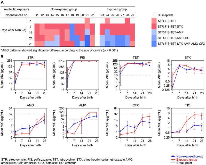
Figure
Figure 2. 2.Characterization
Characterizationof ofresistance
resistance to eight
eight antimicrobials
antimicrobialsininE.E.coli
coliisolated
isolated from
from newborn
newborn dairy
dairy
calves (n = 10) within the first day after birth and at daily intervals for up to 7 days. (A) Antibiogram
calves (n = 10) within the first day after birth and at daily intervals for up to 7 days. (A) Antibiogram
(ABG)
(ABG) ofof fecalE.E.coli
fecal coliisolated
isolatedfrom
fromnewborn
newborn calves.
calves. Each
Eachboxboxrepresents
representsa asingle
singleisolate onon
isolate a given
a given
sampling day. Colors represent ABG patterns, and unfilled boxes indicate
sampling day. Colors represent ABG patterns, and unfilled boxes indicate that the isolates that the isolates were
were
susceptibletotoallalltested
susceptible testedantimicrobials.
antimicrobials. Black
Black boxes
boxesindicate
indicatethat
thatnonoisolates
isolateswere
were obtained
obtainedfrom
from
maternal colostrum samples. (B) Distribution of ARGs associated with streptomycin
maternal colostrum samples. (B) Distribution of ARGs associated with streptomycin (strA and strB), (strA and strB),
sulfa-drugs (sulI and sulII), and tetracycline (tetA and tetB) in E. coli isolates from each calf and the
sulfa-drugs (sulI and sulII), and tetracycline (tetA and tetB) in E. coli isolates from each calf and the
corresponding maternal colostrum.
corresponding maternal colostrum.
The distributions of transmissible ARGs associated with resistance to streptomycin, sulfonamides,
and tetracyclines in E. coli, isolates from the feces of newborn calves and their maternal colostrum are
shown in Figure 2B. The tetB gene was the most prevalent resistance gene detected (n = 41, 75.9%) in
54 AMR fecal E. coli samples from calves, followed by sulII (63.0%), strB (25.9%), strA (24.1%), and tetAAnimals 2020, 10, x FOR PEER REVIEW 7 of 11
3.2. Experiment 2
We
Animals further
2020, investigated the ABG patterns for fecal E. coli isolated from neonatal calves (≤ 28 7days
10, 1776 of 12
old, n = 19) by performing once-weekly follow-up (Figure 3A). Overall, 79 fecal E. coli (83.2%) were
resistant to at least one antimicrobial, and all isolates were resistant to STR, FIS, and TET. The ABG
(5.6%). Although the distribution of ARGs in fecal E. coli isolates showed different patterns per calf, the
patterns for fecal E. coli isolates showed significant differences among calves according to their age
overall rates of strA, sulII, and tetB were higher in 7-day-old calves. The strB gene was detected only in
distribution according to a simple regression analysis (adjusted R2 = 0.365, F = 53.517, p < 0.001). Most
fecal E. coli from 4–7 days old calves, whereas tetA could be found in fecal E. coli of 1- and 3-day-old
of the fecal E. coli isolates from 1-day-old calves (n = 16, 84.2%) were susceptible to all tested
calves. In the maternal colostrum, one strain (isolated from the dam of calf no. 9) harbored strB, sulII,
antimicrobials. The STR-FIS-TET and STR-FIS-TET-STX ABG patterns were found in 18 of 19 (94.7%)
and tetB, and another (isolated from the dam of calf no. 10) had only tetB.
fecal samples from 7-day-old calves and in 15 of 19 (78.9%) samples from 14-day-old calves. The
frequency of these
3.2. Experiment 2 ABG patterns decreased as the neonatal calves aged (21 days, 47.4%; 28 days,
36.8%). In contrast, the frequencies of beta-lactam–resistant (AMP, AMO, TIO, or CFX) fecal E. coli
We further
increased investigated
as the calves aged (5.3%the ABG patternscalves,
for 7-day-old for fecal E. coli
15.8% isolated
at 14 from neonatal
days, 52.6% at 21 days, calves (≤ 28
and 63.2%
days
at old, n = 19) by performing once-weekly follow-up (Figure 3A). Overall, 79 fecal E. coli (83.2%)
28 days).
wereAresistant
comparativeto at least one antimicrobial,
analysis of mean MICand all isolates
values against were
eightresistant to STR, between
antimicrobials FIS, and TET. The
the non-
ABG patterns for fecal E. coli isolates showed significant differences among calves
exposed group (n = 12) and the exposed group (n = 7) is shown in Figure 3B. Mean MICs of STR, FIS, according to their
birth toR7 = = 53.517, p < 0.001).
age distribution 2
TET, and STX in according
both groups to apeaked
simpleduring
regression analysis
the period (adjusted
from 0.365,
days andF were maintained at
Most of the fecal E. coli isolates from 1-day-old calves (n = 16, 84.2%) were susceptible
high levels until 28 days after birth. The MICs of these agents did not differ significantly between to all tested
the
antimicrobials.
two groups at any Thesampling
STR-FIS-TET point.andTheSTR-FIS-TET-STX ABG patterns(AMO,
average MICs of beta-lactams were found
AMP,in 18 of
CFX, 19 TIO)
and (94.7%)
in
fecal samples from 7-day-old calves and in 15 of 19 (78.9%) samples from 14-day-old
both groups increased gradually from days 1 to 28. Notably, we observed a significant effect of time calves. The
frequency
on the MICofvalues
these ABG
of AMO patterns
(p < decreased
0.001), AMP as the
(pAnimals 2020, 10, 1776 8 of 12
A comparative analysis of mean MIC values against eight antimicrobials between the non-exposed
group (n = 12) and the exposed group (n = 7) is shown in Figure 3B. Mean MICs of STR, FIS, TET, and
STX in both groups peaked during the period from birth to 7 days and were maintained at high levels
until 28 days after birth. The MICs of these agents did not differ significantly between the two groups
at any sampling point. The average MICs of beta-lactams (AMO, AMP, CFX, and TIO) in both groups
increased gradually from days 1 to 28. Notably, we observed a significant effect of time on the MIC
values of AMO (p < 0.001), AMP (p < 0.001), CFX (p = 0.014), and TIO (p = 0.001), and a significant
group effect only on the MIC of TIO (p = 0.024). Furthermore, a significant time-by-group interaction
was noted for the MIC values of AMP (p < 0.001) and TIO (p = 0.023).
4. Discussion
Young dairy calves generally show higher frequencies of AMR bacteria and ARGs than older
cattle [5,19,20]. However, the epidemiology and determinants of these frequencies in young calves
remain unclear, thereby limiting the development of strategies to reduce the burden of AMR bacteria
on dairy farms. In this study, we determined the timing of fecal AMR E. coli shedding from neonatal
dairy calves, and focused on two factors, colostrum and antibiotic use, to explain the elevated AMR
rate in young calves.
Previous studies have suggested that AMR E. coli colonization occurs shortly after birth, typically
between 2 and 4 weeks of age [5–7,20]. However, the specific timing of the emergence of AMR fecal
E. coli in calves is unclear due to the lack of data based on daily sampling from the day of calf birth.
The prevalence of AMR fecal E. coli in dairy calves may be affected by many factors, including the use
of antibiotics, vitamin supplements, and maternal colostrum [1,6,8–10,20–22]. To clarify the precise
underlying factors, it is important to establish the timing of AMR E. coli emergence in calves. Although
the E. coli isolates included in this analysis might not completely represent the entire diversity of E. coli
present in the gut of newborn calves, our results confirm that most fecal AMR E. coli isolates (90.0%)
started being shed from neonatal calves at 2–3 days after birth, and these strains show simultaneous
resistance to STR, FIS, and TET. These findings suggest that the occurrence of AMR fecal E. coli in
newborn calves is not intrinsic and could be acquired shortly after birth. Moreover, as the calves (≤7
days old) and dams included in this experiment had never been administered antibiotics, including
STR, FIS, and TET, the emergence time and early temporal phenotypic shifts of AMR in fecal E. coli in
neonatal calves were intriguing. In our preliminary experiment, we determined that the E. coli isolates
from solid food and water used for feeding calves were not resistant to the eight tested antibiotics.
Moreover, the E. coli isolates from vaginal swab and fecal samples of the four dams (mothers of calf
nos. 1–4) were susceptible for the tested antimicrobials. Therefore, based on our results and earlier
reports [1,11,12], we suspected that the early prevalence of AMR E. coli in the calves is associated with
the colostrum diet, which was provided from birth to day 3 in the farm.
Although we were only able to isolate E. coli strains from two maternal colostrum samples, one E.
coli isolate showed similar ABG patterns to fecal E. coli from their corresponding calves aged 4–7 days.
The calf fed the colostrum with E. coli susceptible to all antibiotics showed identical STR-FIS-TET-STX
ABG patterns from 2 to 7 days of age. Although the number of isolates from colostrum samples was
too small to establish a definitive conclusion, these results suggest the possibility of transmission in
beta-lactams–resistant E. coli via feeding colostrum. However, these results could not explain the
emergence of STR-FIS-TET-STX ABG E. coli from 2- and 3-day-old calves fed with colostrum. Therefore,
another approach, such as genotypic characterization of AMR, is warranted to confirm the potential
role of maternal colostrum on prevalence of AMR fecal E. coli in neonatal calves.
Metagenomics analyses of the gut resistome using fecal samples have recently been conducted to
evaluate ARGs in cattle [1,23,24]. However, the results may not reflect all AMR phenotypes, as DNA
from dead AMR E. coli could be released from the fecal sample. In this study, the presence of ARGs was
investigated in E. coli isolates from feces of neonatal calves which showed various AMR phenotypes.
The high frequency of the tetB efflux gene in the neonatal calves is consistent with previous studies’Animals 2020, 10, 1776 9 of 12
results showing that 64.8% of TET-resistant strains from conventional and organic dairies possess tetB
determinants, reflecting the frequent use of tetracycline drugs on the farm [22,25,26]. Tetracyclines
were widely used for infection control and growth promotion worldwide, including Korea, until they
were banned as feed additives, implying that the ARGs associated with TET have evolved in diverse
dairy farm environments [24,25]. The current findings of a high prevalence of tetB in fecal E. coli from
neonatal calves may also be related to the use of tetracyclines as feed additives until July 2011 (in
accordance with Korean governmental policy), as the withdrawal of antibiotics does not immediately
reduce the prevalence of AMR strains and ARGs [5,27]. Furthermore, tetB has been reported in
other Gram-negative bacteria, implying that gene transfer between nonpathogenic and pathogenic
bacteria could occur in dairy farms [28,29]. Therefore, continuous surveillance of transmissible ARGs
of commensal bacteria in dairy calves is essential in dairy farms to prevent the emergence of AMR
pathogenic bacteria. Although only two E. coli isolates were collected from maternal colostrum samples
in this study, tetB genes were detected in both samples and the corresponding calf feces. Thus, the
current findings suggest the potential for tetB transmission between colostrum and newborn calves,
emphasizing the need for effective hygienic procedures in farm environments. Given that only two
isolates from the colostrum were included in this study, it is essential to elucidate the actual role of the
colostrum in acquiring AMR E. coli in newborn calves with a sufficiently large number of samples or
with a metagenomics analysis of the colostrum. In addition, further studies should focus on finding
other possible factors, including host-specific and environmental factors, responsible for the emergence
of AMR fecal E. coli in young calves.
In weekly fecal sampling from neonatal calves (≤28 days old), all AMR fecal E. coli were resistant to
STR, FIS, and TET, and the prevalence of STR-FIS-TET and STR-FIS-TET-STX ABG fecal E. coli decreased
as calves aged. These findings are inconsistent with those of previous studies, which reported that the
prevalence of STR-, FIS (or sulfadiazine)-, and TET-resistant fecal E. coli increases as calves age until 4
weeks after birth [5,10]. We hypothesize that the discrepancy in these results among studies could
be attributed to the differences in the history of exposure to antibiotics in sampled calves. Indeed,
a previous study suggested that the use of ceftiofur influences the emergence of resistance in E. coli and
reduces antibiotic-susceptible strains shed from dairy calves [30]. In this study, seven sampled calves
had a history of exposure to ceftiofur between 14 and 25 days of age, and fecal E. coli in these neonatal
calves showed additional resistance to beta-lactams as they aged, especially after the age of 14 days.
To elucidate the role of ceftiofur use for increasing AMR prevalence, we performed the comparative
analysis of mean MIC values against eight antimicrobials between the non-exposed group (n = 12)
and the exposed group (n = 7). Our results indicate that prior use of ceftiofur influenced the MICs of
not only TIO but also AMP, implying that the repeated administration of specific antibiotics to farm
animals could affect the MICs of other antibiotics in the same class. Despite previous studies showing
that the effects of antibiotics on AMR are transient [6,30], our findings warn that AMR strains could
become concentrated in feces and the dairy farm environment due to the shedding of AMR fecal E. coli
from neonatal calves. Given that NCD is common in neonatal calves at around 2 weeks of age [31], it is
necessary to establish appropriate antimicrobial stewardship practices for NCD treatment to prevent
the further development of AMR in fecal E. coli from neonatal calves. Moreover, since pathogenic E. coli
is known to be one of the major causes of NCD [32], further studies should investigate the prevalence of
pathotypes in fecal E. coli from diarrheic calves, and their dynamic changes of ABG patterns regarding
with the use of antibiotics. There is another limitation in this study that not all newborn calves—only
19 out of the 24 calves which were born during the weekly sampling period—were included in this
study. Although the required sample size (≥18 newborn calve with a confidence level of 90% and an
accepted error of 10%) could be achieved in this study, further studies with a large sample size are
needed to generate a robust conclusion about the changes in antimicrobial resistance patterns of fecal
E. coli from neonatal dairy calves.Animals 2020, 10, 1776 10 of 12
5. Conclusions
In conclusion, our results show that AMR E. coli isolates begin to shed from 2–3 days old calves, and
most of these isolates are resistant to STR, FIS, and TET. Furthermore, STR-FIS-TET or STR-FIS-TET-STX
ABG patterns were most prevalent in neonatal calves (≤28 days old), and the patterns changed as
calves aged. The distribution of ARGs was investigated in E. coli isolation from the feces of neonatal
calves, and tetracyclines-associated resistance gene (tetB) was most prevalent. Although only two
E. coli were analyzed in this study, tetB gene was also detected in E. coli isolates from the maternal
colostrum samples, implying that the gene transmission mightoccur between neonatal calves and their
maternal colostrum. Moreover, the current findings suggest that the use of antibiotics in neonatal
calves could change the MIC values of multiple antimicrobials within the same class. Further studies
are needed to determine the major risk factors to facilitate the development of strategies to reduce the
burden of AMR E. coli on dairy farms.
Author Contributions: Conceptualization, S.-I.O., S.H., and J.G.Y.; methodology, S.-I.O., S.H., and T.-Y.H.;
software, S.-I.O.; validation, S.-I.O. and J.-H.R.; investigation, S.H. and T.-Y.H.; data curation, S.-I.O. and J.G.Y.;
writing—original draft preparation, S.-I.O. and S.H.; writing—review and editing, J.G.Y.; visualization, S.-I.O.;
supervision, J.G.Y. All authors have read and agreed to the published version of the manuscript.
Funding: This work was carried out with the support of “Cooperative Research Program for Agriculture Science
and Technology Development (Project title: Research for the antimicrobial resistance management of bacteria
from cattle, Project No. PJ01191509)”, Rural Development Administration, Republic of Korea.
Acknowledgments: The authors would like to thank Guntai Noh for technical support.
Conflicts of Interest: The authors declare no conflict of interest.
References
1. Liu, J.; Taft, D.H.; Maldonado-Gomez, M.X.; Johnson, D.; Treiber, M.L.; Lemay, D.G.; DePeters, E.J.; Mills, D.A.
The fecal resistome of dairy cattle is associated with diet during nursing. Nat. Commun. 2019, 10, 1–15.
[CrossRef]
2. Mathew, A.G.; Cissell, R.; Liamthong, S. Antibiotic resistance in bacteria associated with food animals: A
United States perspective of livestock production. Foodborne Pathog. Dis. 2007, 4, 115–133. [CrossRef]
3. Springer, H.R.; Denagamage, T.N.; Fenton, G.D.; Haley, B.J.; Van Kessel, J.A.S.; Hovingh, E.P. Antimicrobial
resistance in fecal Escherichia coli and Salmonella enterica from dairy calves: A systematic review. Foodborne
Pathog. Dis. 2019, 16, 23–34. [CrossRef] [PubMed]
4. DeFrancesco, K.A.; Cobbold, R.N.; Rice, D.H.; Besser, T.E.; Hancock, D.D. Antimicrobial resistance of
commensal Escherichia coli from dairy cattle associated with recent multi-resistant salmonellosis outbreaks.
Vet. Microbiol. 2004, 98, 55–61. [CrossRef]
5. Khachatryan, A.R.; Hancock, D.D.; Besser, T.E.; Call, D.R. Role of calf-adapted Escherichia coli in maintenance
of antimicrobial drug resistance in dairy calves. Appl. Environ. Microbiol. 2004, 70, 752–757. [CrossRef]
6. Berge, A.; Atwill, E.R.; Sischo, W. Animal and farm influences on the dynamics of antibiotic resistance in
faecal Escherichia coli in young dairy calves. Prev. Vet. Med. 2005, 69, 25–38. [CrossRef]
7. Watson, E.; Jeckel, S.; Snow, L.; Stubbs, R.; Teale, C.; Wearing, H.; Horton, R.; Toszeghy, M.; Tearne, O.;
Ellis-Iversen, J. Epidemiology of extended spectrum beta-lactamase E. coli (CTX-M-15) on a commercial dairy
farm. Vet. Microbiol. 2012, 154, 339–346. [CrossRef]
8. Khachatryan, A.R.; Besser, T.E.; Hancock, D.D.; Call, D.R. Use of a nonmedicated dietary supplement
correlates with increased prevalence of streptomycin-sulfa-tetracycline-resistant Escherichia coli on a dairy
farm. Appl. Environ. Microbiol. 2006, 72, 4583–4588. [CrossRef]
9. Edrington, T.; Farrow, R.; Carter, B.; Islas, A.; Hagevoort, G.; Callaway, T.; Anderson, R.; Nisbet, D. Age and
diet effects on fecal populations and antibiotic resistance of a multi-drug resistant Escherichia coli in dairy
calves. Agric. Food Anal. Bacteriol. 2012, 2, 162–174.
10. Cao, H.; Pradhan, A.K.; Karns, J.S.; Hovingh, E.; Wolfgang, D.R.; Vinyard, B.T.; Kim, S.W.; Salaheen, S.;
Haley, B.J.; Van Kessel, J.A.S. Age-associated distribution of antimicrobial-resistant Salmonella enterica and
Escherichia coli isolated from dairy herds in Pennsylvania, 2013–2015. Foodborne Pathog. Dis. 2019, 16, 60–67.
[CrossRef] [PubMed]Animals 2020, 10, 1776 11 of 12
11. Fecteau, G.; Baillargeon, P.; Higgins, R.; Paré, J.; Fortin, M. Bacterial contamination of colostrum fed to
newborn calves in Québec dairy herds. Can. Vet. J. 2002, 43, 523–527. [PubMed]
12. Lima, S.F.; Teixeira, A.G.; Lima, F.S.; Ganda, E.K.; Higgins, C.H.; Oikonomou, G.; Bicalho, R.C. The bovine
colostrum microbiome and its association with clinical mastitis. J. Dairy Sci. 2017, 100, 3031–3042. [CrossRef]
[PubMed]
13. Doyle, C.J.; Gleeson, D.; O’Toole, P.W.; Cotter, P.D. Impacts of seasonal housing and teat preparation on
raw milk microbiota: A high-throughput sequencing study. Appl. Environ. Microbiol. 2017, 83, e02694-16.
[CrossRef] [PubMed]
14. Gebreyes, W.A.; Altier, C. Molecular characterization of multidrug-resistant Salmonella enterica subsp. enterica
serovar typhimurium isolates from swine. J. Clin. Microbiol. 2002, 40, 2813–2822. [CrossRef]
15. Lanz, R.; Kuhnert, P.; Boerlin, P. Antimicrobial resistance and resistance gene determinants in clinical
Escherichia coli from different animal species in Switzerland. Vet. Microbiol. 2003, 91, 73–84. [CrossRef]
16. Guillaume, G.; Verbrugge, D.; Chasseur-Libotte, M.-L.; Moens, W.; Collard, J.-M. PCR typing of tetracycline
resistance determinants (Tet A–E) in Salmonella enterica serotype Hadar and in the microbial community of
activated sludges from hospital and urban wastewater treatment facilities in Belgium. FEMS Microbiol. Ecol.
2000, 32, 77–85.
17. CLSI. Performance Standards for Antimicrobial Susceptibility Testing Twenty-Fifth Informational Supplement; CLSI
Document M100-S25; Clinical and Laboratory Standards Institute: Wayne, PA, USA, 2015.
18. Danish Integrated Antimicrobial Resistance Monitoring and Research Programme, Lyngby, Denmark.
DANMAP 2018-Use of Antimicrobial Agents and Occurrence of Antimicrobial Resistance in Bacteria from
Food Animals, Food and Humans in Denmark. Available online: http://www.danmap.org/downloads/reports
(accessed on 30 September 2019).
19. Mathew, A.; Saxton, A.; Upchurch, W.; Chattin, S. Multiple antibiotic resistance patterns of Escherichia coli
isolates from swine farms. Appl. Environ. Microbiol. 1999, 65, 2770–2772. [CrossRef]
20. Donaldson, S.C.; Straley, B.A.; Hegde, N.V.; Sawant, A.A.; DebRoy, C.; Jayarao, B.M. Molecular epidemiology
of ceftiofur-resistant Escherichia coli isolates from dairy calves. Appl. Environ. Microbiol. 2006, 72, 3940–3948.
[CrossRef]
21. Pereira, R.; Siler, J.; Ng, J.; Davis, M.; Grohn, Y.; Warnick, L. Effect of on-farm use of antimicrobial drugs on
resistance in fecal Escherichia coli of preweaned dairy calves. J. Dairy Sci. 2014, 97, 7644–7654. [CrossRef]
22. Walk, S.T.; Mladonicky, J.M.; Middleton, J.A.; Heidt, A.J.; Cunningham, J.R.; Bartlett, P.; Sato, K.; Whittam, T.S.
Influence of antibiotic selection on genetic composition of Escherichia coli populations from conventional and
organic dairy farms. Appl. Environ. Microbiol. 2007, 73, 5982–5989. [CrossRef]
23. Haley, B.J.; Kim, S.-W.; Salaheen, S.; Hovingh, E.; Van Kessel, J.A.S. Differences in the microbial community
and resistome structures of feces from preweaned calves and lactating dairy cows in commercial dairy herds.
Foodborne Pathog. Dis. 2020, 17. [CrossRef] [PubMed]
24. Lim, S.-K.; Kim, D.; Moon, D.-C.; Cho, Y.; Rho, M. Antibiotic resistomes discovered in the gut microbiomes
of Korean swine and cattle. GigaScience 2020, 9, giaa043. [CrossRef] [PubMed]
25. Skočková, A.; Cupáková, Š.; Karpíšková, R.; Janštová, B. Detection of tetracycline resistance genes in
Escherichia coli from raw cow’s milk. J. Microbiol. Biotechnol. Food Sci. 2020, 9, 777–784.
26. Koo, H.-J.; Woo, G.-J. Distribution and transferability of tetracycline resistance determinants in Escherichia
coli isolated from meat and meat products. Int. J. Food Microbiol. 2011, 145, 407–413. [CrossRef]
27. Enne, V.I.; Livermore, D.M.; Stephens, P.; Hall, L.M. Persistence of sulphonamide resistance in Escherichia coli
in the UK despite national prescribing restriction. Lancet 2001, 357, 1325–1328. [CrossRef]
28. Sawant, A.A.; Hegde, N.V.; Straley, B.A.; Donaldson, S.C.; Love, B.C.; Knabel, S.J.; Jayarao, B.M.
Antimicrobial-resistant enteric bacteria from dairy cattle. Appl. Environ. Microbiol. 2007, 73, 156–163.
[CrossRef]
29. Mirzaagha, P.; Louie, M.; Sharma, R.; Yanke, L.J.; Topp, E.; McAllister, T.A. Distribution and characterization
of ampicillin-and tetracycline-resistant Escherichia coli from feedlot cattle fed subtherapeutic antimicrobials.
BMC Microbiol. 2011, 11, 78. [CrossRef]
30. Singer, R.S.; Patterson, S.K.; Wallace, R.L. Effects of therapeutic ceftiofur administration to dairy cattle on
Escherichia coli dynamics in the intestinal tract. Appl. Environ. Microbiol. 2008, 74, 6956–6962. [CrossRef]Animals 2020, 10, 1776 12 of 12
31. Windeyer, M.; Leslie, K.; Godden, S.M.; Hodgins, D.; Lissemore, K.; LeBlanc, S. Factors associated with
morbidity, mortality, and growth of dairy heifer calves up to 3 months of age. Prev. Vet. Med. 2014, 113,
231–240. [CrossRef]
32. Algammal, A.M.; El-Kholy, A.W.; Riad, E.M.; Mohamed, H.E.; Elhaig, M.M.; Al Yousef, S.A.; Hozzein, W.N.;
Ghobashy, M.O.I. Genes encoding the virulence and the antimicrobial resistance in Enterotoxigenic and
Shiga-toxigenic E. coli isolated from diarrheic calves. Toxins 2020, 12, 383. [CrossRef]
© 2020 by the authors. Licensee MDPI, Basel, Switzerland. This article is an open access
article distributed under the terms and conditions of the Creative Commons Attribution
(CC BY) license (http://creativecommons.org/licenses/by/4.0/).You can also read