EFFECT OF ANTIFREEZE GLYCOPROTEINS ON ORGANOID SURVIVAL DURING AND AFTER HYPOTHERMIC STORAGE - MPG.PURE
←
→
Page content transcription
If your browser does not render page correctly, please read the page content below
biomolecules

Article
Effect of Antifreeze Glycoproteins on Organoid
Survival during and after Hypothermic Storage
Guizela Huelsz-Prince 1 , Arthur L. DeVries 2 , Huib J. Bakker 1 , Jeroen S. van Zon 1 and
Konrad Meister 3, *
 1   AMOLF, Science Park 104, 1098 XG Amsterdam, The Netherlands; g.huelsz@amolf.nl (G.H.-P.);
     h.bakker@amolf.nl (H.J.B.); J.v.Zon@amolf.nl (J.S.v.Z.)
 2   University of Illinois at Urbana-Champaign, Urbana, IL 61801, USA; adevries@life.illinois.edu
 3   Max Planck Institute for Polymer Research, Ackermanweg 10, D-55128 Mainz, Germany
 *   Correspondence: meisterk@mpip-mainz.mpg.de; Tel.: +49-6131-379-157

 Received: 23 January 2019; Accepted: 15 March 2019; Published: 19 March 2019

 Abstract: We study the effect of antifreeze glycoproteins (AFGPs) on the survival of organoids under
 hypothermic conditions. We find that the survival of organoids in cold conditions depends on their
 developmental stage. Mature organoids die within 24 h when being stored at 4 ◦ C, while cystic
 organoids can survive up to 48 h. We find that in the presence of AFGPs, the organoid survival is
 prolonged up to 72 h, irrespective of their developmental stage. Fluorescence microscopy experiments
 reveal that the AFGPs predominately localize at the cell surface and cover the cell membranes.
 Our findings support a mechanism in which the positive effect of AFGPs on cell survival during
 hypothermic storage involves the direct interaction of AFGPs with the cell membrane. Our research
 highlights organoids as an attractive multicellular model system for studying the action of AFGPs
 that bridges the gap between single-cell and whole-organ studies.

 Keywords: antifreeze glycoproteins; organoids; hypothermic storage; fluorescence microscopy

1. Introduction
     Hypothermic preservation is a commonly used method in which cells, tissues, or organs are
maintained at low temperatures (1–10 ◦ C) for short-term storage situations that enable distant transport.
During hypothermic storage, cells encounter cold stress that affects their cell physiology, metabolic
activity, and regulation of ion equilibration across membranes [1]. Much effort has been made in
the optimization of storage solutions that minimize cold-induced damage and that increase the
time interval that cells and organs can be cold-stored [2]. However, up to now, this time interval
remains limited, which significantly restricts the donor organ supply, access, and utility. Numerous
organisms have evolved adaptive mechanisms for their survival in fluctuating cold temperatures
and icy environments [3]. Usually, these mechanisms involve the production of antifreeze proteins
(AFPs) and antifreeze glycoproteins (AFGPs) [3,4]. AF(G)Ps have the unique abilities to inhibit ice
recrystallization, dynamically shape ice crystals, and depress the freezing point of a solution in a
noncolligative manner [5,6]. In addition, several AF(G)Ps have shown promising results in applications
that include the enhanced hypothermic storage of cold-sensitive cells, embryos, and other biological
tissues [7–10]. Rubinsky et al. showed that the fertility of bovine oocytes [11] could be significantly
improved by the addition of fish AF(G)Ps, and that rat livers can be stabilized at low temperatures using
AF(G)Ps [12]. The effects of AF(G)Ps on hypothermic storage and cryopreservation have, however,
been discordant, and some studies reported negligible and even adverse effects [13]. Moreover, the
mechanism(s) by which AF(G)Ps may exert a protective effect on cells during cold storage is not

Biomolecules 2019, 9, 110; doi:10.3390/biom9030110                         www.mdpi.com/journal/biomolecules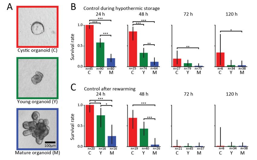
Biomolecules 2019, 9, 110                                                                            2 of 9

understood, and successful cold storage of multicellular system, in particular organs, is still out
of reach.
     So far, the mechanism(s) by which AF(G)Ps exert a protective action of AF(G)Ps is studied in vivo,
mostly in single cells using 2D cell culture [9]. Studies on multicellular systems are typically performed
on whole embryos or organs [12,14], whose large sizes pose severe challenges in terms of the amount
of AFPs required and imaging the action of AFP on the cellular and molecular level.
     Organoids are self-organizing three-dimensional structures that are grown from stem cells in vitro,
which recapitulate the essential features of organ architecture and function [15]. Organoids bridge
the gap between traditional 2D cell cultures and organs, since they are stable cultures that allow for
differentiation, cell–cell and cell–matrix interactions, and 3D organization; yet they remain affordable
and easily accessible for manipulation and experimentation. Since their introduction nearly a decade
ago, organoids from several different tissues have been developed, starting a revolution in both the
biological and medical fields. Organoids provide previously inaccessible insights into morphogenesis,
stem cell biology, and disease, and hold great promise for the future of personalized medicine [16,17].
     Here, we study the survival of intestinal organoids under hypothermic conditions, and how this
survival is affected by the addition of AFGPs. We observe that AFGPs have a clear life-prolonging
effect on organoids in all different developmental stages.

2. Materials and Methods

2.1. AFGP Purification
     Antifreeze glycoproteins were purified from the Antarctic toothfish Dissostichus mawsoni and
fluorescently labeled using fluorescein isothiocyanate (FITC) as described previously [18].

2.2. Organoid Culture
      Intestinal organoids isolated from C57BL/6 mice were a gift from Norman Sachs (Hubrecht
Institute, The Netherlands). Organoids were cultured according to previously described protocols with
minor adjustments [19]. In short, organoids were embedded in basement membrane extract (BME,
Trevingen, Gaithersburg, USA) droplets and overlaid with IntestiCult Organoid Growth Medium
(STEMCELL Technologies, Vancouver, Canada), which was changed every two to three days. Organoid
passaging was performed by mechanically dissociating crypts using a narrowed glass pipette.

2.3. Cold Storage Experiements
      Cold storage experiments were performed with organoids that were embedded in a mixture of
two-thirds BME and one-third 0.5% agarose (VWR Chemicals, Radnor, USA). We added agarose
to prevent BME droplets from disintegrating, as BME becomes liquid at temperatures ~4 ◦ C.
The BME/agarose mixture supported organoid growth, and normal development was observed
for up to three passages. The cold storage experiments were started two to three days after plating
the organoids in the BME/agarose mixture. The plates were sealed with Parafilm and placed in an
air-tight bag containing 5% CO2 and moved to 4 ◦ C. Organoids were stored in cold conditions for
different time periods, after which the plates were returned to an incubator set at 37 ◦ C. The color of
the medium was assessed throughout the procedure to ensure normal pH values and that a lack of
CO2 was not affecting the organoid viability.
      In the survival experiments, AFGP and α-lactalbumin were added to the medium one hour
before the organoids were transferred to a 4 ◦ C fridge. The concentration of the added proteins in
the medium was 10 mg/mL [9]. The experimental design is outlined in Supplementary Figure S1.
For each condition, individual plates with organoids were prepared, and each plate was left at 4 ◦ C for
different periods of time ranging from 24 to 120 h. During hypothermic storage, individual organoids
were followed and visually assessed every 24 h to determine their viability. At each time point, an
organoid was classified as alive or dead. Dead organoids were easily recognizable by an overall lack
Biomolecules 2019, 9, 110 3 of 9
of structure and the presence of dark debris where the organoid was previously located, as shown in
Supplementary Figure S2. Data were collected from the number of independent experiments presented
in Supplementary Figure S1. After cold storage, the organoids were returned to the 37 ◦ C incubator
and their growth and viability were assessed again after 2–3 days. Living organoids grew considerably,
while dead debris remained the same. Hence, the visual distinction between alive and dead organoids
was straightforward.
2.4. Fluorescein Diacetate Test
Viability tests using fluorescein diacetate (FDA) were performed to test the accuracy of the visual
viability assessment [20].
FDA was added to the medium at a concentration of 10 µg/mL, and the medium was removed
after 5 min and replaced with phosphate-buffered saline. After 15 min, the wells were washed twice,
and the organoids were imaged using a widefield fluorescence microscope (Axio Vert.A1, Zeiss). FDA
tests were not used to completely quantify organoid viability, due to an observed inability of the FDA
to completely penetrate BME droplets. The dye showed false negatives when inspecting organoids that
were deeply embedded in the BME/agarose gel, either because the FDA was unable to fully penetrate
the gel, or because it was taken up by the organoids in the periphery of the gel more rapidly than
it could reach the more deeply embedded ones. As a result, fluorescence could only be observed in
healthy organoids close to the edges of the droplets, while live organoids located in central regions did
not show any fluorescence that could be distinguished from the background. The visual assessment of
organoids in the periphery of the gel coincided with the FDA staining, which supports the validity of
our visual protocol.
2.5. Imaging of FITC-AFGP
Organoids were mechanically dissociated, embedded in BME, and plated in 8-well imaging
chambers. Two to three days after passaging, 0.5 mg/mL of FITC-AFGP was added to the medium
and, after one hour, the plates were imaged using a scanning confocal microscope (Eclipse Ti; Nikon).
2.6. Statistical Analysis
We performed a statistical analysis of the data using the two-sided Fisher’s exact test, p < 0.05
was considered statistically significant. In all plots (Figure 1B,C, Figure 2), the error bars represent
95% Clopper–Pearson confidence intervals. In cases where we performed multiple independent
experiments, the data were pooled and plotted together.
3. Results
We investigated the survival rate of intestinal organoids under hypothermic conditions at time
periods spanning from 24 to 120 h. The organoids were categorized into cystic, young, or mature, which
correspond to different developmental stages. Cystic organoids are spherical structures consisting of
stretched-out undifferentiated cells forming a large lumen with thin walls (left panel of Figure 1a).
Young organoids are no longer spherical, have a small lumen, thick walls, and small buds without
pronounced crypts, as shown in the middle panel of Figure 1a. Mature organoids possess clearly
visible crypt structures as shown in the right panel of Figure 1a. Every 24 h, we took a selection of
organoids for which we performed a visual assessment of their viability. Subsequently, the assessed
organoids were returned in the incubator and evaluated on how they recovered from the hypothermic
exposure. We observed considerable growth of live organoids after they were being rewarmed, and we
used this property to distinguish between the live and dead organoids. In fact, we also observed that
healthy-looking crypts were able to bud from such organoids, suggesting that stem cells are present,
which would allow for organoid passaging.Biomolecules 2019, 9, 110 4 of 9
Biomolecules 2019, 9, 110 4 of 9
Figure 1. Survival of organoids under hypothermic conditions with no added proteins. (a) Classification
Figure 1. Survival of organoids under hypothermic conditions with no added proteins. (a)
of organoids according to their developmental stage. Cystic organoids (left panel, red) are spherical
Classification of organoids according to their developmental stage. Cystic organoids (left panel, red)
structures with a large lumen and thin walls. Young organoids are no longer spherical, have a small
are spherical structures with a large lumen and thin walls. Young organoids are no longer spherical,
lumen, thick walls, and small buds rather than crypts (middle panel, green). Mature organoids
have a small lumen, thick walls, and small buds rather than crypts (middle panel, green). Mature
clearly have grown crypt structures (right panel, blue). (b) Survival rates of organoids at different
organoids clearly have grown crypt structures (right panel, blue). (b) Survival rates of organoids at
developmental stages obtained from visually assessing single cells every 24 h during hypothermic
different developmental stages obtained from visually assessing single cells every 24 h during
storage periods ranging from 24 to 120 h. Bars show results pooled from different independent
hypothermic storage periods ranging from 24 to 120 h. Bars show results pooled from different
experiments. (c) Corresponding survival rates obtained from a visual assessment performed two to
independent experiments. (c) Corresponding survival rates obtained from a visual assessment
three days after the organoids were returned to the incubator. For each organoid class and experimental
performed two to three days after the organoids were returned to the incubator. For each organoid
condition, the number (n) of organoids examined is indicated. Asterisks denote significant differences
class and experimental condition, the number (n) of organoids examined is indicated. Asterisks
(* p < 0.05, ** p < 0.01, *** p < 0.001).
denote significant differences (* p < 0.05, ** p < 0.01, *** p < 0.001).
The visual cell viability assessment was supported by fluorescein diacetate fluorescence within
The visualFDA
the cytoplasm. cell serves
viability
as assessment
a viability probewas supported
that measures by fluorescein diacetate
enzymatic activity, fluorescence
which is obligatorywithinto
the cytoplasm. FDA serves as a viability probe that measures enzymatic activity,
activate its fluorescence, and cell membrane integrity, which is necessary for intracellular retention which is obligatory
to the
of activate its fluorescence,
fluorescent product.and FDA cellwas
membrane
chosen integrity,
due to itswhich is necessary
low toxicity, which for allows
intracellular
for anretention
in situ
viability assessment throughout the timespan of the experiments. The use of live/deadfor
of the fluorescent product. FDA was chosen due to its low toxicity, which allows an in
stains situ
is not
viability assessment throughout the timespan of the experiments. The use
straightforward for organoids as the lumen of healthy organoids usually contains dead cell material of live/dead stains is not
straightforward
because old cells for
areorganoids
shed into theas the lumen
lumen, of healthythe
mimicking organoids usually
cell shedding contains
that occursdeadat thecell material
tips of the
because old cells are shed into the lumen, mimicking the cell shedding that occurs
villi in the intestine. As a result, stains that target dead cells are not appropriate as they would show at the tips of the
villi in
false the intestine. As a result, stains that target dead cells are not appropriate as they would show
positives.
falseInpositives.
Figure 1B, we present the results of the survival of the hypothermic organoids without added
In Figure
proteins. We find1B,that
weafter
present
24 hthe results of to
of exposure the4 survival
◦ C, 100%of ofthe
thehypothermic
cystic and 58% organoids
of the young without added
organoids
proteins. while
survived, We find onlythat
20%after 24 mature
of the h of exposure to 4 °C,After
ones survived. 100%48ofh the cystic andwe
of exposure, 58%
findofthat
the84%young of
organoids survived, while only 20% of the mature ones survived. After 48 h
cystic organoids were still alive, and the rate dropped to 32% and 11% for young and mature organoids,of exposure, we find that
84% of cysticAt
respectively. organoids
72 and 120were stillsurvival
h, the alive, and thedecreased
rates rate dropped to 32% and
dramatically, 11%a for
with young and
maximum mature
of 33% for
organoids, respectively. At 72 and 120 h, the survival rates decreased
cystic and 8% for young and mature organoids. Low variability between independent experimentsdramatically, with a maximum
of 33%
was for cystic
observed and 8% for young
(Supplementary and mature organoids. Low variability between independent
Figure S3).
experiments was observed (Supplementary
In Figure 1C, we show the results of organoid Figuresurvival
S3). after we returned them to the incubator at
37 ◦ C.InInFigure 1C, we
agreement show
with thethe results
visual of organoid
inspection survival
results shownafter we returned
in Figure 1B, we them to the
find that mostincubator
cystic
organoids survived hypothermic exposures of up to 48 h, and could readily be brought backcystic
at 37 °C. In agreement with the visual inspection results shown in Figure 1B, we find that most into
culturing. By contrast, a significant portion of the larger organoids did not survive rewarming and into
organoids survived hypothermic exposures of up to 48 h, and could readily be brought back the
culturing. By contrast, a significant portion of the larger organoids did not survive rewarming and
the return to the incubator. We further observe that no organoids survived rewarming after being
exposed to hypothermic storage periods of 72 h or longer.Biomolecules 2019, 9, 110 5 of 9
return to the incubator. We further observe that no organoids survived rewarming after being exposed
Biomolecules 2019, 9, 110 5 of 9
to hypothermic storage periods of 72 h or longer.
3.1. Antifreeze Glycoproteins
Antifreeze Glycoproteins
In order to evaluate whether the addition of AFGP influences organoid survival under
In order to evaluate whether the addition of AFGP influences organoid survival under
hypothermic conditions, we repeated the previous experiments after adding AFGP to the growth
hypothermic conditions, we repeated the previous experiments after adding AFGP to the growth
medium. For cold-storage experiments, an AFGP concentration of 10 mg/mL was chosen, as this
medium. For cold-storage experiments, an AFGP concentration of 10 mg/mL was chosen, as this
concentration showed protective effects in a previous study on cells [9]. We also performed control
concentration showed protective effects in a previous study on cells [9]. We also performed control
experiments in which we added the non-antifreeze protein α-lactalbumin to the growth medium. We
experiments in which we added the non-antifreeze protein α-lactalbumin to the growth medium.
chose α-lactalbumin as the control protein as this protein has a similar size, shows no antifreeze
We chose α-lactalbumin as the control protein as this protein has a similar size, shows no antifreeze
activity, and albumins have been used as control proteins in previous AF(G)P studies [9].
activity, and albumins have been used as control proteins in previous AF(G)P studies [9].
In Figure 2, we present the survival rates of organoids at different developmental stages in the
In Figure 2, we present the survival rates of organoids at different developmental stages in the
presence of AFGP and α-lactalbumin. After 24 and 48 h, we find that 100% of the organoids appeared
presence of AFGP and α-lactalbumin. After 24 and 48 h, we find that 100% of the organoids appeared
alive in the presence of AFGPs (Figure 2a). At 72 and 120 h, the survival rates remained above 80%
alive in the presence of AFGPs (Figure 2a). At 72 and 120 h, the survival rates remained above 80% for
for cystic and above 90% for young and mature organoids. We thus find that the addition of AFGP
cystic and above 90% for young and mature organoids. We thus find that the addition of AFGP to the
to the medium has a beneficial effect on organoid survival at all developmental stages. We also
medium has a beneficial effect on organoid survival at all developmental stages. We also observed low
observed low variability between independent experiments (Supplementary Figure S3).
variability between independent experiments (Supplementary Figure S3).
Figure 2. Survival of organoids under hypothermic conditions with antifreeze glycoprotein (AFGP)
Figure 2. Survival of
and α-lactalbumin organoids
present. under hypothermic
(a) Survival conditions
rates of organoids at the with antifreeze
different glycoprotein
developmental (AFGP)
stages when
and α-lactalbumin present. (a) Survival rates of organoids at the different developmental
AFGP was added to the growth medium. Rates were obtained from visually assessing the organoids stages when
AFGP was
every 24 added hypothermic
h during to the growthstorage
medium. Ratesranging
periods were obtained
from 24 to from
120 visually assessing
h. Bars show resultsthe organoids
pooled from
every 24 independent
different h during hypothermic
experiments.storage periods ranging
(b) Corresponding from rates
survival 24 toobtained
120 h. Bars
fromshow results
a visual pooled
assessment
from different
performed twoindependent
to three daysexperiments. (b) Corresponding
after the organoids were returnedsurvival rates obtained
to the incubator. from arates
(c) Survival visual
of
assessment performed two to three days after the organoids were returned to
organoids at the different developmental stages when α-lactalbumin was added to the growth medium. the incubator. (c)
Survival rates of
Black asterisks organoids
denote at the differences
significant different developmental stages when
between developmental α-lactalbumin
stages, was added
and red asterisks to
below
the
barsgrowth medium. Black
denote differences from asterisks
controls indenote
Figuresignificant
1 (* p < 0.05,differences
** p < 0.01,between developmental
*** p < 0.001, stages,
ns: not significant).
and red asterisks below bars denote differences from controls in Figure 1 (* p < 0.05, ** p < 0.01, *** p <
Uponns:
0.001, return to the incubator (Figure 2B), 100% of organoids were alive after hypothermic storage
not significant).
of 24 h. The positive effect of AFGP on the viability of the organoids was also observed after 48 h
where 96% of
Upon organoids
return to thesurvived.
incubatorAfter 72 h,2B),
(Figure the survival
100% ofrates remained
organoids high
were for after
alive younghypothermic
and mature
organoids
storage (~93%),
of 24 h. The while
positivetheeffect
rate of
dropped
AFGP on to the
69%viability
for cystic organoids.
of the organoidsEven afterobserved
was also remaining in
after
hypothermic
48 h where 96% storage for 120 h,survived.
of organoids most organoids
After 72survive. However,
h, the survival none
rates of the organoids
remained that were
high for young and
hypothermically
mature organoids stored for while
(~93%), 120 h survived after being
the rate dropped towarmed
69% for up in the
cystic incubator.Even
organoids. In strong contrast to
after remaining
AFGP, α-lactalbumin
in hypothermic didfor
storage not120
provide
h, mostany protection
organoids againstHowever,
survive. hypothermicnonestress,
of theasorganoids
shown in Figure 2C.
that were
hypothermically stored for 120 h survived after being warmed up in the incubator. In strong contrast
to AFGP, α-lactalbumin did not provide any protection against hypothermic stress, as shown in
Figure 2C. We find that after 24 h of cold exposure 100% of cystic, 77% of young and 17% of mature
organoids survived. After 48 h, 83% of cystic and 56% of young organoids are still alive, but only 9%Biomolecules 2019, 9, 110 6 of 9
Biomolecules
We find that 2019, 9, 110
after 6 of 9
24 h of cold exposure 100% of cystic, 77% of young and 17% of mature organoids
survived. After 48 h, 83% of cystic and 56% of young organoids are still alive, but only 9% of mature
of mature organoids survived. The survival rates in the presence of α-lactalbumin are comparable to
organoids survived. The survival rates in the presence of α-lactalbumin are comparable to the survival
the survival rates observed when no proteins are added (Figure 1b).
rates observed when no proteins are added (Figure 1b).
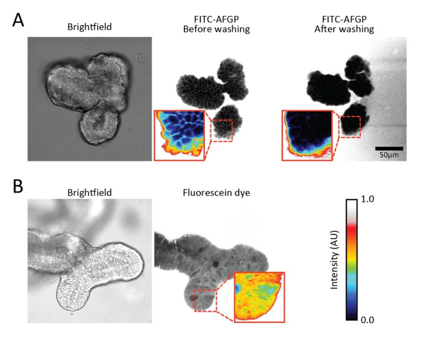
Figure 3 shows fluorescence images of organoids in the presence of FITC-AFGP. We find that
Figure 3 shows fluorescence images of organoids in the presence of FITC-AFGP. We find that
the labeled AFGPs are predominately localized on the cell surface. This finding suggests that AFGPs
the labeled AFGPs are predominately localized on the cell surface. This finding suggests that AFGPs
preferably localize at cell membranes. Control experiments using solely fluorescein dye showed no
preferably localize at cell membranes. Control experiments using solely fluorescein dye showed no
fluorescent pattern (Figure 3b), indicating that AFGP (and not the fluorescent label) was responsible
fluorescent pattern (Figure 3b), indicating that AFGP (and not the fluorescent label) was responsible
for the membrane localization. We observe that washing the organoids with PBS buffer resulted in
for the membrane localization. We observe that washing the organoids with PBS buffer resulted in
disappearance of the observed fluorescent pattern, as shown in Figure 3a. This observation suggests
disappearance of the observed fluorescent pattern, as shown in Figure 3a. This observation suggests
that AFGPs were only loosely bound to the membrane and were not inserted into the membrane.
that AFGPs were only loosely bound to the membrane and were not inserted into the membrane.
Figure 3. Fluorescence images of organoids in the presence of FITC-labeled AFGP. (a) Brightfield and
Figure 3. Fluorescence
fluorescence images of animages of organoids
organoid in the presence
with FITC-AFGP of FITC-labeled
in the medium reveal thatAFGP. (a) Brightfield
fluorescence and
is observed
on the cell outlines. Washing of the imaging wells with buffer eliminated the fluorescence pattern;is
fluorescence images of an organoid with FITC-AFGP in the medium reveal that fluorescence
observed
(b) on the
Brightfield andcell outlines. Washing
fluorescence images ofofanthe imagingwith
organoid wells with buffer
fluorescein eliminated
diacetate in thethe fluorescence
medium show
pattern;
that (b) Brightfield
fluorescence is presentandinfluorescence
the cytoplasm images of an
of most organoid
cells. with fluorescein
Pixel intensities diacetate
are colored in the
in insets to
highlight
medium theshowobserved fluorescence
that fluorescence patterns.
is present inColors represent
the cytoplasm ofthe same
most intensities
cells. in all images.
Pixel intensities are colored
in insets to highlight the observed fluorescence patterns. Colors represent the same intensities in all
4. Discussion
images.
The hypothermic storage of cells causes cold stress that affects the cell physiology, metabolic
4. Discussion
activity, and regulation of ion equilibration across membranes [1]. We observed a statistically significant
dependence of organoid storage
The hypothermic survivalof
after hypothermic
cells causes coldstorage
stress on theaffects
that developmental stage of the organoid.
the cell physiology, metabolic
More developed mature organoids showed much lower viability
activity, and regulation of ion equilibration across membranes [1]. We observed rates compared to cystic organoids.
a statistically
Hypothermic storage likely
significant dependence activates different
of organoid survival sets
afterofhypothermic
stress pathways thaton
storage arethe
cell- and tissue-specific.
developmental stage
Cystic
of the organoid. More developed mature organoids showed much lower viability organoids.
organoids are composed of cells that strongly differ from the cells of mature rates comparedCystic
to
organoids, sometimes
cystic organoids. referred to storage
Hypothermic as “enterospheres”, consist
likely activates of undifferentiated
different sets of stress cells with stem
pathways thatcell-like
are cell-
properties. They differ
and tissue-specific. fromorganoids
Cystic mature intestinal stem cells
are composed of in having
cells a distinctdiffer
that strongly set of from
gene expression
the cells of
patterns as well as different sets of active signaling pathways [21–23].
mature organoids. Cystic organoids, sometimes referred to as “enterospheres”, consist These characteristics enableof
cystic organoids to
undifferentiated recapitulate
cells with stemthe essential
cell-like features They
properties. of intestinal tissue
differ from underintestinal
mature a state of stem
repaircells
after
in
injury [21,22]. By contrast, mature organoids have only a small percentage of stem
having a distinct set of gene expression patterns as well as different sets of active signaling pathways and progenitor
cells, andThese
[21–23]. are mostly composed
characteristics of fully
enable differentiated
cystic organoids to secretory and absorptive
recapitulate the essentialcells which
features ofresemble
intestinal
the
tissue under a state of repair after injury [21,22]. By contrast, mature organoids have only acystic
intestinal tissue in a homeostatic state. The distinct cell identities and functions in both small
percentage of stem and progenitor cells, and are mostly composed of fully differentiated secretory
and absorptive cells which resemble the intestinal tissue in a homeostatic state. The distinct cell
identities and functions in both cystic and mature organoids likely result in differences in their abilityBiomolecules 2019, 9, 110 7 of 9
and mature organoids likely result in differences in their ability to respond to stress during and
after hypothermic storage. Young organoids are likely to contain subsets of both cystic and mature
properties, thus explaining their intermediate survival rates.
The characteristic shape and behavior of cystic organoids (a round and enlarged lumen, thin
walls formed by stretched-out cells, and occasional rapid contractions observed after expulsion of
material from the lumen (data not shown) suggest that they are under high intraluminal pressure
due to accumulation of fluid within the lumen. This cystic morphology has been documented when
organoids swell as a result of adding forskolin [24] or cholera toxin [25] to the medium. Furthermore,
addition of the signaling factor Wnt3a resulted in organoids adopting a cystic morphology as well as
their cells assuming an undifferentiated state [26]. In all cases, the cystic organoid morphology has
been linked to increased chloride secretion by the cystic fibrosis transmembrane conductance regulator
(CFTR) ion channel which, in turn, causes increased fluid secretion into the lumen. This suggests
that ion transportation in cystic organoids is highly dynamic compared to more mature organoids.
We speculate that as a result, ion leakage caused by hypothermic storage has a smaller impact on cystic
organoids in comparison to young and mature organoids, accounting for their higher survival rate.
We find that the addition of AFGPs to the medium has a statistically significant positive effect
on the cold survival of organoids, which is in line with previous studies [10]. Current mechanisms
that explain the positive effect of AFPs on the cold survival of cells involve the blockage or alteration
of the flow of ions into cells [27,28] and the protection of cell membranes as they pass through their
phase transition temperatures [29,30]. Tomczak et al. proposed that AFGPs may form a monolayer
covering the membrane surface, thereby reducing the leakage of ions across the membrane as it
is cooled through its thermal transition temperature [30]. Using fluorescence microscopy, we find
strong evidence that AFGPs localize at the cell membranes, suggesting that the protection mechanism
of AFGPs is indeed closely connected to their interaction with cell membranes. From the obtained
fluorescence data, we cannot infer whether AFGPs target specific ion channels. We observe that AFGPs
loosely interact with the cell membranes of the organoids and find that AFGPs do not only cover
model membranes but entire multicell systems.
Interestingly, we further observe that upon rewarming, the survival rate remains high (93%) for
young and mature organoids while it drops to 69% for cystic organoids in the presence of AFGPs.
Garner et al. showed that AFGPs interact with a model membrane at both 5 and 30 ◦ C, but that the
interaction at 30 ◦ C is much weaker [31]. We speculate that the distinct cell identities in cystic and
more mature organoids do not only affect their ability to respond to stress during hypothermic storage
but also to stress experienced during rewarming. More mature organoids could, for instance, have
a different membrane composition with more phosphate groups. Such groups could enable a better
interaction with AFGPs and a prolonged protection. Clearly, the response to rewarming stress must
also be considered when investigating potential AFGP applications for the storage of cells and tissues
in the cold for medical purposes.
5. Conclusions
In conclusion, organoids provide a flexible and previously inaccessible method of studying
the effects of cryopreservation on multicellular systems, which could bring us a step closer to
the cryopreservation of entire organs. We find clear evidence that AFGPs have a strong positive
effect on the survival of organoids during and after hypothermic storage at 4 ◦ C. If this protection
against hypothermia-related perturbations could be extended for even longer periods and to complete
intestinal organs, then it would have enormous practical implications for the transfer and storage
of organs.
Supplementary Materials: The following are available online at http://www.mdpi.com/2218-273X/9/3/110/s1,
Figure S1: Design of the Hypothermic storage experiments. Figure S2: Optical images of live and dead organoids.
Figure S3: Average survival rates from independent organoid experiments.Biomolecules 2019, 9, 110 8 of 9
Author Contributions: K.M. and G.H.-P. designed the experimental strategy and analyzed the data. G.H.-P.
performed the experiments. J.S.v.Z. and H.J.B. assisted in designing the experiments. A.L.D. provided novel
reagents and labeled the Antifreeze Glycoproteins. K.M., G.H.-P., J.S.v.Z., H.J.B. and A.L.D. all wrote the
manuscript together.
Funding: This research received no external funding.
Conflicts of Interest: The authors declare no conflict of interest.
References
1. Rubinsky, B. Principles of Low Temperature Cell Preservation. Heart Failure Rev. 2003, 8, 277–284. [CrossRef]
2. Mathew, A.J.; Baust, J.M.; Van Buskirk, R.G.; Baust, J.G. Cell preservation in reparative and regenerative
medicine: Evolution of individualized solution composition. Tissue Eng. 2004, 10, 1662–1671. [CrossRef]
[PubMed]
3. DeVries, A.L.; Wohlschlag, D.E. Freezing resistance in some Antarctic fishes. Science 1969, 163, 1073–1075.
[CrossRef] [PubMed]
4. Duman, J.G. Antifreeze and Ice Nucleator Proteins in Terrestrial Arthropods. Annu. Rev. Physiol. 2001, 63,
327–357. [CrossRef] [PubMed]
5. Olijve, L.L.C.; Meister, K.; DeVries, A.L.; Duman, J.G.; Guo, S.; Bakker, H.J.; Voets, I.K. Blocking rapid ice
crystal growth through nonbasal plane adsorption of antifreeze proteins. Proc. Natl. Acad. Sci. USA 2016,
113, 3740–3745. [CrossRef]
6. Raymond, J.A.; DeVries, A.L. Adsorption inhibition as a mechanism of freezing resistance in polar fishes.
Proc. Natl. Acad. Sci. USA 1977, 74, 2589–2593. [CrossRef] [PubMed]
7. Amir, G.; Horowitz, L.; Rubinsky, B.; Yousif, B.S.; Lavee, J.; Smolinsky, A.K. Subzero nonfreezing
cryopresevation of rat hearts using antifreeze protein I and antifreeze protein III. Cryobiology 2004, 48,
273–282. [CrossRef]
8. Lee, H.H.; Lee, H.J.; Kim, H.J.; Lee, J.H.; Ko, Y.; Kim, S.M.; Lee, J.R.; Suh, C.S.; Kim, S.H. Effects of antifreeze
proteins on the vitrification of mouse oocytes: Comparison of three different antifreeze proteins. Hum. Reprod.
2015, 30, 2110–2119. [CrossRef]
9. Kamijima, T.; Sakashita, M.; Miura, A.; Nishimiya, Y.; Tsuda, S. Antifreeze protein prolongs the life-time of
insulinoma cells during hypothermic preservation. PLoS ONE 2013, 8, e73643. [CrossRef]
10. Tablin, F.; Oliver, A.E.; Walker, N.J.; Crowe, L.M.; Crowe, J.H. Membrane phase transition of intact human
platelets: Correlation with cold-induced activation. J. Cell. Physiol. 1996, 168, 305–313. [CrossRef]
11. Rubinsky, B.; Arav, A.; Fletcher, G.L. Hypothermic protection–a fundamental property of “antifreeze”
proteins. Biochem. Biophys. Res. Commun. 1991, 180, 566–571. [CrossRef]
12. Rubinsky, B.; Arav, A.; Hong, J.S.; Lee, C.Y. Freezing of mammalian livers with glycerol and antifreeze
proteins. Biochem. Biophys. Res. Commun. 1994, 200, 732–741. [CrossRef] [PubMed]
13. Wang, T.; Zhu, Q.; Yang, X.; Layne, J.R., Jr.; Devries, A.L. Antifreeze glycoproteins from antarctic notothenioid
fishes fail to protect the rat cardiac explant during hypothermic and freezing preservation. Cryobiology 1994,
31, 185–192. [CrossRef] [PubMed]
14. Martinez-Paramo, S.; Barbosa, V.; Perez-Cerezales, S.; Robles, V.; Herraez, M.P. Cryoprotective effects of
antifreeze proteins delivered into zebrafish embryos. Cryobiology 2009, 58, 128–133. [CrossRef] [PubMed]
15. de Souza, N. Organoids. Nat. Methods 2018, 15, 23. [CrossRef]
16. Fatehullah, A.; Tan, S.H.; Barker, N. Organoids as an in vitro model of human development and disease.
Nat. Cell Biol. 2016, 18, 246. [CrossRef] [PubMed]
17. Drost, J.; Clevers, H. Organoids in cancer research. Nat. Rev. Cancer 2018, 18, 407–418. [CrossRef]
18. Evans, C.W.; Gubala, V.; Nooney, R.; Williams, D.E.; Brimble, M.A.; Devries, A.L. How do Antarctic
notothenioid fishes cope with internal ice? A novel function for antifreeze glycoproteins. Antarct. Sci. 2011,
23, 57–64. [CrossRef]
19. Mahe, M.M.; Aihara, E.; Schumacher, M.A.; Zavros, Y.; Montrose, M.H.; Helmrath, M.A.; Sato, T.; Shroyer, N.F.
Establishment of Gastrointestinal Epithelial Organoids. Curr. Protocols Mouse Biol. 2013, 3, 217–240.
[CrossRef] [PubMed]
20. Widholm, J.M. The use of fluorescein diacetate and phenosafranine for determining viability of cultured
plant cells. Stain Technol. 1972, 47, 189–194. [CrossRef] [PubMed]Biomolecules 2019, 9, 110 9 of 9
21. Yui, S.; Azzolin, L.; Maimets, M.; Pedersen, M.T.; Fordham, R.P.; Hansen, S.L.; Larsen, H.L.; Guiu, J.;
Alves, M.R.P.; Rundsten, C.F.; et al. YAP/TAZ-Dependent Reprogramming of Colonic Epithelium Links
ECM Remodeling to Tissue Regeneration. Cell Stem Cell 2018, 22, 35–49.e37. [CrossRef] [PubMed]
22. Gjorevski, N.; Sachs, N.; Manfrin, A.; Giger, S.; Bragina, M.E.; Ordonez-Moran, P.; Clevers, H.; Lutolf, M.P.
Designer matrices for intestinal stem cell and organoid culture. Nature 2016, 539, 560–564. [CrossRef]
[PubMed]
23. Smith, N.R.; Swain, J.R.; Davies, P.S.; Gallagher, A.C.; Parappilly, M.S.; Beach, C.Z.; Streeter, P.R.;
Williamson, I.A.; Magness, S.T.; Wong, M.H. Monoclonal Antibodies Reveal Dynamic Plasticity Between
Lgr5- and Bmi1-Expressing Intestinal Cell Populations. Cell. Mol. Gastroenterol. Hepatol. 2018, 6, 79–96.
[CrossRef] [PubMed]
24. Dekkers, J.F.; Wiegerinck, C.L.; de Jonge, H.R.; Bronsveld, I.; Janssens, H.M.; de Winter-de Groot, K.M.;
Brandsma, A.M.; de Jong, N.W.M.; Bijvelds, M.J.C.; Scholte, B.J.; et al. A functional CFTR assay using
primary cystic fibrosis intestinal organoids. Nat. Med. 2013, 19, 939. [CrossRef] [PubMed]
25. Zomer-van Ommen, D.D.; Pukin, A.V.; Fu, O.; Quarles van Ufford, L.H.C.; Janssens, H.M.; Beekman, J.M.;
Pieters, R.J. Functional Characterization of Cholera Toxin Inhibitors Using Human Intestinal Organoids.
J. Med. Chem. 2016, 59, 6968–6972. [CrossRef] [PubMed]
26. Strubberg, A.M.; Liu, J.; Walker, N.M.; Stefanski, C.D.; MacLeod, R.J.; Magness, S.T.; Clarke, L.L. Cftr
Modulates Wnt/beta-Catenin Signaling and Stem Cell Proliferation in Murine Intestine. Cell. Mol.
Gastroenterol. Hepatol. 2018, 5, 253–271. [CrossRef]
27. Rubinsky, B.; Mattioli, M.; Arav, A.; Barboni, B.; Fletcher, G.L. Inhibition of Ca2+ and K+ currents by
antifreeze proteins. Am. J. Physiol. 1992, 262, R542–R545. [CrossRef]
28. Rubinsky, B.; Arav, A.; Mattioli, M.; Devries, A.L. The effect of antifreeze glycopeptides on membrane
potential changes at hypothermic temperatures. Biochem. Biophys. Res. Commun. 1990, 173, 1369–1374.
[CrossRef]
29. Hays, L.M.; Feeney, R.E.; Crowe, L.M.; Crowe, J.H.; Oliver, A.E. Antifreeze glycoproteins inhibit leakage
from liposomes during thermotropic phase transitions. Proc. Natl. Acad. Sci. USA 1996, 93, 6835–6840.
[CrossRef]
30. Tomczak, M.M.; Hincha, D.K.; Estrada, S.D.; Wolkers, W.F.; Crowe, L.M.; Feeney, R.E.; Tablin, F.; Crowe, J.H.
A mechanism for stabilization of membranes at low temperatures by an antifreeze protein. Biophys. J. 2002,
82, 874–881. [CrossRef]
31. Garner, J.; Inglis, S.R.; Hook, J.; Separovic, F.; Harding, M.M. A solid-state NMR study of the interaction
of fish antifreeze proteins with phospholipid membranes. Eur. Biophys. J. 2008, 37, 1031–1038. [CrossRef]
[PubMed]
© 2019 by the authors. Licensee MDPI, Basel, Switzerland. This article is an open access
article distributed under the terms and conditions of the Creative Commons Attribution
(CC BY) license (http://creativecommons.org/licenses/by/4.0/).You can also read