Molecular Dynamics Simulation Study of the Interaction between Human Angiotensin Converting Enzyme 2 and Spike Protein Receptor Binding Domain of ...
←
→
Page content transcription
If your browser does not render page correctly, please read the page content below
biomolecules

Article
Molecular Dynamics Simulation Study of the Interaction between
Human Angiotensin Converting Enzyme 2 and Spike Protein
Receptor Binding Domain of the SARS-CoV-2 B.1.617 Variant
Priya Antony and Ranjit Vijayan *

                                          Department of Biology, College of Science, United Arab Emirates University,
                                          Al Ain P.O. Box 15551, United Arab Emirates; 201990021@uaeu.ac.ae
                                          * Correspondence: ranjit.v@uaeu.ac.ae

                                          Abstract: The COVID-19 pandemic, caused by the SARS-CoV-2 virus, has had a significant impact
                                          on people’s daily lives. The rapidly spreading B.1.617 lineage harbors two key mutations—L452R
                                          and E484Q—in the receptor binding domain (RBD) of its spike (S) protein. To understand the
                                          impact and structural dynamics of the variations in the interface of S protein and its host factor, the
                                          human angiotensin-converting enzyme 2 (hACE2), triplicate 500 ns molecular dynamics simulations
                                          were performed using single (E484Q or L452R) and double (E484Q + L452R) mutant structures
                                          and compared to wild type simulations. Our results indicate that the E484Q mutation disrupts the
                                          conserved salt bridge formed between Lys31 of hACE2 and Glu484 of S protein. Additionally, E484Q,

                                   which could favor the up conformation of the RBD, may help in enhanced hACE2 binding and
                                          immune escape. L452R introduces a charged patch near the binding surface that permits increased
Citation: Antony, P.; Vijayan, R.
                                          electrostatic attraction between the proteins. An improved network of intramolecular interactions
Molecular Dynamics Simulation
Study of the Interaction between
                                          observed is likely to increase the stability of the S protein and conformational changes may prevent
Human Angiotensin Converting              the binding of neutralizing antibodies. The results obtained from the molecular dynamics simulations
Enzyme 2 and Spike Protein Receptor       suggest that structural and dynamic changes introduced by these variations enhance the affinity of
Binding Domain of the SARS-CoV-2          the viral S protein to hACE2 and could form the basis for further studies.
B.1.617 Variant. Biomolecules 2021, 11,
1244. https://doi.org/10.3390/            Keywords: SARS-CoV-2; spike protein; B.1.617 variant; delta variant; E484Q; L452R; molecular dynamics
biom11081244

Academic Editors: Vladimir N.
Uversky and Daniele Di Marino             1. Introduction
                                               The coronavirus disease 2019 (COVID-19) pandemic, caused by the severe acute respi-
Received: 18 July 2021
                                          ratory syndrome coronavirus 2 (SARS-CoV-2), has had a catastrophic impact worldwide.
Accepted: 17 August 2021
Published: 20 August 2021
                                          Since its emergence in December 2019, the total number of confirmed cases has exceeded
                                          200 million and it has caused over 4 million deaths. The year 2020 was a challenging one,
Publisher’s Note: MDPI stays neutral
                                          and one of the highlights was the expedited development of several COVID-19 vaccines
with regard to jurisdictional claims in
                                          with impressive efficacy [1]. However, strategies to contain the pandemic have been af-
published maps and institutional affil-
                                          fected by the emergence of new viral variants possessing increased transmissibility and/or
iations.                                  capability to escape the immune system.
                                               SARS-CoV-2 is an RNA virus and its genome encodes 16 non-structural (nsp1–16) and
                                          four structural proteins, namely spike (S), nucleocapsid (N), membrane (M), and envelope
                                          (E). The S protein, a type 1 fusion protein, is primarily involved in the invasion of the
                                          host cell by SARS-CoV-2 [2]. Several studies have reported that the human angiotensin-
Copyright: © 2021 by the authors.
Licensee MDPI, Basel, Switzerland.
                                          converting enzyme (hACE2) serves as a high-affinity receptor for the receptor-binding
This article is an open access article
                                          domain (RBD) of the S protein (Figure 1A) [3].
distributed under the terms and
                                               Like other viruses, SARS-CoV-2 mutates and evolves as it replicates. A large number
conditions of the Creative Commons        of SARS-CoV-2 genomes have now been sequenced. This provides impressive insights
Attribution (CC BY) license (https://     into viral variants and their spread over time. These variations are routinely monitored by
creativecommons.org/licenses/by/          sequencing studies, epidemiological investigations and molecular studies. Even though
4.0/).                                    thousands of variants emerged in the early phase of the infection, most of these did not

Biomolecules 2021, 11, 1244. https://doi.org/10.3390/biom11081244                                      https://www.mdpi.com/journal/biomolecules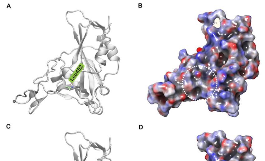
Biomolecules 2021, 11, 1244 2 of 10
have a significant impact on viral spread and infectivity [2]. However, in April 2020, the
original SARS-CoV-2 virus acquired the D614G mutation in the S protein [4]. The now
ubiquitous D614G variant exhibited higher transmissibility and infectivity with efficient
replication [5]. As the pandemic rages on, multiple genomic variants of SARS-CoV-2 have
emerged in different parts of the world including the United Kingdom (UK), South Africa,
Brazil, and India. The dramatic rise in COVID-19 cases in the UK has been attributed to a
variant named B.1.1.7 or 20I/501Y.V1. Sequencing analysis identified several mutations
in the spike region including D614G. Of these, N501Y appeared to be the major mutation
in the RBD of the S protein [6]. Epidemiological studies indicated that this variant has
up to 80% higher transmissibility and was associated with an increased risk of death
when compared to previously reported variants [7]. The South African variant B.1.351 or
20H/501Y is characterized by three mutations in the RBD region of the S protein—K417N,
E484K, and N501Y [8]. Both variants (B.1.351 and B.1.1.7) share the N501Y mutation
that aids in increased binding affinity and immune escape [9]. The variant P.1, originally
reported in Brazil, harbors N501Y, E484K, K417T mutations in the S protein. These variants
were classified as ‘variants of concern’ (VOCs) due to their higher transmissibility, immune
escape capability, lower response to vaccines and severity of the disease. To disassociate
variants and country names, a new naming system was recently devised by the World
Health Organization (WHO). Under this naming convention, the B.1.1.7 variant, originally
Biomolecules 2021, 11, x 2 of 11
identified in the UK, was called alpha, the B.1.351 variant was named beta, and the
P.1 variant as gamma [10].
Figure
Figure 1. Structures
1. Structures of of SARS-CoV-2spike
SARS-CoV-2 spike(S)
(S)protein
proteinreceptor-binding
receptor-binding domain
domain (RBD)
(RBD)bound
boundtotohuman
humanACE2 ACE2 (hACE2).
(hACE2).
(A) SARS-CoV-2 S protein RBD (grey) bound to hACE2
(A) SARS-CoV-2 S protein RBD (grey) bound to hACE2 (pink) based (pink) based on Protein Data Bank (PDB) structure with
Protein Data Bank (PDB) structure with PDB
PDBID:ID:
6M0J;
6M0J; (B)(B)
TheThe boxed
boxed region
region ininAAisisenlarged
enlargedshowing
showingthethewild
wild type
type residues
residues Leu452
Leu452and
andGlu484
Glu484ininstick
stickrepresentation.
representation.
(C)(C)
TheThe
boxed region
boxed regioninin
AA is isenlarged
enlargedshowing
showingthe the mutated residues Arg452
mutated residues Arg452andandGln484.
Gln484.
Since
Like early
other MarchSARS-CoV-2
viruses, 2021, the numbers
mutatesof reported
and evolves COVID-19 cases andAdeaths
as it replicates. have
large number
ofrisen quite sharply
SARS-CoV-2 in India.
genomes havePhylogenetic analysis revealed
now been sequenced. that the major
This provides viral variant
impressive insights
circulating in India belonged to the newly identified lineage B.1.617. This lineage includes
into viral variants and their spread over time. These variations are routinely monitored
three main subtypes (B1.617.1, B.1.617.2, and B.1.617.3) harboring several mutations in the
by sequencing studies, epidemiological investigations and molecular studies. Even
S protein, including the synonymous D111D variation and the nonsynonymous G142D,
though thousands of variants emerged in the early phase of the infection, most of these
L452R, E484Q, D614G and P681R variations. Of these, the combination of E484Q and L452R
did notparticular
is of have a significant
concern as impact
they areon viral spread
positioned in theand infectivity [2].motif
receptor-binding However,
(RBM) in April
of the
2020, the original
S protein (Figure SARS-CoV-2 virus crystal
1B,C). Even though acquired the D614G
structures mutation
of both RBDs ofinSARS-CoV-2
the S proteinand[4].
The now ubiquitous D614G variant exhibited higher transmissibility and
SARS-CoV share the same scaffold, sequence differences in the RBM region of SARS-CoV-2 infectivity with
efficient replication [5]. As the pandemic rages on, multiple genomic variants
promote greater electrostatic complementarity with hACE2. This in turn provides enhanced of SARS-
CoV-2 have
binding emerged
affinity in differenttoparts
of SARS-CoV-2 hACE2of the world including
compared to SARS-CoV the [11].
United TheKingdom
combination(UK),
South Africa,
of these Brazil, andalong
two mutations, India. Theother
with dramatic rise in
mutations, COVID-19
gives the viruscases in the
survival UK hasand
advantage been
the abilitytotoa spread
attributed variantrapidly.
namedThe B.1.617
B.1.1.7 lineage has received
or 20I/501Y.V1. particular
Sequencing analysisattention due to
identified sev-
eral mutations in the spike region including D614G. Of these, N501Y appeared to be the
major mutation in the RBD of the S protein [6]. Epidemiological studies indicated that this
variant has up to 80% higher transmissibility and was associated with an increased risk of
death when compared to previously reported variants [7]. The South African variant
B.1.351 or 20H/501Y is characterized by three mutations in the RBD region of the S pro-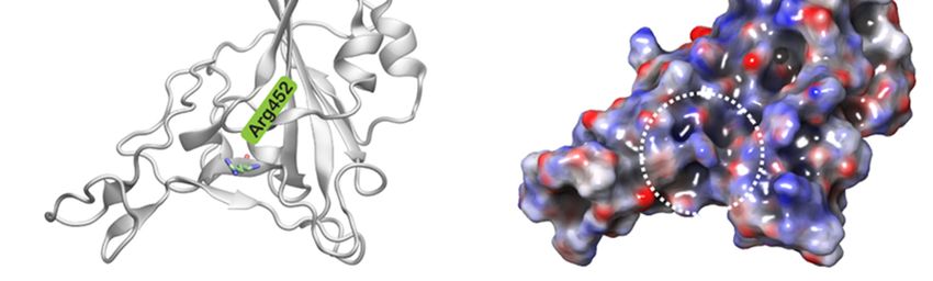
Biomolecules 2021, 11, 1244                                                                                        3 of 10

                              its increased infectivity, high virulence capacity, and potential immune escape. Currently,
                              while vaccination programs have been implemented in several nations to a limited extent,
                              this wave of COVID-19 infections is creating serious global health concerns.
                                    Gaining a deeper understanding of the interactions between the viral S protein and
                              hACE2 is crucial for developing drugs and vaccinations as well as for ascertaining the
                              efficacy of existing vaccinations against novel SARS-CoV-2 variants. Our previous study
                              evaluated the dynamic interactions of SARS-CoV-2/SARS-CoV RBD with hACE2 and
                              identified important interactions that assist the stable binding of SARS-CoV-2 when
                              compared to SARS-CoV [12]. Here, we extend this to investigate the structural stabil-
                              ity, binding affinity and intermolecular polar and hydrophobic contacts in the S pro-
                              tein RBD of the B.1.617 variant—E484Q and L452R independently and in combination
                              (E484Q + L452R)—using multiple 500 ns molecular dynamics (MD) simulations and bind-
                              ing free energy calculations.

                              2. Materials and Methods
                                   Coordinates of the three-dimensional X-ray crystal structures of the SARS-CoV-2
                              RBD bound to hACE2 were obtained from the Protein Data Bank (PDB; PDB ID: 6M0J).
                              Single and double mutant complexes were created by mutating the amino acids Glu484
                              to Gln484 and Leu452 to Arg452 using Schrödinger Maestro 2019-4 (Schrödinger, LLC,
                              New York, NY, USA). These mutant complexes were first pre-processed using the Protein
                              Preparation Wizard of Schrödinger (Schrödinger, LLC, New York, NY, USA). The protein
                              preparation process involved assigning the correct bond order, creating disulfide bonds,
                              adjusting ionization states, removing unwanted water molecules, metals and cofactors,
                              correcting the orientation of groups, capping the termini, adding missing atoms and
                              sidechains and assigning partial charges. Hydrogen atoms were incorporated, and a
                              standard protonation state at pH 7 was used. The structures of the single and double
                              mutant spike protein RBD bound to ACE2 were placed in orthorhombic boxes of size
                              125 Å × 125 Å × 125 Å and solvated with single point charge (SPC) water molecules using
                              the Desmond System Builder (Schrödinger, LLC, New York, NY, USA). All simulation
                              systems were neutralized with counter ions and a salt concentration of 0.15 M NaCl was
                              maintained. The simulations were performed for 500 ns using Desmond [13] in triplicate
                              with different set of initial velocities assigned to each atom. The systems were described
                              using the OPLS forcefield. Before the production run, all simulation systems were subjected
                              to Desmond’s default eight-stage relaxation protocol. The isotropic Martyna–Tobias–Klein
                              barostat and the Nose–Hoover thermostat were used to maintain the pressure at 1 atm
                              and temperature at 300 K, respectively [14,15]. Long-range coulombic interactions were
                              evaluated using the smooth particle mesh Ewald method and the short-range cutoff was set
                              as 9.0 Å [16]. A time-reversible reference system propagator algorithm (RESPA) integrator
                              was employed with an inner time step of 2.0 fs and an outer time step of 6.0 fs [17]. The
                              binding free energy of all three mutant complexes was evaluated using the molecular
                              mechanics generalized Born surface area (MM-GBSA) approach. Frames were extracted
                              every 10 ns from MD simulation trajectories and MM-GBSA based binding free energy
                              was computed using Schrödinger Prime employing the VSGB 2.0 solvation model [18].
                              The data obtained from the simulations were analyzed using packaged and in-house
                              scripts. MD trajectories were analyzed to identify critical interactions that were formed,
                              retained and disrupted in the interface between S-RBD and hACE2. Structural stability of
                              the single mutant and double mutant complexes were examined using root mean square
                              deviation (RMSD), root mean square fluctuation (RMSF) and radius of gyration (Rg).
                              Surface potential of the wild type and L452R mutants was calculated using the Adaptive
                              Poisson–Boltzmann Solver (APBS) in Schrödinger Maestro. Graphs were plotted using R
                              version 3.6.3 (https://www.r-project.org, accessed on 8 July 2020) and images of structures
                              were generated using Visual Molecular Dynamics version 1.9.3 [19].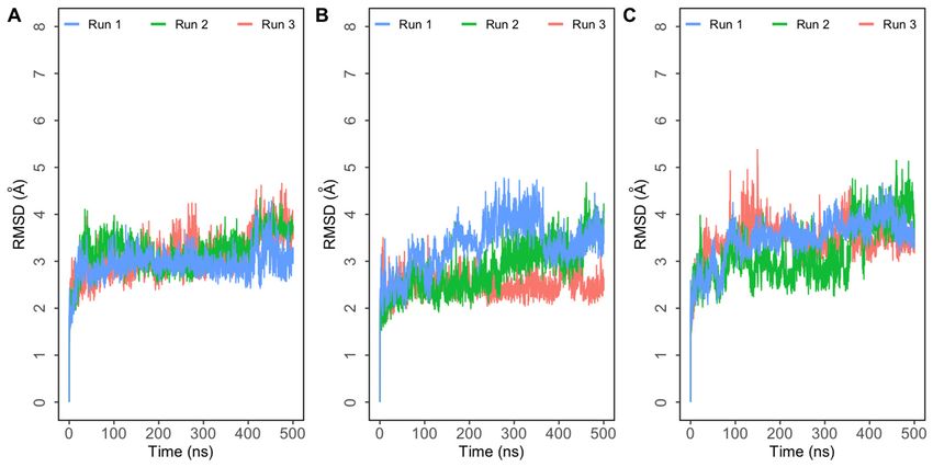
son–Boltzmann Solver (APBS) in Schrödinger Maestro. Graphs were plotted using R ver-
sion 3.6.3 (https://www.r-project.org, accessed on 8 July 2020) and images of structures
Biomolecules 2021, 11, 1244 4 of 10
were generated using Visual Molecular Dynamics version 1.9.3 [19].
3. Results and Discussion
3. Results and Discussion
Molecular dynamics simulations trajectories of three systems—single mutants E484Q
Molecular dynamics simulations trajectories of three systems—single mutants E484Q
and L452R, and double mutant E484Q + L452R—in triplicate were analyzed. RMSD plots
and L452R, and double mutant E484Q + L452R—in triplicate were analyzed. RMSD plots
of the three
of the complexes
three complexesindicated thatthese
indicated that these systems
systems reached
reached equilibrium
equilibrium quicklyquickly
and the and the
RMSDRMSDof the complexes
of the complexesstabilized around
stabilized around 4Å4 in
Å all
in simulations
all simulations (Figure
(Figure 2). 2).
Figure
Figure 2. Root mean
Root2.mean square
square deviation(RMSD)
deviation (RMSD) of ofprotein
proteinbackbone
backboneatoms with respect
atoms to the initial
with respect structure
to the initialobtained fromobtained
structure
three independent 500 ns simulations of SARS-CoV-2 spike (S) protein bound to human ACE2 (hACE2).
from three independent 500 ns simulations of SARS-CoV-2 spike (S) protein bound to human ACE2 (hACE2). (A) (A) Simulations of Simu-
double mutant E484Q + L452R complex; (B) simulations of E484Q complex; (C) simulations of L452R
lations of double mutant E484Q + L452R complex; (B) simulations of E484Q complex; (C) simulations of L452R complex.complex.
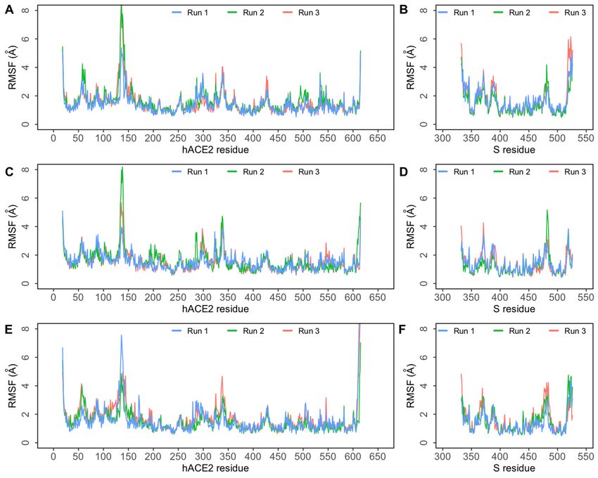
To assess residue-level protein fluctuations and backbone flexibility RMSF of backbone
Toatoms
Cα assess residue-level
were computed andprotein fluctuations
plotted (Figure 3). Thisand backbone
is particularly flexibility
relevant RMSFinof back-
for residues
bonetheCα atomsinterface.
S-hACE2 were computed
The bindingand plotted
interface (Figure
of S RBD 3). This
consists of fourisloop
particularly
regions—loop relevant
1: for
residues 438–450, loop 2: residues 455–470, loop 3: residues 471–491,
residues in the S-hACE2 interface. The binding interface of S RBD consists of four loop and loop 4: residues
495–508. These
regions—loop provide the
1: residues necessary
438–450, loopflexibility while455–470,
2: residues binding to hACE2.
loop Studies471–491,
3: residues have and
suggested that the loop 3 and loop 4 regions are the most flexible regions in the RBD [20].
loop 4: residues 495–508. These provide the necessary flexibility while binding to hACE2
Closer observation of the RMSF plot (Figure 3) revealed that in all three complexes, higher
Studies have was
flexibility suggested
observed that theloop
in the loop 3 andbetween
region loop 4 470–490.
regions Inareallthe
threemost flexiblethe
complexes, regions in
the RBD
fluctuation of the ACE2 backbone was comparable. As indicated by the flat Rg plotsall three
[20]. Closer observation of the RMSF plot (Figure 3) revealed that in
complexes, higher Figure
(Supplementary flexibility wasoverall
S1), the observed in the loop
compactness region between
and secondary structure 470–490.
elementsIn ofall three
the protein complexes were found to be preserved throughout the
complexes, the fluctuation of the ACE2 backbone was comparable. As indicated by the simulation.
Throughout
flat Rg plots the simulation,
(Supplementary several
Figure S1),intermolecular polar and hydrophobic
the overall compactness and secondarycontactsstructure
were observed to form, break and reform in the protein complexes. To elucidate how the
elements of the protein complexes were found to be preserved throughout the simulation
single mutant and double mutant affects the interaction pattern of SARS-CoV-2 RBD and
hACE2 interaction at the molecular level, these data were compared to our previously
reported wildtype data [12]. Residue level interactions sustained for at least 50% of
simulation time in at least one simulation are presented in Figure 4. The data revealed
that, in both single mutant complexes, intermolecular interactions observed were similar
to wildtype data. Four interfacial residues of spike protein—Lys417, Gln493, Tyr449,
and Gln498—in the single mutant complexes interacted with Asp30, Glu35, Asp38 and
Lys353 of hACE2, as reported previously [12]. Apart from these conserved interactions, in
the E484Q mutant complex, the backbone and sidechain of Lys353 in hACE2 interacted
with Gly502 and Gly496 of S RBD (Figure 4B). Additionally, the sidechain of Lys31 of
hACE2 consistently interacted with Gln493 of S protein in all three simulations, while the
sidechain of Gln498 in S protein also showed consistent interaction with Asp38 of hACE2.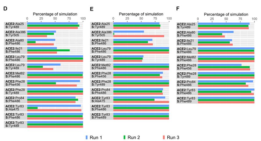
Biomolecules 2021, 11, 1244                                                                                                           5 of 10

                                         Simulation data revealed that in both single mutant complexes Thr27 of hACE2 interacted
                                         with Tyr489 of S protein. Besides these interactions, residues of single mutant L452R,
                                         Tyr489 and Gly496 of S protein, interacted with Gln24 and Lys353 of hACE2, respectively
                                         (Figure 4A). Normally, Lys417, on the S-RBD surface, forms a salt bridge with Asp30 of
                                         hACE2. Unlike the single mutant complexes, this conserved interaction was notably absent
                                         in the double mutant complex (Figure 4C). Additionally, in both single mutants, Asp38 of
                                         hACE2 interacted with Tyr449 of the S protein. However, in the double mutant complex
Biomolecules 2021, 11, x
                                         Asp38 was observed to interact with Gln498 of S protein (Figure 4C). Similar to the E484Q,5 of 11
                                         in the double mutant complex, the sidechain of Lys31 of hACE2 consistently interacted
                                         with Gln493 of the S protein in all simulations (Figure 4C).

           Figure
      Figure        3. Root
              3. Root  mean  mean  square
                                square      fluctuation
                                       fluctuation      (RMSF)ofofprotein
                                                      (RMSF)        protein Cα
                                                                            Cα atoms
                                                                                atomsobtained
                                                                                       obtainedfrom
                                                                                                  fromthree independent
                                                                                                        three  independent 500 ns
                                                                                                                               500simulations
                                                                                                                                    ns simulations
      of SARS-CoV-2 spike (S) protein bound to human ACE2 (hACE2). (A) RMSF of Cα atoms of hACE2 protein ininthe
           of  SARS-CoV-2      spike (S)  protein bound    to  human  ACE2   (hACE2).   (A) RMSF    of Cα   atoms  of  hACE2   protein     theE484Q
           E484Q
      + L452R      + L452R
                complex;      complex;
                            (B)  RMSF of (B)CαRMSF  of Cα
                                                atoms       atoms of SARS-CoV-2
                                                        of SARS-CoV-2     S RBD in S RBD in the E484Q
                                                                                     the E484Q   + L452R+ L452R   complex;
                                                                                                            complex;         (C) RMSF
                                                                                                                        (C) RMSF        of atoms
                                                                                                                                    of Cα  Cα     of
      hACE2atoms   of hACE2
              protein    in theprotein
                                 E484Q in   the E484Q
                                          complex;   (D)complex;
                                                          RMSF of(D) CαRMSF
                                                                         atomsofof
                                                                                 CαSARS-CoV-2
                                                                                     atoms of SARS-CoV-2
                                                                                                   S RBD in StheRBD   in thecomplex;
                                                                                                                  E484Q      E484Q complex;
                                                                                                                                       (E) RMSF of
           (E) RMSF
      Cα atoms          of Cα protein
                  of hACE2      atoms ofinhACE2    protein
                                            the L452R        in the L452R
                                                        complex;          complex;
                                                                    (F) RMSF   of Cα(F) RMSF
                                                                                      atoms  ofof  Cα atoms ofSSARS-CoV-2
                                                                                                SARS-CoV-2                      S RBD in
                                                                                                                  RBD in the L452R         the
                                                                                                                                        complex.
            L452R complex.
                                           Throughout the simulation, several intermolecular polar and hydrophobic contacts
                                      were observed to form, break and reform in the protein complexes. To elucidate how the
                                      single mutant and double mutant affects the interaction pattern of SARS-CoV-2 RBD and
                                      hACE2 interaction at the molecular level, these data were compared to our previously
                                      reported wildtype data [12]. Residue level interactions sustained for at least 50% of simu-
                                      lation time in at least one simulation are presented in Figure 4. The data revealed that, in
                                      both single mutant complexes, intermolecular interactions observed were similar to
                                      wildtype data. Four interfacial residues of spike protein—Lys417, Gln493, Tyr449, and
                                      Gln498—in the single mutant complexes interacted with Asp30, Glu35, Asp38 and Lys353
                                      of hACE2, as reported previously [12]. Apart from these conserved interactions, in the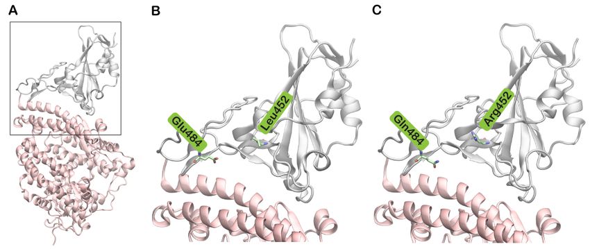
Biomolecules 2021, 11, x                                                                                                              6 of 11

                                    complex Asp38 was observed to interact with Gln498 of S protein (Figure 4C). Similar to
Biomolecules 2021, 11, 1244                                                                                           6 of 10
                                    the E484Q, in the double mutant complex, the sidechain of Lys31 of hACE2 consistently
                                    interacted with Gln493 of the S protein in all simulations (Figure 4C).

       Figure4.4.The
      Figure      Thepercentage
                        percentage of
                                   of simulation
                                       simulation time
                                                    time during
                                                         during which
                                                                which intermolecular   contacts were retained
                                                                       intermolecular contacts        retained between
                                                                                                               between human
                                                                                                                           human ACE2
                                                                                                                                   ACE2
       (hACE2) and
      (hACE2)    and SARS-CoV-2
                       SARS-CoV-2 spike (S) (S) protein
                                                proteinreceptor-binding
                                                        receptor-bindingdomain
                                                                          domain(RBD)
                                                                                   (RBD)residues. (A)
                                                                                          residues.   Intermolecular
                                                                                                    (A) Intermolecular  polar contacts
                                                                                                                          polar        in
                                                                                                                                contacts
       the  L452R   mutant   complex;   (B) Intermolecular  polar contacts  in the E484Q  mutant   complex;  (C) Intermolecular
      in the L452R mutant complex; (B) Intermolecular polar contacts in the E484Q mutant complex; (C) Intermolecular polar         polar
       contacts in
      contacts   in the
                     the double
                         double mutant
                                mutant E484Q
                                          E484Q ++ L452R
                                                     L452R complex;
                                                           complex; (D)
                                                                     (D) Intermolecular
                                                                         Intermolecular hydrophobic
                                                                                          hydrophobic contacts
                                                                                                        contacts in
                                                                                                                  in the
                                                                                                                      the L452R
                                                                                                                          L452R mutant
                                                                                                                                 mutant
       complex; (E) Intermolecular hydrophobic contacts in the E484Q mutant complex; (F) Intermolecular hydrophobic contacts
      complex; (E) Intermolecular hydrophobic contacts in the E484Q mutant complex; (F) Intermolecular hydrophobic contacts
       in the double mutant E484Q + L452R complex.
      in the double mutant E484Q + L452R complex.
                                        To gain
                                        To  gain aa deeper
                                                    deeperunderstanding
                                                           understandingofofthe effect
                                                                              the      of of
                                                                                  effect  thethe
                                                                                              mutated residues,
                                                                                                 mutated        the the
                                                                                                         residues,  interaction
                                                                                                                        interac-
                                   pattern  of the three complexes was evaluated   and  compared  to the wild type simulation.
                                   tion pattern of the three complexes was evaluated and compared to the wild type sim-
                                   ulation. Glu484 (E484), situated on a flexible loop of spike RBD, forms a salt-bridge
                                   with Lys31 of hACE2. Lys31 and Lys353 on the surface of hACE2 are regarded as virus
                                   binding hotspots [21]. In the double mutant, residues Lys31 and Glu35 of hACE2 con-
                                   sistently interacted with Gln493, while Asp38 and Lys353 formed hydrogen bonds with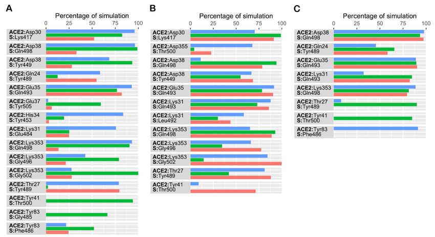
Biomolecules 2021, 11, 1244                                                                                          7 of 10

                              Gln498 of the spike protein (Figure 4C). The formation of a salt bridge between Lys31 of
                              hACE2 and Glu484 of S enhances the affinity of SARS-CoV-2 to hACE2 when compared to
                              SARS-CoV [22]. After Glu484 was mutated to Gln484, this salt bridge was disrupted in the
                              E484Q and double mutant complexes. However, as expected, this salt bridge was observed
                              in the L452R single mutant complex (Figure 4A). Hence, the simulation data suggests that
                              the E484Q mutation could play a role in disrupting the interfacial interaction. However,
                              studies indicate an increased fitness of the E484Q mutant when compared to the wild
                              type [23]. This paradox may be explained by considering the dynamic state of the RBD. The
                              RBD of SARS-CoV-2 exhibits two conformational states—a down/closed conformation,
                              in which the receptor binding region is not exposed, and an up/open state that allows
                              receptor binding [24]. Cryo-EM studies have suggested that the RBD of SARS-CoV-2 exists
                              mostly in the down state [25]. Apart from forming the intermolecular salt bridge, Glu484
                              also stabilizes the RBD down conformation by forming an intramolecular hydrogen bond
                              with Phe490 [26]. In the present analysis, Glu484 showed strong interactions with Phe490,
                              while the mutant Gln484 formed fewer interactions with Phe490. Since this interaction is
                              weaker in the E484Q mutant complex, it could favor the up conformation of the RBD and
                              result in higher hACE2 binding affinity and immune escape. In agreement with this, Gobeil
                              et al. reported that the E484K mutation causes conformational changes in the RBD that
                              favors the up state and reduces antibody binding [26]. Thus, conformational changes in
                              the RBD, induced by the E484Q mutation, could impact the stability of the hACE2 binding
                              surface. Furthermore, Glu484 is as an important antibody escape site of SARS-CoV-2 and
                              mutations at this location could impact the binding and neutralization by antibodies [27].
                              Studies have also demonstrated that the E484Q mutation could reduce the neutralization
                              by plasma and antibodies by 10-fold [28]. This variant is also resistant to bamlanivimab, a
                              recombinant antibody approved for the COVID-19 treatment, by failing to block viral entry
                              mediated by glycoproteins [29].
                                    It has been reported that Leu452, in spite of being located in the RBM region, does
                              not directly interact with hACE2 [30]. However, Leu452 (Figure 5A), together with Phe490
                              and Leu492, forms a hydrophobic patch on the surface of the S protein (Figure 5B) [31]. A
                              mutation to a highly polar and hydrophilic arginine (Figure 5C) could potentially introduce
                              local perturbations (Figure 5D) that could affect how it interacts with a complementary
                              surface. Additionally, Leu452 is a hotspot located in close proximity to the negatively
                              charged residues Glu35, Glu27 and Asp38 of hACE2 [32]. The incorporation of additional
                              charged residues in the vicinity of the binding interface could increase the electrostatic
                              attraction between two proteins. Hence, the mutation of leucine to a positively charged
                              arginine enhances electrostatic complementarity in the interface (Figure 5D). Compared
                              to Leu452, Arg452 was observed to interact more with nearby residues including Ser349,
                              Tyr351, Phe490, Leu492 and Ser494. The increased intramolecular interactions could
                              thus increase the stability of the S protein. The role of this mutation in neutralizing
                              antibodies and sera from convalescent patients has been investigated [28]. The changes
                              induced by L452R mutation prevents the binding of neutralizing antibodies and promotes
                              high infectivity.
                                    The stability of hydrogen bond interactions in the single and double mutant complexes
                              was also monitored (Supplementary Figure S2). The number of intermolecular hydrogen
                              bonds is also higher in the mutant complexes (mean ± SD for E484Q+L452R simulations:
                              10.8 ± 2.1, 10.4 ± 1.9, 10.3 ± 2.0; E484Q simulations: 11.4 ± 2.2, 11.6 ± 1.9, 14.4 ± 2.1;
                              L452R simulations: 11.9 ± 2.0, 13 ± 1.9, 8.9 ± 2.2).
                                    In support of our results, Cherian and colleagues reported that the L452R mutation,
                              and E484Q, could reduce intramolecular and intermolecular interactions and disrupts an
                              electrostatic bond with Lys31 of hACE2 [27]. A change from a hydrophobic leucine on the
                              protein surface to arginine (L452R) also increases its interactions with water molecules that
                              could further stabilize the protein. The double mutant complex, harboring a combination
                              of these two mutations, is capable of escaping neutralizing antibodies, increasing viral
                              infectivity and viral replication.
protein surface to arginine (L452R) also increases its interactions with water molecules
that could further stabilize the protein. The double mutant complex, harboring a combi-
Biomolecules 2021, 11, 1244 8 of 10
nation of these two mutations, is capable of escaping neutralizing antibodies, increasing
viral infectivity and viral replication.
(A)Cartoon
Figure5.5.(A)
Figure Cartoon representation
representation ofofthe
theSARS-CoV-2
SARS-CoV-2 spike (S)(S)
spike protein receptor-binding
protein domain
receptor-binding (RBD)
domain of wild
(RBD) of type
wildwith
type
Leu452
with shown
Leu452 in stick
shown representation.
in stick (B) The
representation. (B)electrostatic potential,
The electrostatic calculated
potential, using Adaptive
calculated Poisson–Boltzmann
using Adaptive Solver
Poisson–Boltzmann
Solver (APBS),
(APBS), mappedmapped on surface
on to the to the surface of the
of the wild wild
type type protein.
protein. (C) Cartoon
(C) Cartoon representation
representation of the SRBD
of the S protein protein RBD
of the of the
mutated
mutated
Arg452 shown in stick representation. (D) The electrostatic potential, calculated using APBS, mapped on to the surface the
Arg452 shown in stick representation. (D) The electrostatic potential, calculated using APBS, mapped on to of
surface of theprotein
the mutant mutant(L452R).
protein (L452R).
Residues
Residues that were involved
that were involvedininhydrophobic
hydrophobic interactions
interactions in the
in the S-hACE2
S-hACE2 interface
interface are
included
are in Figure
included in Figure4. All
4. three complexes
All three complexesformed consistent
formed hydrophobic
consistent contacts
hydrophobic with
contacts
hACE2
with but when
hACE2 but whencompared to the to
compared double mutant,
the double single mutants
mutant, exhibit slightly
single mutants higher
exhibit slightly
numbers
higher of hydrophobic
numbers contacts. contacts.
of hydrophobic In all threeInprotein complexes,
all three proteinIle21, Leu79, Met82,
complexes, Tyr83,
Ile21, Leu79,
and Pro84
Met82, of and
Tyr83, hACE2 Pro84exhibited
of hACE2consistent
exhibited contact with Phe486
consistent contact of thePhe486
with S protein whereas
of the S pro-
Tyr489 interacted with Ala25, Phe28 and Tyr83 of hACE2 in all three runs.
tein whereas Tyr489 interacted with Ala25, Phe28 and Tyr83 of hACE2 in all three runs. Compared to the
wild type data, additional hydrophobic contacts involving Ile21, Ala25,
Compared to the wild type data, additional hydrophobic contacts involving Ile21, Ala25,and Pro84 of hACE2
were
and observed
Pro84 in thewere
of hACE2 mutant complexes
observed in the(Figure
mutant 4D–F). All three
complexes protein
(Figure mutant
4D–F). Allcomplexes
three pro-
were observed to form similar hydrophobic interactions in
tein mutant complexes were observed to form similar hydrophobic interactions the simulations. Phe28 of
in the
hACE2 was observed to form sustained contacts with Phe456 in all
simulations. Phe28 of hACE2 was observed to form sustained contacts with Phe456 in all three complexes. In
both single mutant complexes, Tyr83 formed hydrophobic contacts with Ala475 of hACE2
three complexes. In both single mutant complexes, Tyr83 formed hydrophobic contacts
(Figure 4D,E). This interaction was not observed in any of the double mutant simulations.
with Ala475 of hACE2 (Figure 4D,E). This interaction was not observed in any of the dou-
To compare how mutations in S RBD affect the binding free energy (∆Gbind ), it was esti-
ble mutant simulations.
mated, using frames extracted from all MD simulations, based on the MM-GBSA approach.
The binding free energy of the double mutant simulations were −147.55 ± 16.22 kcal/mol,
−132.83 ± 18.71 kcal/mol, and −127.80 ± 21.30 kcal/mol, respectively. In the single mutant
complex involving the E484Q mutant, ∆Gbind values computed were −131.25 ± 20.95 kcal/mol,
−143.47 ± 14.96 kcal/mol, and −149.14 ± 20.98 kcal/mol, and for the L452R mutant the val-Biomolecules 2021, 11, 1244 9 of 10
ues were −146.58 ± 19.70 kcal/mol, −148.60 ± 17.67 kcal/mol, and −117.22 ± 22.15 kcal/mol.
Importantly, compared to the wild type, all the mutant complexes exhibited a more favor-
able ∆Gbind . This supports the higher binding affinity of the mutants compared to the wild
type virus [33].
In summary, this study looks at the structural dynamics of the S protein-hACE2
interface of the rapidly spreading B.1.617 variant using multiple molecular dynamics
simulations. Single and double mutant simulations were used to identify the differences
in interactions and structural conformations of the spike protein when compared to the
wild type. Overall, the data provide deeper insights into the higher affinity of the B.1.617
variant of SARS-CoV-2 and could form the basis for further studies.
4. Conclusions
Even though a conserved salt bridge is absent, the E484Q mutation could favor the
open conformation of RBD that aids in enhanced hACE2 binding and immune escape. The
L452R mutation enhances the electrostatics of the binding surface and aids electrostatic
attraction between hACE2 and the S protein. The enhanced network of intramolecular
interactions increases the stability of the S protein and the conformational changes induced
by these mutations could prevent the binding of neutralizing antibodies and promote
higher infectivity. The results presented here are based on molecular dynamics simulations.
Further, wet-lab studies are essential to fully validate these observations.
Supplementary Materials: The following are available online at https://www.mdpi.com/article/
10.3390/biom11081244/s1, Figure S1: Radius of gyration (Rg) of human ACE2 (hACE2) and spike
(S) protein of SARS-CoV-2 receptor-binding domain (RBD) from three 500 ns simulations, Figure S2:
Hydrogen bonds between human ACE2 (hACE2) and spike (S) protein of SARS-CoV-2 receptor-
binding domain (RBD) from three 500 ns simulations.
Author Contributions: R.V. conceived the idea and performed the experiments. R.V. and P.A.
analyzed the data and wrote the manuscript. Both authors have read and agreed to the published
version of the manuscript.
Funding: This work was supported by a UPAR grant (12S006) to R.V. and a graduate fellowship
(12S071) to P.A. from the United Arab Emirates University.
Institutional Review Board Statement: Not applicable.
Informed Consent Statement: Not applicable.
Conflicts of Interest: The authors declare no conflict of interest.
References
1. Yan, Z.-P.; Yang, M.; Lai, C.-L. COVID-19 Vaccines: A Review of the Safety and Efficacy of Current Clinical Trials. Pharmaceuticals
2021, 14, 406. [CrossRef] [PubMed]
2. Hu, B.; Guo, H.; Zhou, P.; Shi, Z.-L. Characteristics of SARS-CoV-2 and COVID-19. Nat. Rev. Microbiol. 2021, 19, 141–154. [CrossRef]
3. Hoffmann, M.; Kleine-Weber, H.; Schroeder, S.; Krüger, N.; Herrler, T.; Erichsen, S.; Schiergens, T.S.; Herrler, G.; Wu, N.-H.;
Nitsche, A.; et al. SARS-CoV-2 Cell Entry Depends on ACE2 and TMPRSS2 and Is Blocked by a Clinically Proven Protease
Inhibitor. Cell 2020, 181, 271–280. [CrossRef]
4. Korber, B.; Fischer, W.M.; Gnanakaran, S.; Yoon, H.; Theiler, J.; Abfalterer, W.; Hengartner, N.; Giorgi, E.E.; Bhattacharya, T.; Foley,
B.; et al. Tracking Changes in SARS-CoV-2 Spike: Evidence that D614G Increases Infectivity of the COVID-19 Virus. Cell 2020,
182, 812–827. [CrossRef] [PubMed]
5. Mascola, J.R.; Graham, B.S.; Fauci, A.S. SARS-CoV-2 Viral Variants—Tackling a Moving Target. JAMA 2021, 325, 1261. [CrossRef]
6. Wise, J. COVID-19: New coronavirus variant is identified in UK. BMJ 2020, 371, m4857. [CrossRef] [PubMed]
7. Davies, N.G.; Abbott, S.; Barnard, R.C.; Jarvis, C.I.; Kucharski, A.J.; Munday, J.D.; Pearson, C.A.B.; Russell, T.W.; Tully, D.C.;
Washburne, A.D.; et al. Estimated transmissibility and impact of SARS-CoV-2 lineage B.1.1.7 in England. Science 2021, 372,
eabg3055. [CrossRef]
8. Tang, J.W.; Toovey, O.T.R.; Harvey, K.N.; Hui, D.D.S. Introduction of the South African SARS-CoV-2 variant 501Y.V2 into the UK.
J. Infect. 2021, 82, e8–e10. [CrossRef]
9. Gómez, C.E.; Perdiguero, B.; Esteban, M. Emerging SARS-CoV-2 Variants and Impact in Global Vaccination Programs against
SARS-CoV-2/COVID-19. Vaccines 2021, 9, 243. [CrossRef] [PubMed]Biomolecules 2021, 11, 1244 10 of 10
10. Konings, F.; Perkins, M.D.; Kuhn, J.H.; Pallen, M.J.; Alm, E.J.; Archer, B.N.; Barakat, A.; Bedford, T.; Bhiman, J.N.; Caly, L.; et al.
SARS-CoV-2 Variants of Interest and Concern naming scheme conducive for global discourse. Nat. Microbiol. 2021, 6, 821–823.
[CrossRef] [PubMed]
11. Tai, W.; He, L.; Zhang, X.; Pu, J.; Voronin, D.; Jiang, S.; Zhou, Y.; Du, L. Characterization of the receptor-binding domain (RBD)
of 2019 novel coronavirus: Implication for development of RBD protein as a viral attachment inhibitor and vaccine. Cell. Mol.
Immunol. 2020, 17, 613–620. [CrossRef] [PubMed]
12. Ali, A.; Vijayan, R. Dynamics of the ACE2–SARS-CoV-2/SARS-CoV spike protein interface reveal unique mechanisms. Sci. Rep.
2020, 10, 14214. [CrossRef] [PubMed]
13. Bowers, K.J.; Chow, D.E.; Xu, H.; Dror, R.O.; Eastwood, M.P.; Gregersen, B.A.; Klepeis, J.L.; Kolossvary, I.; Moraes, M.A.; Sacerdoti,
F.D.; et al. Scalable Algorithms for Molecular Dynamics Simulations on Commodity Clusters. In Proceedings of the ACM/IEEE
SC 2006 Conference (SC’06), Tampa, FL, USA, 11–17 November 2006; p. 43.
14. Martyna, G.J.; Klein, M.L.; Tuckerman, M. Nosé–Hoover chains: The canonical ensemble via continuous dynamics. J. Chem. Phys.
1992, 97, 2635–2643. [CrossRef]
15. Martyna, G.J.; Tobias, D.J.; Klein, M.L. Constant pressure molecular dynamics algorithms. J. Chem. Phys. 1994, 101, 4177–4189.
[CrossRef]
16. Essmann, U.; Perera, L.; Berkowitz, M.L.; Darden, T.; Lee, H.; Pedersen, L.G. A smooth particle mesh Ewald method. J. Chem.
Phys. 1995, 103, 8577–8593. [CrossRef]
17. Tuckerman, M.; Berne, B.J.; Martyna, G.J. Reversible multiple time scale molecular dynamics. J. Chem. Phys. 1992, 97, 1990–2001.
[CrossRef]
18. Li, J.; Abel, R.; Zhu, K.; Cao, Y.; Zhao, S.; Friesner, R.A. The VSGB 2.0 model: A next generation energy model for high resolution
protein structure modeling: The VSGB 2.0 Energy Model. Proteins Struct. Funct. Bioinform. 2011, 79, 2794–2812. [CrossRef] [PubMed]
19. Humphrey, W.; Dalke, A.; Schulten, K. VMD: Visual molecular dynamics. J. Mol. Graph. 1996, 14, 33–38. [CrossRef]
20. Williams, J.K.; Wang, B.; Sam, A.; Hoop, C.L.; Case, D.A.; Baum, J. Molecular Dynamics Analysis of a Flexible Loop at the Binding
Interface of the SARS-CoV-2 Spike Protein Receptor-Binding Domain. bioRxiv 2021. [CrossRef]
21. Wan, Y.; Shang, J.; Graham, R.; Baric, R.S.; Li, F. Receptor Recognition by the Novel Coronavirus from Wuhan: An Analysis Based
on Decade-Long Structural Studies of SARS Coronavirus. J. Virol. 2020, 94, e00127-20. [CrossRef]
22. Wrobel, A.G.; Benton, D.J.; Xu, P.; Roustan, C.; Martin, S.R.; Rosenthal, P.B.; Skehel, J.J.; Gamblin, S.J. SARS-CoV-2 and bat RaTG13
spike glycoprotein structures inform on virus evolution and furin-cleavage effects. Nat. Struct. Mol. Biol. 2020, 27, 763–767. [CrossRef]
23. Yi, C.; Sun, X.; Ye, J.; Ding, L.; Liu, M.; Yang, Z.; Lu, X.; Zhang, Y.; Ma, L.; Gu, W.; et al. Key residues of the receptor binding motif
in the spike protein of SARS-CoV-2 that interact with ACE2 and neutralizing antibodies. Cell. Mol. Immunol. 2020, 17, 621–630.
[CrossRef] [PubMed]
24. Henderson, R.; Edwards, R.J.; Mansouri, K.; Janowska, K.; Stalls, V.; Gobeil, S.M.C.; Kopp, M.; Li, D.; Parks, R.; Hsu, A.L.; et al.
Controlling the SARS-CoV-2 spike glycoprotein conformation. Nat. Struct. Mol. Biol. 2020, 27, 925–933. [CrossRef]
25. Shang, J.; Wan, Y.; Luo, C.; Ye, G.; Geng, Q.; Auerbach, A.; Li, F. Cell entry mechanisms of SARS-CoV-2. Proc. Natl. Acad. Sci. USA
2020, 117, 11727–11734. [CrossRef] [PubMed]
26. Gobeil, S.M.-C.; Janowska, K.; McDowell, S.; Mansouri, K.; Parks, R.; Stalls, V.; Kopp, M.F.; Manne, K.; Saunders, K.; Edwards, R.J.;
et al. Effect of natural mutations of SARS-CoV-2 on spike structure, conformation and antigenicity. Microbiology 2021, 373, eabi6226.
27. Cherian, S.; Potdar, V.; Jadhav, S.; Yadav, P.; Gupta, N.; Das, M.; Rakshit, P.; Singh, S.; Abraham, P.; Panda, S.; et al. Convergent
evolution of SARS-CoV-2 spike mutations, L452R, E484Q and P681R, in the second wave of COVID-19 in Maharashtra, India.
Microorganisms 2021, 9, 1542. [CrossRef]
28. Greaney, A.J.; Starr, T.N.; Gilchuk, P.; Zost, S.J.; Binshtein, E.; Loes, A.N.; Hilton, S.K.; Huddleston, J.; Eguia, R.; Crawford, K.H.D.;
et al. Complete Mapping of Mutations to the SARS-CoV-2 Spike Receptor-Binding Domain that Escape Antibody Recognition.
Cell Host Microbe 2021, 29, 44–57. [CrossRef]
29. Hoffmann, M.; Hofmann-Winkler, H.; Krüger, N.; Kempf, A.; Nehlmeier, I.; Graichen, L.; Sidarovich, A.; Moldenhauer, A.-S.;
Winkler, M.S.; Schulz, S.; et al. SARS-CoV-2 variant B.1.617 is resistant to Bamlanivimab and evades antibodies induced by
infection and vaccination. Cell Rep. 2021, 36, 109415. [CrossRef]
30. Lan, J.; Ge, J.; Yu, J.; Shan, S.; Zhou, H.; Fan, S.; Zhang, Q.; Shi, X.; Wang, Q.; Zhang, L.; et al. Structure of the SARS-CoV-2 spike
receptor-binding domain bound to the ACE2 receptor. Nature 2020, 581, 215–220. [CrossRef]
31. Deng, X.; Garcia-Knight, M.A.; Khalid, M.M.; Servellita, V.; Wang, C.; Morris, M.K.; Sotomayor-González, A.; Glasner, D.R.;
Reyes, K.R.; Gliwa, A.S.; et al. Transmission, infectivity, and neutralization of a spike L452R SARS-CoV-2 variant. Cell 2021, 184,
3426–3437.e8. [CrossRef]
32. Verkhivker, G.M.; Agajanian, S.; Oztas, D.Y.; Gupta, G. Comparative Perturbation-Based Modeling of the SARS-CoV-2 Spike
Protein Binding with Host Receptor and Neutralizing Antibodies: Structurally Adaptable Allosteric Communication Hotspots
Define Spike Sites Targeted by Global Circulating Mutations. Biochemistry 2021, 60, 1459–1484. [CrossRef] [PubMed]
33. Forouzesh, N.; Mishra, N. An Effective MM/GBSA Protocol for Absolute Binding Free Energy Calculations: A Case Study on
SARS-CoV-2 Spike Protein and the Human ACE2 Receptor. Molecules 2021, 26, 2383. [CrossRef] [PubMed]You can also read