Roots Structure and Development of Austrobaileya scandens (Austrobaileyaceae) and Implications for Their Evolution in Angiosperms - MDPI
←
→
Page content transcription
If your browser does not render page correctly, please read the page content below
Article

Roots Structure and Development of Austrobaileya
scandens (Austrobaileyaceae) and Implications for
Their Evolution in Angiosperms
Julien B. Bachelier 1,*, Imran Razik 2, Maria Schauer 1 and James L. Seago Jr. 2,3
 1 Structural and Functional Plant Diversity Group, Institute of Biology, Freie Universität Berlin,
   Altensteinstrasse 6, 14195 Berlin, Germany; maria.schauer@fu-berlin.de
 2 Department of Biological Sciences of State University of New York (SUNY) at Oswego, 30 Centennial

   Drive, NY 13126, USA; razik.2@buckeyemail.osu.edu (I.R.); jseago@twcny.rr.com (J.L.S.J.)
 3 Seago Botanical Consulting, Minetto, NY 13115, USA

 * Correspondence: julien.bachelier@fu-berlin.de

 Received: 21 November 2019; Accepted: 23 December 2019; Published: 1 January 2020

 Abstract: Since the resolution of the ANA grade [Amborellales, Nymphaeales, Austrobaileyales] as
 sister to all other flowering plants, a few comparative studies of root structure have suggested that
 some of their anatomical traits could be of importance to understanding root evolutionary
 development and angiosperm phylogeny. However, there is still a paucity of information on root
 structure and apical meristems (RAMs) in these lineages and especially the sister to all other
 Austrobaileyales, Austrobaileya scandens. We used microtome sections and bright field,
 epifluorescence, laser confocal, and scanning electron microscopy to study adventitious root RAMs
 and tissues of A. scandens. Our results indicate that root structure is relatively simple in A. scandens.
 The epidermis has a thick cuticle and lacks root hairs. The stele is typically diarch, or some
 modification thereof, and surrounded by a cortex differentiated into a uniseriate endodermis, a
 middle region sometimes packed with starch, some oil cells, and colonized by arbuscular
 mycorrhizal fungi, and a multiseriate exodermis. Secondary growth produced many vessel
 elements in the secondary xylem and scattered sclerenchymatous fibers in secondary phloem. The
 absence of distinct patterning within the RAM and between the RAM and derivative differentiating
 tissues shows that the RAM is open and characterized by common initials. Roots structure and
 anatomy of A. scandens are thus essentially similar to some previously described in Amborella or
 Illicium in the ANA grade and many magnoliids, and suggest that the first woody flowering plants
 likely had an open RAM with common initials. Their functional and evolutionary significance in
 woody early-diverging and basal lineages of flowering plants and gymnosperms remains unclear,
 but they are clearly ancestral traits.

 Keywords: ANA grade; Austrobaileyales; common initials; magnoliids; open meristem

1. Introduction
      Since the resolution of the ANA grade [Amborellales, Nymphaeales, Austrobaileyales] as sister
to all other flowering plants, most studies of anatomical traits that might be relevant to understanding
the evolutionary development and phylogeny of angiosperms have been limited to multiple floral
reproductive characters and to certain leaf and xylem traits [1,2]. However, it has been suggested that
certain root traits might also be important to elucidate their elusive origin and rapid diversification
[3–5].

Plants 2020, 9, 54; doi:10.3390/plants9010054                                  www.mdpi.com/journal/plants
Plants 2020, 9, 54                                                                                   2 of 11

      One significant root character is the occurrence of common initials, or CI, in root apical
meristems, or RAM. In CIs, there is no clear and distinct boundary among the meristematic cells and
tissues at the base of the root cap and the tips of the main root tissues, protoderm, ground meristem,
and procambium, in the open RAMs of Amborellales and other woody species of the ANA grade
(Austrobaileyales) and magnoliids (Laurales, Magnoliales, Canellales) [4]. Furthermore, the RAMs of
basal angiosperms turn out to be very similar to those in many gymnosperms, which are also known
for having common initials for most tissues of the root apex [6–8]. Indeed, Heimsch and Seago [4]
noted that gymnosperm CI RAMs, as reported by Pillai [8], are also similar, when present, to those
found in the more variable RAMs of Amborellales and Schisandraceae of the Austrobaileyales.
However, with the exception of Illicium, there is relatively little or no observation on the RAM and
root developmental anatomy of other members of the Schisandraceae or Austrobaileyales, despite its
position as sister to all other members of the order. Austrobaileya scandens roots have received little
attention. Bailey and Swamy [9] studied A. scandens but not its roots. Metcalfe and Chalk [10] included
A. scandens as a member of the Schisandraceae and reported very little on it and nothing on its roots.
Dickison and Endress [11], Baranova [12,13], Feild et al. [14], and Carpenter [15,16] dealt with various
aspects of shoot anatomy, and Srivastava [17] characterized gymnospermous features in its stem
secondary phloem. It was not until 2001 that Carlquist [18] reported on root wood, with vessel
elements, tracheids, and fiber-tracheids, and bark structure. He touched upon the cortex, which he
stated had compound starch grains and some oil cells, and secondary phloem, which was comprised
of sieve cells and parenchyma, but lacked sieve tube elements and sclerenchyma.
      Since nothing is known about Austrobaileya early root development and structure and RAM
organization, our goals were to characterize them and the differentiation of primary tissues and
secondary tissues during primary and early secondary root growth, and to evaluate if Austrobaileya
shares more traits with other members of early-diverging lineages like Amborella, the closely related
Illicium (Austrobaileyales), or the magnoliids. We were also interested to ascertain if they may still
have some potentially new ancestral traits shared with extant and extinct gymnosperms (or other
vascular plant lineages).

2. Materials and Methods
     Material—About 30 adventitious and lateral roots of Austrobaileya scandens were collected from
three 12-year old plants, which had been grown from seeds and cultivated in pots at the Weld Hill
research building greenhouse in the Arnold Arboretum of Harvard University, Boston,
Massachusetts. All roots were fixed in summer 2012 with 4% acrolein (Polysciences Inc., Warrington,
PA, USA) in a modified PIPES buffer adjusted to pH 6.8 (50 mM PIPES and 1 mM MgSO4, both from
BDH, London, UK; and 5 mM EGTA from Research Organics Inc., Cleveland, OH, USA) for 24 h.
After rinsing with the same buffer, root material was dehydrated using an increasing ethanol dilution
series and stored in 70% ethanol.
     Scanning Electron microscopy (SEM) —Five roots were rehydrated and treated with 2%
Osmium tetroxide for 2 h before complete re-dehydration using an increasing ethanol dilution series
up to 100% and a transfer in acetone overnight. All material was further processed with an
AutoSAMDRI-815 automatic critical point dryer (Tousimis, Rockville, MD, US) and specimens were
mounted on aluminum stubs before observations.
     Histological sections—About half of the roots were sectioned by hand using double-edged
Wilkinson razor feather blades, while 10 were further dehydrated using dilutions up to a 100%
ethanol solution, infiltrated and embedded in methacrylate resins (JB-4, Polysciences, Warrington,
PA, USA, or Kulzer’s Technovit 7100, Electron Microscopy Sciences, Hatfield, PA, USA), and cut in
4–10 µm thick serial sections using a rotary microtome (Thermo Fisher Scientific, Waltham, MA,
USA) and disposable blades (Leica Biosystems, Wetzlar, Germany). All sections were stained
generally following Jensen [19], either with saturated phloroglucinol-HCl for detection of lignin,
berberine hemisulfate counterstained with 0.05% toluidine blue O (TBO) for detection of suberin,
fluorol yellow for detection of lipids, calcofluor (i.e., Fluorescent Brightener 28) for detection of plant
cell walls and fungal hyphae, Iodine Potassium (IKI) for detection of starch grains, or 0.05% TBO and
Plants 2020, 9, 54                                                                                          3 of 11

1.0% ruthenium red for general staining (all Sigma-Aldrich, Saint Louis, MO, USA). Resin sections
were permanently mounted on glass slides using Citrisolv cleaning solution (Decon Labs, Inc., King
of Prussia, PA, USA) and Permount (also Sigma-Aldrich) or Histomount (Agar Scientific, Stansted,
United Kingdom) mounting media, and stored permanently at FU Berlin.
     Image acquisition, processing and plates editing—Digital pictures of the roots in Figure 1 were
taken using the camera of a Samsung Galaxy S7 cell phone (Samsung Inc., Maetan-dong, South
Korea). In SEM images, acquired with an Hitachi100 Scanning Electron Microscope (Hitachi Ltd,
Tokyo, Japan), a black background has been edited to provide better contrast around the smooth
contour of the roots. All hand and microtome sections were studied using a Zeiss LSM700 confocal
microscope mounted with a Zeiss MRc Axiocam camera, and using brightfield, Differential
Interference Contrast (DIC), or epifluorescence illumination, and the 405 nm and 488 nm solid-state
lasers (Carl Zeiss AG, Oberkochen, Germany). All pictures and plates were edited and arranged using
Adobe Creative Suite (Adobe, San Jose, California, USA).

3. Results

3.1. Root System and Apex Structure
     Multiple adventitious roots arose from basal regions of stems and yielded numerous lateral
roots, forming a homogeneous orange and glabrous root system (Figure 1A). All adventitious roots
and lateral roots were very similar (Figure 1B), and both had tips varying in shape from elongated to
slightly truncated (Figures 1C, 1D, 2A, 2B).

      Figure 1. Root systems of Austrobaileya scandens. (A) root system with adventitious roots (inset: young
      adventitious root indicated by white arrows). (B) adventitious root bearing laterals, with old part
      covered by phellem, and young part still covered by epidermis. C–D: SEM pictures of roots with
      elongated (C) and truncated (D) tips, indicating continuous and arrested growth, respectively. Scale
      bars: A, B = 5 cm (1 cm in inset); C, D = 1 mm.

      In elongated root tips (Figure 2A), median longitudinal sections showed RAM with a root cap
and its columella region not completely distinguishable from peripheral root cap tissues and common
initials for the other associated tissues, protoderm, ground meristem, and procambium that were not
traceable to the columellar region base (Figure 2A). While the procambium narrowed at its tip, it was
not clearly separated by distinct cell walls or cell files from cells at the tip of the ground meristem or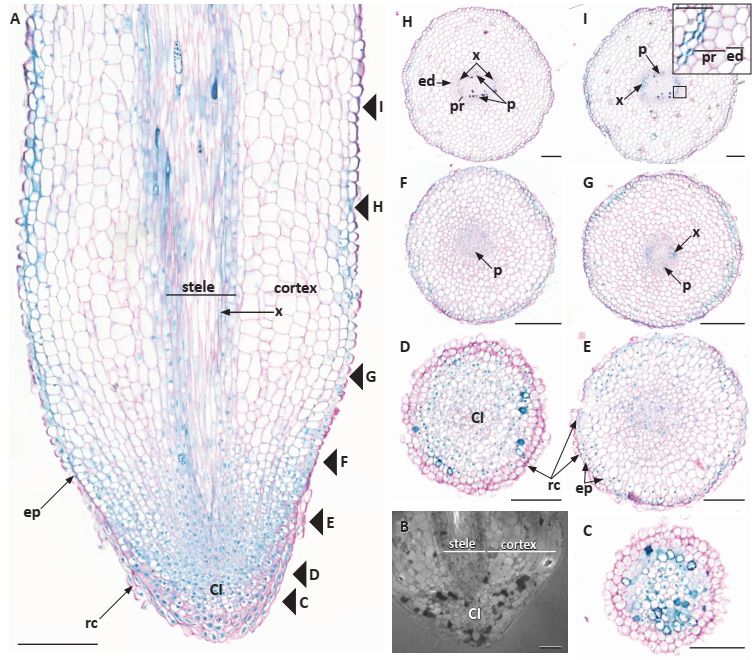
Plants 2020, 9, 54                                                                                             4 of 11

base of the columella region of the RAM, even though the procambium tended to stain more intensely
than the surrounding ground meristem because its cells appeared to have a denser cytoplasm. The
ground meristem was indistinguishable from the other common initials and, near its tip, did not have
orderly files of cells emanating from its tip-most region. The same RAM structure was observed in
truncated root tips (Figure 2B).
      Figure 2. Root primary anatomy of Austrobaileya scandens. A: median longitudinal section of elongated
      root tip showing RAM with common initials and the major tissues of the central cylinder or stele,
      cortex, epidermis, and root cap. Arrowheads and letters designate positions of images in 5C–I. B:
      truncated root tip with arrested growth, common initials, and stele and cortex differentiated close to
      the RAM. C–I: transverse section series from root tip with root cap through common initials and
      tissues differentiating into epidermis, cortex with endodermis, and stele with pericycle, primary
      phloem, and primary xylem with diarch stele. C: < 0.2 mm, level of the root cap through the columella.
      D: at 0.2–0.4 mm, level of CI region in center. E: at 0.4–0.5 mm, level immediately proximal to CI with
      procambium region more densely stained in center. F: at 0.5–1 mm, level of first protophloem
      differentiation. G: at 1–1.5 mm, level of first xylem tracheid. H: at 1.5–3 mm, distinct diarch stele with
      multiple protophloem elements at p, pericycle and endodermis now evident. I: at 3–4 mm, level of
      diarch stele with multiple tracheids at x and protophloem at p, surrounded with 2–3 layers of
      pericycle and a layer endodermis (inset, with pericycle and endodermis). Abbreviations: CI: common
      initials; ed: endodermis; ep: epidermis; p: phloem; pr: pericycle; rc: root cap; x: xylem. Scale bars: A =
      250 µm; B–I = 100 µm (50 µm in inset).

    In transverse sections, there was no distinct columella (Figure 2C), even though the procambium
narrowed at its tip, its boundaries with the ground meristem could not be determined (Figure 2D).
Beginning in the RAM (Figure 2D) and proceeding proximally, cell files could be seen longitudinally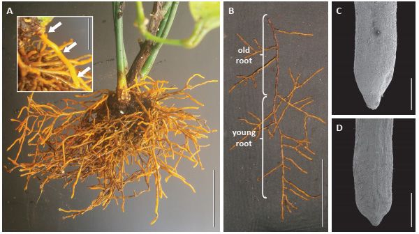
Plants 2020, 9, 54                                                                                        5 of 11

in the ground meristem (Figure 2A), but not radially, even though there were some extending from
the periphery of the CI into nearby ground meristem (Figure 2A, E). However, the common initials
in the RAM did not contribute to an orderly structure initiating the tissues, and a distinction between
the procambium, ground meristem, and protoderm could not be made up to 1.0 mm behind the tip
(Figure 2F). Between 0.5 and 1.5 mm behind the tip the first phloem elements appear (Figure 2F)
followed by the differentiation of the first protoxylem tracheids (Figure 2G), and the premises of a
stele surrounded by a fairly thick cortex (Figure 2F).
     In the truncated tip cells broadened out immediately behind or proximal to the almost
pyramidal-shaped root cap (Figures 1D, 2B). Their ground meristem cells had swollen and the
vascular tissues were differentiated almost down to the tip, indicating that they had stopped
elongating. Such roots also appeared to be more likely to have a cortex packed with starch, sometimes
with aseptate or septate inter- and intracellular hyphae, indicating that the roots may be mycorrhizal
and/or harboring an endophytic fungus (Figure 3).

      Figure 3. Transition zone between younger and older part of Austrobaileya scandens roots. Transverse
      SEM section and inset of root with glabrous epidermis covered with a thick cuticle and hyphae (white
      arrows), and surrounding a cortex with a three-cell-layer thick, multiseriate hypodermis surrounding
      a middle cortex colonized by arbuscular mycorrhizae. Oblong-shaped diarch stele, including
      tracheids and vessel elements (*), two protoxylem and protophloem poles, delimited by uniseriate
      endodermis and two–three cell-layers thick pericycle. Abbreviations: cu: cuticle; ed: endodermis; ep:
      epidermis; H: hyphae; hyp: hypodermis; p: phloem; pr: pericycle; x: xylem. Scale bar = 200 µm (50 µm
      in inset).

3.2. Primary Root Growth and Tissue Differentiation
     The first protophloem cells typically differentiated and matured prior to protoxylem tracheid
differentiation and maturation (Figures 2F, 4A). More protophloem elements were produced just
laterally to the first element and the first protoxylem cells became prominent across each broad xylem
pole (Figures 2G, 4B, C). Diarchy was characteristic (Figures 2F–I, 4B–H). Sometimes, there appeared
to be asymmetric xylem patterns with one xylem pole exhibiting separated protoxylem cells or
clusters of cells (Figure 4B, D, E), or even both xylem poles with slightly separate protoxylem cells in
Plants 2020, 9, 54 6 of 11
each pole, in which the protoxylem tracheids had not differentiated and matured uniformly across a
pole (Figure 4C–E). However, the protoxylem production revealed that there were typically still only
two xylem poles with numerous tracheids in each (Figure 4D, F–H) and a vessel element or two in a
pole (Figure 4H). Primary xylem gradually filled in the center of the vascular cylinder (Figures 3, 4I,
J) and vessel elements could be identified (e.g., Figures 3, 4H, I). While we did not specifically test for
phloem elements, we could identify them by cell position and shape (e.g., Figures 2H, I, 4A–C).
The pericycle early in primary growth became multiple cell layers (Figure 4F, I–K), and although
it remained unstained with phloroglucinol-HCl (Figure 4J), it can be viewed in SEM (Figure 3). At
first, it was also difficult to discern a discrete endodermal layer (Figures 2E, G, 4A). However, the
cortex became delimited by an endodermis with distinct Casparian bands, which covered over half
of the radial walls (Figure 4D, F, H, I). Suberin lamellae were then produced in most endodermal cells
opposite the two phloem poles of the stele, leaving all endodermis cells opposite the large protoxylem
poles as passage cells (Figure 4H, I). In the endodermis most cells developed suberin lamellae (Figure
4I).
The mid cortex comprised many cells that varied depending on the sizes of adventitious and
lateral roots, and there were oil cells (Figure 4G, H), small air spaces (Figure 4I), and many cells that
contained multiple, compound starch grains (Figure 3). A hypodermis generally differentiated into a
three-cell-layer thick exodermis with fewer cytoplasmic contents and with Casparian bands
developing on its radial (and transverse) cell walls (Figures 3, 4E, F, I, L). The exodermis also had thin
suberin lamellae, which accounted for the fluorescence on the tangential cell walls (Figure 4F, I), in
addition to the radial and transverse cell walls. There were also mycorrhizal hyphae in the mid cortex
cells of many roots (Figure 3).
Outer cell walls in the root epidermis had a very thick cuticle of two distinct layers (Figures 3,
4E, G, I, L), even down to the proximity of the root tip under peripheral root cap cells (Figure 2A, E–
H). There were neither trichoblasts nor root hairs, but mycorrhizae could be observed on the
epidermis (Figure 3).
3.3. Secondary Root Growth and Tissue Differentiation
As noted above, the pericycle stained and photographed differently from the internal
procambium, primary xylem and phloem (Figures 3, 4I–K). Procambial cells between primary xylem
and phloem and inner pericycle cells over the xylem poles became vascular cambium (Figure 4I–K).
Secondary xylem was somewhat asymmetrically produced with vessel elements being formed
outside protophloem (Figure 4M–O). The cork cambium or phellogen differentiated from the outer
layers of redivided pericycle (Figure 4I–L) and started to produce the first phellem cells under the
endodermis and just outside phloem (Figures 4L and inset, 5). As the cork cambium proliferated
(Figures 4L, N, O, 5A), multiple layers of phelloderm were produced to the interior of the phellogen
(Figure 5B, C). While the root expanded secondarily, more vessel elements were produced along with
tracheids, parenchyma, and rays, and eventually became distributed throughout the secondary
xylem (Figures 4N, 5B, C), as first described by Carlquist [18]. Secondary phloem (Figures 4N, O, 5B),
with sieve cells, parenchyma and wider rays with starch (Figures 4O, 5C), were also as described in
more detail by Carlquist [18]. We also detected phloem fibers (Figures 4L, N, 5C) and noted that
starch accumulated in parenchyma cells and phloem ray cells. The cortex, with its starch, oil cells,
and mycorrhizae, and the epidermis remained around the root for a considerable distance behind the
root tip (Figures 4N, O, 5A, B), but eventually are pushed outward, disintegrated, and sloughed off
(Figure 5).Plants 2020, 9, 54 7 of 11
Figure 4. Primary and secondary tissue differentiation in Austrobaileya scandens roots. Transverse
sections of different specimens imaged with laser (A–F, I, K, L, N), epifluorescence (G, H, M), and
bright field (J, O) illumination, arranged in ontogenic series, with the youngest stage on the left of
each row. A–D: microtome sections stained with berberine and TBO showing the successive
differentiation of two phloem and two (more or less symmetrical) xylem poles, and endodermis. A:
note protophloem elements. B: note separated protoxylem elements in one pole. C: note xylem poles
with arc of tracheids in each pole. D: first endodermis with Casparian bands evident, one xylem pole
with many tracheary elements and other xylem pole still with separate regions of tracheary cells. E–
H: hand sections stained with fluorol yellow showing epidermis and endodermis, and diarch stele. E:
diarch stele with separated protoxylem in each pole, epidermis with thick two-layered cuticle (inset).
F: endodermis with Casparian bands (inset), three-layered exodermal hypodermis, epidermis with
thick cuticle. G: diarch stele and oil cells. H: diarch stele with many tracheids and some vessel
elements, endodermis, oil cells. I–L: hand sections stained with calcofluor (I) and phloroglucinol-HCl
(J–L) showing transition from primary to secondary growth and successive differentiation of a
multiseriate pericycle, with inner portion opposite protoxylem becoming vascular cambium andPlants 2020, 9, 54 8 of 11
outer portion cork cambium, while the cortex and epidermis are still present. I: note metaxylem in
center of stele and hyphae in inner cortex, and pericycle and endodermis in inset. J: full protoxylem
and metaxylem, phloem, pericycle, and endodermis. K: primary xylem surrounded by wide pericycle.
L: early stage of cork cambium and phellem development, oil cells still evident. M: hand section
stained with berberine showing primary xylem in center, vessels (some marked by asterisks *), few
vessels in secondary xylem outside protoxylem poles in early secondary growth. N: hand section
stained with calcofluor showing more secondary growth, vessels (*) mostly throughout secondary
xylem, secondary phloem fibers under arrows at sp, cork cambium with phellem (inset). O: hand
section stained with IKI showing primary and secondary xylem with wood rays with starch grains,
secondary phloem, cork cambium, phellem, and remnant cortex. Abbreviations: Cb: Casparian bands;
cc: cork cambium; co: cortex; cu: cuticle; ed: endodermis; ep: epidermis; hyp: hypodermis;; o: oil cells;
p: phloem; pl: phellem; pr: pericycle; sp; secondary phloem; sx: secondary xylem; vc: vascular
cambium; wr, wood rays; x: primary xylem. Scale bars = 100 µm (50 µm in insets).
Figure 5. Secondary tissue differentiation in Austrobaileya scandens roots. Transverse sections of one
specimen arranged in ontogenic series, with the youngest stage on the left and transition from
primary to secondary growth (A), to those of early (B) and later (C) secondary growth, stained with
TBO and ruthenium red. A: beginning of secondary growth, vascular cambium, early secondary
phloem, remnant primary phloem, cork cambium, and phellem. Note the innermost part of the cortex
with hyphae is crushed, while the outermost cortex and epidermis with a thick two-layered cuticle
are still present. B: secondary xylem with vessel elements (*), vascular cambium, secondary phloem,
layer of phelloderm at arrow, cork cambium, phellem, and remnant cortex. C: secondary growth with
primary and secondary phloem and xylem (vessels at *), wood and phloem rays, phelloderm, cork
cambium and multiseriate phellem, and remnant cortex. Abbreviations: cc: cork cambium; co:
remnant cortex; cu: cuticle; ep: epidermis; H: hyphae; pd: phelloderm; pl: phellem; pp: primary
phloem; sp: secondary phloem; sx: secondary xylem; vc: vascular cambium; wr: wood ray; . Scale bars
= 100 µm.Plants 2020, 9, 54 9 of 11
4. Discussion
We started this project because we were interested in possible pleisiomorphic characteristics in
this representative of the ANA grade and basal angiosperms after the broad comparative studies of
Clowes [20] with 157 angiosperm species (including five Nymphaeales [Nymphaeaceae: Nymphaea],
two Austrobaileyales [Schisandraceae: Schisandra, Kadsura], 15 magnoliids, 65 monocots, and 70
eudicots), Groot et al. [21] with 66 species (including two Nymphaeales [Nymphaeaceae: Nymphaea],
three magnoliids, and 61 eudicots), and Heimsch and Seago [4] with 425 species (including Amborella
of the Amborellales, three Nymphaeales [Nymphaeaceae: Nuphar and Nymphaea], two
Austrobaileyales [Schisandraceae: Illicium, Schisandra], eight magnoliids, 45 monocots, and 368
eudicots).
RAMs were consistently comprised of common initials in A. scandens, whereas considerable
variability was reported in roots of Amborella and in Illicium (Schisandraceae), the only other member
of the Austrobaileyales that has been investigated to date [4]. Nevertheless, Amborella and Illicium
share some characteristics with Austrobaileya in that some of their roots with open RAMs occasionally
have a stronger resemblance to coniferous gymnosperm RAMs [6,8] than to other gymnosperms [7].
Manifestations of the RAM with common initials in gymnosperms and some early-diverging lineages
angiosperms, including members of the ANA grade and magnoliids, which also exhibit RAM
common initials, must reflect the ancestral condition of this structural feature of roots and may be
pleisiomorphic for seed plants as a whole [4,22].
In many respects, A. scandens roots also exhibited primary growth generally similar to Amborella
and Illicium roots [5], although A. scandens, along with Illicium but unlike Amborella, had tracheids
and vessel elements [18,23,24]. In A. scandens, primary phloem with sieve cells was more prominent,
and there was no triarchy or tetrarchy as observed in some Amborella or Illicium roots, respectively.
Yet, all exhibit a cortex illustrative of differentiation from common initials and endodermis and
exodermis [5]. Although it has vessel elements, unlike Amborella, the presence of common initials in
the RAM and of phloem with sieve cells [18] provides evidence of a pleisiomorphic state for A.
scandens.
Early secondary growth in A. scandens had vessel elements somewhat restricted to regions
opposite the broad swath of the phloem poles [18], whereas in I. floridanum, solitary vessel elements
were scattered throughout [5]. In some ways, roots of A. scandens were similar to the roots of Amborella
trichopoda, with a simple diarchy in which some protoxylem in each pole is slightly separated from
other protoxylem of the same pole, probably by means of delayed maturation. Phloem was
characterized by sieve cells and not sieve tube elements as found in most angiosperms, and this trait
in A. scandens was similar to the phloem in Amborella [18]. Additionally, the endodermis had similar
Casparian bands and early production of suberin lamellae opposite broad phloem poles [5]. Carlquist
[18] had previously identified tracheids and vessel elements in these roots, and as Bailey and Swamy
[9] had positively identified sieve cells. While Carlquist [18] did not describe any sclerenchymatous
cells in root secondary phloem in A. scandens, we found scattered fibrous cells in the secondary
phloem, similar to those found in stems. Similar to our findings, Carlquist [18] demonstrated a
multiple-layered phelloderm under the phellogen and phellem.
Another fascinating feature of adventitious roots in A. scandens was the presence and thickness
of a cuticle over the outer walls of epidermis and even its precursor protoderm and total absence of
root hairs. The thick cuticle, as well as the presence of mycorrhizae, probably precluded the
production of root hairs [20,25] and maybe a distally produced cuticle in roots contributed to or was
a result of slow growth. However, that there were no trichoblasts does not mean that root hairs cannot
be produced, like in roots of magnoliids where as Baylis [26] noted, they often tend to be absent or, if
present, appear to be slow-growing or stunted. Our study also revealed that starch grains
accumulated during early primary root growth in the cortex and in secondary phloem, including
phloem rays of older roots. Furthermore, since the cells of the middle cortex were pushed out beyond
the secondary growth periderm, they in effect deposited outside the root any remnant starch, oil, and
mycorrhizae still in those cells. While the presence of starch in roots of A. scandens had already been
reported in the field [14], the presence of mycorrhizae was reported and illustrated here for the firstPlants 2020, 9, 54 10 of 11
time and needs to be confirmed in its natural habitat. Oil cells were found in the cortex of young
roots, and Srivastava [17] and Carlquist [18] reported them in periderm of older roots. Oil droplets
were also recently documented in the starchy endosperm of the seeds of A. scandens [27], which are
by far the largest of the ANA grade and among the largest of magnoliids. Their composition and
function remain unclear, although the presence of ethereal oils has been reported earlier in Illicium
and many other magnoliids, such as Aristolochiaceace or Piperaceae, and were easily perceived when
crushing the roots of A. scandens.
Two final points should be made. The distally thick cuticle may also prevent organisms in the
rhizosphere from access to substances in the root during early root growth, and one of the possible
outcomes of the disintegration of cortical cells with their contents of starch, oil, and mycorrhizae
could be their availability to microorganisms in the soil. Lastly, as per Baylis [25, see also 14], maybe
this developmental trait of early angiosperms facilitated their survival and evolution.
5. Conclusions and Perspectives
We suggest that the characteristics we report for A. scandens are consistent with shared
plesiomorphic traits reported for other members of the ANA grade of basal angiosperms and that
these anatomical traits should aid scholars to better understand the phylogenetic and evolutionary
relations of early-diverging and basal lineages of extant and extinct angiosperms.
Previous studies also identify different types of RAM, from open meristem with CI as found in
early-diverging and basal angiosperms, to main types of open and closed meristems in monocots and
eudicots. However, these types can sometimes shift during the ontogeny of a plant, as suggested by
the co-occurrence of open RAM with CI and different types of closed meristems in Amborella and
Illicium, and their evolutionary relationships and developmental transitions which may bind them
remain to be resolved. Given the rather stable and robust current phylogeny of angiosperms, a re-
evaluation of RAM types and their correlation with other root traits could contribute to reconstruct
the root traits of the angiosperm common ancestor and shed a new light on their ecological origin
and rapid subsequent radiation.
Author Contributions: Conceptualization: JBB with assistance from IR and JLS; performed the work and
analyzed the data: JBB, IR, JLS, MS; wrote the manuscript: JBB, JLS, MS. All authors have read and agreed to the
published version of the manuscript.
Funding: This work was supported by SUNY, College at Oswego, the Arnold Arboretum at Harvard University,
and Freie Universitãt Berlin.
Acknowledgments: The authors thank Ned Friedman, Director of the Arnold Arboretum at Harvard University,
for giving J.B.B. the opportunity to care for his plants of A. scandens and notice and access their unique root
systems. The authors also thank the Department of Biology at SUNY Oswego, NY, and the FB BCP and Institute
of Biology of the Freie Universität, Berlin, for their support, and two anonymous reviewers for their kind
comments and suggestions which contributed to improve our manuscript.
Conflicts of Interest: The authors declare no conflict of interest.
References
1. Sauquet, H. von Balthazar, M.; Doyle, J.A.; Endress, P.K.; Bailes, E.J.; Carrive, L.; Chartier, M.; Chomicki,
G.; Little, S.A.; Luna, J.A.; Massoni, J.; et al. The ancestral flower of angiosperms and its early diversification.
Nat. Commun. 2017, 16047, 1–10.
2. Soltis, D.; Soltis, P.; Endress, P.K.; Chase, M.; Manchester, S.; Judd, W.; Majure, L.; Mavrodiev, E. Phylogeny
and Evolution of the Angiosperms; The University of Chicago Press: Chicago, IL, USA, 2018.
3. Seago, J.L., Jr.; Marsh, L.C.; Stevens, K.J.; Soukup, A.; Votrubová, O.; Enstone, D.E. A re-examination of the
root cortex in wetland flowering plants with respect to aerenchyma. Ann. Bot. 2005, 96, 565–579.
4. Heimsch, C.; Seago, J.L., Jr. Organization of the root apical meristem in angiosperms. Am. J. Bot. 2008, 95,
1–21.
5. Seago, J.L., Jr.; Fernando, D.D. Anatomical aspects of angiosperm root evolution. Ann. Bot. 2013, 112, 223–
238.Plants 2020, 9, 54 11 of 11
6. Esau, K. Plant Anatomy; John Wiley & Sons: New York, NY, USA, 1953.
7. Pillai, A. Root apical organization in gymnosperms—Some cycads and Ginkgo biloba. Proc. Indian Acad. Sci.
B 1963, 57, 211–222.
8. Pillai, A. Root apical organization in gymnosperms—Some conifers. Bull. Torrey Bot. Club 1964, 91, 1–13.
9. Bailey, I.W.; Swamy, B.G.L. The morphology and relationships of Austrobaileya. J. Arnold Arbor. 1949, 30,
211–226.
10. Metcalfe, C.R.; Chalk, L. Anatomy of the Dicotyledons: Leaves, Stem, and Wood in Relation to Taxonomy with
Notes on Economic Uses; Oxford University Press: London, UK, 1950.
11. Dickison, W.C.; Endress, P.K. Ontogeny of the stem-node-leaf vascular continuum of Austrobaileya. Am. J.
Bot. 1983, 70, 906–911.
12. Baranova, M.A. The epidermal structures and systematic position of the Austrobaileyaceae. Bot. Zhurnal
1992, 77, 1–17.
13. Baranova, M.A. The epidermal structure of Austrobaileya (Austrobaileyaceae): A further comment. Kew Bull.
2004, 59, 489–491.
14. Feild, T.S.; Franks, P.J.; Sage, T.L. Ecophysiological shade adaptation in the basal angiosperm Austrobaileya
scandens (Austrobaileyaceae). Int. J. Plant Sci. 2003, 164, 313–324.
15. Carpenter, K.J. Stomatal architecture and evolution in basal angiosperms. Am. J. Bot. 2005, 92, 1595–1615.
16. Carpenter, K.J. Specialized structures in the leaf epidermis of basal angiosperms: Morphology, distribution,
and homology. Am. J. Bot. 2006, 93, 665–681.
17. Srivastava, L.M. The secondary phloem of Austrobaileya scandens. Can. J. Bot. 1970, 48, 341–359.
18. Carlquist, S. Observations on the vegetative anatomy of Austrobaileya: Habital, organographic and
phylogenetic conclusions. Bot. J. Linn. Soc. 2001, 135, 1–11.
19. Jensen, W.A. Botanical Histochemistry; W. H. Freeman: San Francisco, CA, USA, 1962.
20. Clowes, F.A.L. Pattern in root meristem development in angiosperms. New Phytol. Trust 2000, 146, 83–94.
21. Groot, E.P.; Doyle, J.A; Nichol, S.A.; Rost, T.L. Phylogenetic distribution and evolution of root apical
meristem organization in dicotyledonous angiosperms. Int. J. Plant Sci. 2004, 165, 87–105.
22. Clowes, F.A.L. The difference between open and closed meristems. Ann. Bot. 1981, 48, 761–767.
23. Bailey, I.W; Swamy, B.G.L. Amborella trichopoda Baill, a new morphological type of dicotyledon. J. Arnold
Arbor. 1948, 29, 245–254.
24. Carlquist, S.; Schneider, E.L. Vegetative anatomy of the New Caledonian endemic Amborella trichopoda:
Relationships with the Illiciales and implications for vessel origin. Pac. Sci. 2001, 55, 305–312.
25. Clowes, F.A.L. The structure of mycorrhizal roots of Fagus sylvatica. New Phytol. 1951, 50, 1–16.
26. Baylis, G.T.S. The Magnoliid mycorrhiza and mycotrophy in root systems derived from it. In
Endomycorrhizas; Sanders, E.R., Mosse, B., Tinker, P.B., Eds.; Academic Press: London, UK, 1975; pp. 373–
389.
27. Losada, J.M.; Bachelier, J.B.; Friedman, W.E. Prolonged embryogenesis in Austrobaileya scandens
(Austrobaileyaceae): Its ecological and evolutionary significance. New Phytol. 2017, 215, 851–864.
© 2020 by the authors. Licensee MDPI, Basel, Switzerland. This article is an open access
article distributed under the terms and conditions of the Creative Commons Attribution
(CC BY) license (http://creativecommons.org/licenses/by/4.0/).You can also read