Long non coding RNA HOTAIR knockdown alleviates lipopolysaccharide induced acute respiratory distress syndrome and the associated inflammatory ...
←
→
Page content transcription
If your browser does not render page correctly, please read the page content below
EXPERIMENTAL AND THERAPEUTIC MEDICINE 22: 1160, 2021

               Long non‑coding RNA HOTAIR knockdown alleviates
               lipopolysaccharide‑induced acute respiratory distress
                syndrome and the associated inflammatory response
                  by modulating the microRNA‑30a‑5p/PDE7A axis
                                 HONGRONG WANG*, SHASHA SONG* and XIANYU MU

                   Department of Emergency, Yantai Yuhuangding Hospital, Yantai, Shandong 264000, P.R. China

                                       Received August 20, 2020; Accepted May 7, 2021

                                                 DOI: 10.3892/etm.2021.10594

Abstract. Acute respiratory distress syndrome (ARDS) is a        in vitro. HOTAIR knockdown also attenuated ARDS and the
severe pulmonary disease, which can be modulated by certain      inflammatory response induced by LPS in vivo. The suppression
long non‑coding (lnc)RNAs. The present study aimed to inves‑     of HOTAIR alleviated ARDS and the inflammatory response
tigate the regulatory mechanism of lncRNA HOTAIR in ARDS         induced by LPS by modulating the miR‑30a‑5p/PDE7A axis.
and the inflammatory response induced by lipopolysaccharide      These results provide a potential therapeutic strategy for
(LPS). The mRNA expression levels of HOTAIR, microRNA            ARDS.
(miR)‑30a‑5p and PDE7A were determined using reverse
transcription‑quantitative PCR, while a MTT assay was used       Introduction
to assess the viability of the MLE‑12 cells and ELISA was
used to determine the concentration of different inflamma‑       Acute respiratory distress syndrome (ARDS) is the
tory factors [tumor necrosis factor (TNF)‑α, IL‑1β and IL‑6].    extreme manifestation of acute lung injury (ALI) (1). In
The interactions between miR‑30a‑5p and HOTAIR/PDE7A             2016, estimates of the incidence of ARDS in high‑ and
were predicted using TargetScan and StarBase databases and       middle‑income countries varied between 10.1 and 86.2 per
verified using a dual‑luciferase reporter assay. The protein     100,000 individuals per year in the general population (2).
expression levels of PDE7A were determined using western         As a pulmonary disease that occurs in response to stimuli
blot analysis. Mouse models of LPS‑induced ARDS were             targeting the alveolar‑capillary membrane, ALI/ARDS can
established to investigate the suppressive effect of HOTAIR      lead to increased vascular permeability and subsequently,
knockdown on ARDS in vivo. lncRNA HOTAIR was                     pulmonary oedema (3). Lipopolysaccharide (LPS) is
increased in LPS‑treated MLE‑12 cells and in a ARDS              considered to be a major active factor during the inflam‑
mouse model. HOTAIR knockdown decreased the concentra‑           matory response (4,5). LPS‑induced inflammation has been
tion of TNF‑α, IL‑1β and IL‑6, and increased cell viability      reported to play a critical role in the pathogenesis of ALI (6).
in vitro. miR‑30a‑5p upregulation decreased TNF‑ α ,             Consequently, further molecular research on the potential
IL‑1β and IL‑6 concentrations, and increased cell viability      mechanisms of LPS‑induced inflammatory factor release in
in vitro. HOTAIR targeted miR‑30a‑5p and miR‑30a‑5p              ALI/ARDS and the development of new therapeutic targets
targeted PDE7A. miR‑30a‑5p downregulation and PDE7A              for ARDS are urgently required.
upregulation reversed the suppressive effect of HOTAIR               There is increasing evidence that long non‑coding (lnc)
knockdown on the concentrations of TNF‑α, IL‑1β and IL‑6,        RNAs have been associated with regulating pulmonary
and the positive effect of HOTAIR knockdown on cell viability    diseases, including ALI/ARDS (7‑9). Dai et al (7) showed
                                                                 that MALAT1 overexpression increased the concentration of
                                                                 LPS‑induced inflammatory factors in mouse alveolar cells;
                                                                 therefore, accelerating the progression of LPS‑induced ALI.
                                                                 Wang et al (8) revealed that THRIL mRNA expression level
Correspondence to: Dr Xianyu Mu, Department of Emergency,
Yantai Yuhuangding Hospital, 20 Yuhuangding East Road, Zhifu,
                                                                 was positively correlated with IL‑1β and TNF‑α concentration
Yantai, Shandong 264000, P.R. China                              in the tissues of patients with ARDS. Zhou et al (9) found
E‑mail: muxianyu270@163.com                                      that NEAT1 expression was markedly elevated in a mouse
                                                                 model of ALI and NEAT1 knockdown inhibited the release
*
    Contributed equally                                          of inflammatory factors from mouse alveolar epithelial cells.
                                                                 Notably, HOTAIR has been associated with regulating the
Key words: acute respiratory distress syndrome, inflammatory     LPS‑induced inflammatory response in different aspects, such
response, HOTAIR, miR‑30a‑5p, PDE7A                              as LPS‑induced inflammatory injury in mouse chondrogenic
                                                                 cells (10), and in the LPS‑induced inflammatory response
                                                                 in mice macrophages (11) and in cardiomyocytes from mice
2 WANG et al: REGULATORY MECHANISM OF HOTAIR ON LPS-INDUCED ARDS
with sepsis (12). However, the detailed mechanism of HOTAIR 45 min using a First‑Strand cDNA Synthesis kit (APeXBIO
in the LPS‑induced inflammatory response in ALI/ARDS Technology LLC) and qPCR was performed with SYBR
remains unknown. Green FAST MasterMix kit (Qiagen GmbH), according to
It is widely known that microRNAs (miRNA/miR) have the manufacturer's instructions. The following thermocycling
been associated with LPS‑induced ALI and the release of conditions were used for qPCR: Initial denaturation at 94˚C
inflammatory mediators (13‑15). Li et al (13) revealed that for 10 min, followed by 40 cycles at 94˚C for 10 sec, 60˚C for
the overexpression of miR‑150 decreased the concentration 20 sec and 72˚C for 1 min. GADPH and U6 were used as the
of IL‑6, IL‑1β and tumor necrosis factor (TNF)‑ α in mice internal references. The respective sequences of primers were
with LPS‑induced ALI. Cheng et al (14) demonstrated as follows: HOTAIR forward, 5'‑GGTAGAA AAAGCA AC
that miR‑424 upregulation inhibited the release of inflam‑ CACGAAGC‑3' and reverse, 5'‑ACATAAACCTCTGTCTGT
matory factors from LPS‑induced alveolar epithelial cells. GAGTGCC‑3'; miR‑30a‑5p forward, 5'‑AACGAGACGACG
He et al (15) found that lung injury and the inflammatory ACAGAC‑3' and reverse, 5'‑TGTA AACATCCTCGAC TG
response induced by LPS were attenuated by miR‑146b GAAG‑3'; PDE7A forward, 5'‑GGAA ATAGTCTAGTAAGC
overexpression; therefore, ameliorating LPS‑induced ALI. TTAACC‑3' and reverse, 5'‑GGCAGATGTGAGA ATA AG
Notably, miR‑30a‑5p has been shown to have a protec‑ CCTG ‑3'; GAPDH forward, 5'‑TCCG CCCCTTCCG CTGA
tive role in LPS‑induced ALI in children (16). In addition, TG‑3' and reverse, 5'‑CACGGAAGGCCATGCCAGTGA‑3';
lncRNAs can act as competing endogenous RNAs or U6 forward, 5'‑CTCG CTTCGG CAG CACA‑3' and reverse
sponges of miRNAs. HOTAIR has been reported to regu‑ 5'‑AACG CTTCAGAAT TTG CGT‑3'. Gene expression was
late numerous miRNAs in several types of cancer, such quantified using the 2‑ΔΔCq method (22).
as miR‑138‑5p in ovarian cancer (17), miR‑1277‑5p in gastric
cancer (18), miR‑449b‑5p (19) or miR‑601 (20) in breast MTT assay. The viability of the MLE‑12 cells was determined
cancer and miR‑203 in lung cancer (21). However, the mecha‑ using a MTT assay. Transfected and LPS‑treated MLE‑12 cells
nisms whereby lncRNA HOTAIR regulates miR‑30a‑5p in were seeded into a 96‑well plate, at 2x10⁵ cells per well and
ARDS/ALI have not yet been reported. incubated at 37˚C. After incubation for 24 h, 20 µl MTT
In the present study the effects of lncRNA HOTAIR knock‑ (Shanghai GeneChem, Co., Ltd.) was added to each well. After
down on the concentrations of inflammatory factors in MLE‑12 incubation for 2 h at 37˚C, cell viability (optical density at
cells, the regulatory mechanism of how HOTAIR regulates 570 nm) was analyzed using a Multiskan Spectrum microplate
the miR‑30a‑5p/PDE7A axis in LPS‑induced MLE‑12 cells reader (Thermo Fisher Scientific, Inc.).
and the effect of HOTAIR knockdown on LPS‑induced ARDS
in vivo were investigated. The results are aimed to provide a ELISA. The concentration of the inflammatory factors [TNF‑α
promising therapeutic target for ARDS. (cat. no. 70‑EK182HS‑96), IL‑1β (cat. no. 70‑EK101BHS‑96)
and IL‑6 (cat. no. 70‑EK106/2‑24)] in the MLE‑12 cells or
Materials and methods mouse bronchoalveolar lavage fluid (BALF) were measured
using specific ELISA kits (Multisciences Biotech Co., Ltd.),
Cell culture, grouping and transfection. The murine alveolar according to the manufacturer's protocol. A Multiskan
epithelial cell line, MLE‑12, was purchased from Baiye Spectrum microplate reader (Thermo Fisher Scientific, Inc.)
Biotech, Ltd. The MLE‑12 cells were cultured at 37˚C in a was used to determine the absorbance at 450 nm.
humidified incubator with 5% CO2, in DMEM (Invitrogen;
Thermo Fisher Scientific, Inc.), supplemented with 10% fetal Target prediction. The miRNA targets of HOTAIR were
bovine serum (Gibco; Thermo Fisher Scientific, Inc.) and 0.1% predicted using StarBase database (http://starbase.sysu.edu.cn/).
penicillin/streptomycin. In addition, the mRNA targets of miR‑30a‑5p were predicted
A short hairpin (sh)RNA‑non‑targeting negative control using TargetScan database (http://www.targetscan.org).
(sh‑NC) and shRNA targeting HOTAIR (sh‑HOTAIR)
were purchased from Sangon Biotech, Co., Ltd. A Dual luciferase reporter (DLR) assay. The 3'‑untranslated
PDE7A‑overexpression plasmid (pcDNA‑PDE7A) and its NC region sequences containing the HOTAIR binding site were
(pcDNA‑NC), miR‑30a‑5p mimics/miR‑NC and miR‑30a‑5p cloned into the pGL3 vector (Promega Corporation) to estab‑
inhibitor/inhibitor NC were all purchased from Guangzhou lish the wild‑type (WT) vector. The Phusion Site‑Directed
RiboBio Co., Ltd. The aforementioned molecules (all at Mutagenesis kit (Thermo Fisher Scientific, Inc.) was used to
20 nM) were transfected into the MLE‑12 cells using construct the mutant vector. The WT or mutant vectors and
Lipofectamine® 3000 (Invitrogen; Thermo Fisher Scientific, the miR‑146a mimics or miR‑NC were co‑transfected into the
Inc.) at 37˚C for 48 h. Subsequently, the transfected cells were MLE‑12 cells using Lipofectamine 3000 (Invitrogen; Thermo
induced with LPS (1 µg/ml; Sigma‑Aldrich; Merck KGaA). Fisher Scientific, Inc.) at 37˚C for 48 h. A DLR Assay System
MLE‑12 cells without any treatments were served as the (Promega Corporation) was used to determine relative lucif‑
controls. The next day, the MLE‑12 cells were harvested for erase activity. The activity of firefly luciferase was normalized
the following experiments. to the activity of Renilla luciferase.
Reverse transcription‑quantitative PCR (RT‑qPCR). Total Western blot analysis. RIPA buffer (Beyotime Institute of
RNA was extracted from the mouse cells or lung tissues using Biotechnology), containing protease inhibitors was used to
TRIzol® (Invitrogen; Thermo Fisher Scientific, Inc.). Total extract total protein from the MLE‑12 cells. Protein concen‑
RNA (500 ng) was reverse transcribed into cDNA at 42˚C for tration was then determined using a BCA Protein Assay kitEXPERIMENTAL AND THERAPEUTIC MEDICINE 22: 1160, 2021 3 (Abcam). A total of 50 µg protein/lane was separated using the mean ± standard deviation. A Student's t‑test (unpaired) 10% SDS‑PAGE and the resolved proteins were transferred was used to assess the differences between two groups, while onto PVDF membranes. The membranes were blocked with one‑way ANOVA was used to evaluate the differences among 5% bovine serum albumin (Thermo Fisher Scientific, Inc.) for multiple groups, followed by a Tukey's multiple comparisons 2 h at room temperature. Following which, the membranes test. P
4                                WANG et al: REGULATORY MECHANISM OF HOTAIR ON LPS-INDUCED ARDS

Figure 1. lncRNA HOTAIR knockdown decreases the concentration of inflammatory factors in LPS‑induced MLE‑12 cells. (A) The mRNA expression level of
lncRNA HOTAIR in the MLE‑12 cells was detected using RT‑qPCR. **P
EXPERIMENTAL AND THERAPEUTIC MEDICINE 22: 1160, 2021                                                         5

Figure 3. Overexpression of miR‑30a‑5p reduces the concentration of the inflammatory factors in LPS‑induced MLE‑12 cells. (A) The mRNA expression level
of miR‑30a‑5p in the MLE‑12 cells was detected using RT‑qPCR. **P
6                               WANG et al: REGULATORY MECHANISM OF HOTAIR ON LPS-INDUCED ARDS

Figure 5. lncRNA HOTAIR knockdown decreases the concentration of the inflammatory factors in LPS‑induced MLE‑12 cells by regulating the
miR‑30a‑5p/PDE7A axis. (A) The protein expression level of PDE7A in the MLE‑12 cells was determined using a western blot assay. **P
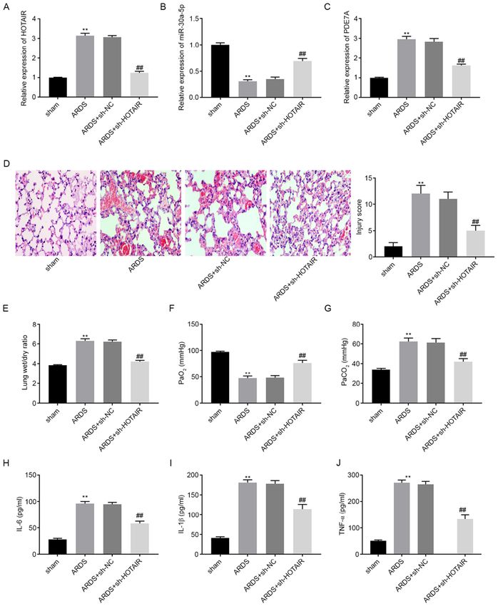
EXPERIMENTAL AND THERAPEUTIC MEDICINE 22: 1160, 2021                                                         7

Figure 6. lncRNA HOTAIR knockdown attenuates LPS‑induced ARDS and ARDS‑related inflammatory response in vivo. The mRNA expression level of
(A) HOTAIR, (B) miR‑30a‑5p and (C) PDE7A in the lung tissues of mice was detected using reverse transcription‑quantitative PCR. (D) Histopathological
change and injury score were detected using H&E staining. Magnification, x200. (E) Lung wet/dry weight ratio, (F) PaO2 and (G) PaCO2 was measured using
an automatic blood gas analyzer. The concentration of (H) IL‑6, (I) IL‑1β and (J) TNF‑α in BALF of mice was measured using ELISA. **P
8 WANG et al: REGULATORY MECHANISM OF HOTAIR ON LPS-INDUCED ARDS
ARDS (25). Several lncRNAs have been associated with the LPS‑treated alveolar epithelial cells. Guo et al (30) showed
progression of ALI and ARDS (7‑9). Dai et al (7) found high that miR‑497a‑5p overexpression decreased the concentration
protein expression levels of MALAT1 in the lung tissues of IL‑6, IL‑1β and TNF‑α in LPS‑treated RAW264.7 cells.
from an LPS‑induced rat ALI model. Wang et al (8) showed Li et al (13) reported that high expression levels of miR‑150
that THRIL expression was upregulated in lung tissues inhibited the secretion of IL‑6, IL‑1β and TNF‑ α from
from patients with ARDS. Zhou et al (9) demonstrated that LPS‑treated human pulmonary epithelial cells. In the present
NEAT1 was highly expressed in the lung tissues of mice with study, it was found that miR‑30a‑5p upregulation suppressed
LPS‑induced ALI and in LPS‑treated mouse alveolar epithelial the secretion of IL‑6, IL‑1β and TNF‑ α from LPS‑treated
cells. Consistent with the aforementioned results, the present MLE‑12 cells. These results suggested that miR‑30a‑5p may
study showed that HOTAIR mRNA expression levels were be a suppressor of the inflammatory response. Consistent with
increased in the lung tissues from mice with LPS‑induced these results, miR‑30a‑5p inhibition has been shown to promote
ARDS and in LPS‑treated MLE‑12 cells. Therefore, the the release of IL‑6, IL‑1β and TNF‑α from PC‑12 cells (31). In
results from the present study suggested that HOTAIR may act addition, miR‑30a‑5p was shown to be the target of HOTAIR in
as a pathogenic factor in ARDS. the present study. Downregulation of miR‑30a‑5p reversed the
Previous studies have reported that lncRNAs act as positive effect of sh‑HOTAIR on MLE‑12 cell viability and the
crucial regulators in hypoxic lung diseases and inflamma‑ inhibitory effect of sh‑HOTAIR on inflammatory factor release
tory epithelial injury (26,27), by affecting processes, such as from MLE‑12 cells. This suggested that HOTAIR knockdown
inflammation and interstitial oedema (7,28,29). Dai et al (7) alleviated the LPS‑induced inflammatory response by modu‑
have shown that MALAT1 inhibition led to a marked decrease lating miR‑30a‑5p.
in the concentration of IL‑6, IL‑1β and TNF‑ α in rat lung En z ymes wit h i n t he phospho d iest era se ( PDE)
tissues. In addition, histopathological examination showed family are found in most proinflammatory and immune
that MALAT1 inhibition distinctly attenuated lung tissue cells (24). As a member of the PDE family, PDE7A has
injury. Jiang et al (29) showed that the PaO2/FiO2 ratio was been associated with the inflammatory response (24,32-
decreased and the lung wet/dry weight ratio was increased in 34). Goto et al (32) reported that PDE7A downregulation
ARDS. Kcnq1ot1 knockdown in mice was shown to reverse ameliorated concanavalin A‑induced hepatitis in mice
the suppressive effect of LPS on the PaO2/FiO2 ratio and the by suppressing the concentration of IL‑4 and TNF‑ α.
positive effect of LPS on the lung wet/dry weight ratio (29). Kadoshima‑Yamaoka et al (33) showed that ASB16165, a
Consistent with the aforementioned results, in the present PDE7A inhibitor, reduced cutaneous TNF‑ α concentration in
study, it was found that injury scores, lung wet/dry weight 12‑o‑tetradecanoylphorbol‑13‑acetate‑induced skin inflam‑
ratios, PaCO2 levels and the concentration of IL‑6, IL‑1β mation in mice. Yamamoto et al (34) demonstrated that
and TNF‑α were increased, and PaO2 levels were decreased YM‑393059, an inhibitor of PDE7A, inhibited LPS‑induced
in a mouse model of ARDS. HOTAIR knockdown using a TNF‑ α production in mice. In the present study, it was found
sh HOTAIR lentivirus injection reversed the positive effect of that PDE7A protein expression levels were upregulated
LPS on injury scores, lung wet/dry weight ratios, PaCO2 levels, in LPS‑treated MLE‑12 cells and PDE7A overexpression
and the concentration of IL‑6, IL‑1β and TNF‑ α, and the accelerated inflammatory factor release from the MLE‑12
negative effect of LPS on PaO2 levels. These results suggested cells. These results suggested that PDE7A was associated
that HOTAIR knockdown attenuated the LPS‑induced inflam‑ with promoting the secretion of the inflammatory factors,
matory response and LPS‑induced ALI. To further verify whereas inhibiting PDE7A may be beneficial in preventing
this hypothesis, further experiments were performed using inflammation in ARDS. In addition, PDE7A overexpression
the MLE‑12 cells and it was found that sh‑HOTAIR reversed reversed the positive effect of sh‑HOTAIR on cell viability
the negative effect of LPS on cell viability and the positive and the inhibitory effect of sh‑HOTAIR on inflammatory
effect of LPS on the concentration of IL‑6, IL‑1β and TNF‑α. factor release from the MLE‑12 cells. This suggested
The results from the present study are consistent with those that HOTAIR knockdown attenuated the LPS‑induced
of other studies (10‑12), suggesting that HOTAIR knockdown inflammatory response by regulating PDE7A. At the same
attenuated the LPS‑induced inflammatory response and time, PDE7A was the target gene of miR‑30a‑5p. We further
LPS‑induced ALI in vivo and in vitro. hypothesized that knockdown of HOTAIR ameliorated
Increasing evidence has indicated that miRNA expres‑ the LPS‑induced inflammatory response by regulating
sion levels were downregulated in LPS‑induced rat models the miR‑30a‑5p/PDE7A axis.
of ALI or LPS‑treated cells. For example, miR‑497a‑5p (30) Taken together, the results of the current study showed
and miR‑381‑3p (29) expression levels were downregulated that HOTAIR knockdown ameliorated the LPS‑induced
in an LPS‑induced mouse model of ALI, while miR‑424 (14) inflammatory response and ARDS in vivo and reduced
expression levels were downregulated in alveolar epithelial LPS‑induced inflammatory factor production by regu‑
cells, and miR‑146b (15) expression levels were downregulated lating the miR‑30a‑5p/PDE7A axis in vitro. However, there
in murine lung alveolar macrophages. Similarly, in the present were also some limitations. First, the detailed mechanism
study it was found that miR‑30a‑5p expression levels were involving HOTAIR, miR‑30a‑5p and PDE7A was not
downregulated in both an LPS‑induced mouse model of ARDS confirmed in vivo. Second, the detailed mechanism of this axis
and in LPS‑treated MLE‑12 cells. In addition, previous studies and the signaling pathways involved remain unclear. Further
have shown that miRNAs have a role in modulating pulmonary experiments will be performed to elucidate these in the future.
inflammation (13,14,30). Cheng et al (14) showed that upregula‑ In conclusion, these findings may contribute to the develop‑
tion of miR‑424 suppressed the secretion of IL‑6 and IL‑8 from ment of a new strategy for treating ARDS.EXPERIMENTAL AND THERAPEUTIC MEDICINE 22: 1160, 2021 9
Acknowledgements 9. Zhou H, Wang X and Zhang B: Depression of lncRNA NEAT1
Antagonizes LPS‑evoked acute injury and inflammatory
response in alveolar epithelial cells via HMGB1‑RAGE signaling.
Not applicable. Mediators Inflamm 2020: 8019467, 2020.
10. Duan G, Song S and Niu S: WITHDRAWN: Long non‑coding
RNA HOTAIR promotes LPS‑induced inflammatory injury
Funding by down‑regulation of microRNA‑124 in murine chondrogenic
ATDC5 cells. Life Sci: July 20, 2018 (Epub ahead of print).
No funding was received. 11. Obaid M, Udden SMN, Deb P, Shihabeddin N, Zaki MH and
Mandal SS: LncRNA HOTAIR regulates lipopolysaccha‑
ride‑induced cytokine expression and inflammatory response in
Availability of data and materials macrophages. Sci Rep 8: 15670, 2018.
12. Wu H, Liu J, Li W, Liu G and Li Z: LncRNA‑HOTAIR promotes
TNF‑ α production in cardiomyocytes of LPS‑induced sepsis
All data generated or analyzed during this study are included mice by activating NF‑ κ B pathway. Biochem Biophys Res
in this published article. Commun 471: 240‑246, 2016.
13. Li P, Yao Y, Ma Y and Chen Y: MiR‑150 attenuates LPS‑induced
acute lung injury via targeting AKT3. Int Immunopharmacol 75:
Authors' contributions 105794, 2019.
14. Cheng D, Zhu C, Liang Y, Xing Y and Shi C: MiR‑424 over‑
XM conceived and designed the present study. HW and SS expression protects alveolar epithelial cells from LPS‑induced
apoptosis and inflammation by targeting FGF2 via the NF‑κ B
performed the experiments, analyzed the data and drafted the pathway. Life Sci 242: 117213, 2020.
article. XM revised the article critically for important intel‑ 15. He R, Li Y, Zhou L, Su X, Pan P and Hu C: miR‑146b overexpres‑
lectual content. XM, HW and SS confirm the authenticity of sion ameliorates lipopolysaccharide‑induced acute lung injury
in vivo and in vitro. J Cell Biochem 120: 2929‑2939, 2019.
all the raw data. All authors have read and approved the final 16. Zhou T and Chen YL: The functional mechanisms of miR‑30b‑5p
manuscript. in acute lung injury in children. Med Sci Monit 25: 40‑51, 2019.
17. Zhang Y, Ai H, Fan X, Chen S, Wang Y and Liu L: Knockdown
of long non‑coding RNA HOTAIR reverses cisplatin resistance
Ethics approval and consent to participate of ovarian cancer cells through inhibiting miR‑138‑5p‑regulated
EZH2 and SIRT1. Biol Res 53: 18, 2020.
All experimental procedures were conducted in agreement 18. Wei Z, Chen L, Meng L, Han W, Huang L and Xu A: LncRNA
HOTAIR promotes the growth and metastasis of gastric cancer
with the principles approved by the ethical committee of Yantai by sponging miR‑1277‑5p and upregulating COL5A1. Gastric
Yuhuangding hospital (Yantai, China; approval no. 2019017). Cancer 23: 1018‑1032, 2020.
19. Zhang S, Wang B, Xiao H, Dong J, Li Y, Zhu C, Jin Y, Li H,
Cui M and Fan S: LncRNA HOTAIR enhances breast cancer
Patient consent for publication radioresistance through facilitating HSPA1A expression via
sequestering miR‑449b‑5p. Thorac Cancer 11: 1801‑1816, 2020.
Not applicable. 20. Wang Y, Gong G, Xu J, Zhang Y, Wu S and Wang S: Long
noncoding RNA HOTAIR promotes breast cancer development
by targeting ZEB1 via sponging miR‑601. Cancer Cell Int 20:
Competing interests 320, 2020.
21. Zhang C, Xu L, Deng G, Ding Y, Bi K, Jin H, Shu J, Yang J,
Deng H, Wang Z and Wang Y: Exosomal HOTAIR promotes
The authors declare that they have no competing interests. proliferation, migration and invasion of lung cancer by sponging
miR‑203. Sci China Life Sci 63: 1265‑1268, 2020.
References 22. Livak KJ and Schmittgen TD: Analysis of relative gene expres‑
sion data using real‑time quantitative PCR and the 2(‑Delta Delta
C(T)) method. Methods 25: 402‑408, 2001.
1. Patel VJ, Roy SB, Mehta HJ, Joo M and Sadikot RT: Alternative 23. Guo Z, Li Q, Han Y, Liang Y, Xu Z and Ren T: Prevention of
and Natural therapies for acute lung injury and acute respiratory LPS‑induced acute lung injury in mice by progranulin. Mediators
distress syndrome. Biomed Res Int 2018: 2476824, 2018. Inflamm 2012: 540794, 2012.
2. Bellani G, Laffey JG, Pham T, Fan E, Brochard L, Esteban A, 24. Smith SJ, Brookes‑Fazakerley S, Donnelly LE, Barnes PJ,
Gattinoni L, van Haren F, Larsson A, McAuley DF, et al: Barnette MS and Giembycz MA: Ubiquitous expression of phos‑
Epidemiology, patterns of care, and mortality for patients with phodiesterase 7A in human proinflammatory and immune cells.
acute respiratory distress syndrome in intensive care units in Am J Physiol Lung Cell Mol Physiol 284: L279‑L289, 2003.
50 countries. JAMA 315: 788‑800, 2016. 25. Petrucci N and De Feo C: Lung protective ventilation strategy for
3. Villar J, Blanco J and Kacmarek RM: Current incidence and the acute respiratory distress syndrome. Cochrane Database Syst
outcome of the acute respiratory distress syndrome. Curr Opin Rev 2013: CD003844, 2007.
Crit Care 22: 1‑6, 2016. 26. Sun H, Chen J, Qian W, Kang J, Wang J, Jiang L, Qiao L,
4. Tsai CL, Lin YC, Wang HM and Chou TC: Baicalein, Chen W and Zhang J: Integrated long non‑coding RNA analyses
an active component of Scutellaria baicalensis, protects identify novel regulators of epithelial‑mesenchymal transition
against lipopolysaccharide‑induced acute lung injury in rats. in the mouse model of pulmonary fibrosis. J Cell Mol Med 20:
J Ethnopharmacol 153: 197‑206, 2014. 1234‑1246, 2016.
5. Opitz B, Van Laak V, Eitel J and Suttorp N: Innate immune 27. Liang H, Gu Y, Li T, Zhang Y, Huangfu L, Hu M, Zhao D, Chen Y,
recognition in infectious and noninfectious diseases of the lung. Liu S, Dong Y, et al: Integrated analyses identify the involvement
Am J Respir Crit Care Med 181: 1294‑1309, 2010. of microRNA‑26a in epithelial‑mesenchymal transition during
6. Han S and Mallampalli RK: The acute respiratory distress syndrome: idiopathic pulmonary fibrosis. Cell Death Dis 5: e1238, 2014.
From mechanism to translation. J Immunol 194: 855‑860, 2015. 28. Dunkel B: Acute lung injury and acute respiratory distress
7. Dai L, Zhang G, Cheng Z, Wang X, Jia L, Jing X, Wang H, syndrome in foals. Equine Vet J 5: 127‑133, 2006.
Zhang R, Liu M, Jiang T, et al: Knockdown of LncRNA 29. Jiang X, Yu M, Zhu T, Lou L, Chen X, Li Q, Wei D and Sun R:
MALAT1 contributes to the suppression of inflammatory Kcnq1ot1/miR‑381‑3p/ETS2 axis regulates inflammation in
responses by up‑regulating miR‑146a in LPS‑induced acute lung mouse models of acute respiratory distress syndrome. Mol Ther
injury. Connect Tissue Res 59: 581‑592, 2018. Nucleic Acids 19: 179‑189, 2020.
8. Wang Y, Fu X, Yu B and Ai F: Long non‑coding RNA THRIL 30. Guo S, Chen Y, Liu J, Yang J, Yang C, Zhang T, Jiang K, Wu Z,
predicts increased acute respiratory distress syndrome risk and Shaukat A and Deng G: miR‑497a‑5p attenuates lipopolysaccha‑
positively correlates with disease severity, inflammation, and ride‑induced inflammatory injury by targeting IRAK2. J Cell
mortality in sepsis patients. J Clin Lab Anal 33: e22882, 2019. Physiol 234: 22874‑22883, 2019.10 WANG et al: REGULATORY MECHANISM OF HOTAIR ON LPS-INDUCED ARDS
31. Zhu S, Zhou Z, Li Z, Shao J, Jiao G, Huang YE and Lin Y: 33. Kadoshima‑Yamaoka K, Goto M, Murakawa M, Yoshioka R,
Suppression of LINC00707 alleviates lipopolysaccha‑ Tanaka Y, Inoue H, Murafuji H, Kanki S, Hayashi Y,
ride‑induced inflammation and apoptosis in PC‑12 cells by Nagahira K, et al: ASB16165, a phosphodiesterase 7A inhibitor,
regulated miR‑30a‑5p/Neurod 1. Biosci Biotechnol Biochem 83: reduces cutaneous TNF‑alpha level and ameliorates skin edema in
2049‑2056, 2019. phorbol ester 12‑O‑tetradecanoylphorbol‑13‑acetate‑induced skin
32. Goto M, Tanaka Y, Murakawa M, Kadoshima‑Yamaoka K, inflammation model in mice. Eur J Pharmacol 613: 163‑166, 2009.
Inoue H, Murafuji H, Nagahira A, Kanki S, Hayashi Y, 34. Yamamoto S, Sugahara S, Naito R, Ichikawa A, Ikeda K, Yamada T
Nagahira K, et al: Inhibition of phosphodiesterase 7A and Shimizu Y: The effects of a novel phosphodiesterase 7A and ‑4
ameliorates Concanavalin A‑induced hepatitis in mice. Int dual inhibitor, YM‑393059, on T‑cell‑related cytokine production
Immunopharmacol 9: 1347‑1351, 2009. in vitro and in vivo. Eur J Pharmacol 541: 106‑114, 2006.You can also read