A Case of Gastric Adenocarcinoma in a Shih Tzu Dog: Successful Treatment of Early Gastric Cancer
←
→
Page content transcription
If your browser does not render page correctly, please read the page content below
NOTE      Internal medicine

A Case of Gastric Adenocarcinoma in a Shih Tzu Dog: Successful Treatment of Early
Gastric Cancer

Hee-Chun LEE1), Ji-Hyun KIM1), Cho-Hee JEE1), Jae-Hoon LEE1), Jong-Hyun MOON1), Na-Hyun KIM2),
Jung-Hyang SUR2), Kyu-Woan CHO1), Byeong-Teck KANG3), Jeongim HA4) and Dong-In JUNG1)*

1)Research Institute of Life Sciences, College of Veterinary Medicine, Gyeongsang National University, Jinju 660–701, South Korea
2)Department  of Pathobiology, Small Animal Tumor Diagnostic Center, College of Veterinary Medicine, Konkuk University, Seoul
  143–701, South Korea
3)Laboratory of Veterinary Dermatology and Neurology, College of Veterinary Medicine, Chungbuk National University, Cheongju,

  Chungbuk, 361–763, South Korea
4)Department of Cell and Developmental Biology, School of Dentistry, DRI and Brain Korea 21 Program, Seoul National University,

  Seoul 110–749, South Korea

(Received 17 June 2013/Accepted 5 March 2014/Published online in J-STAGE 20 March 2014)

ABSTRACT. A 9-year-old castrated male Shih Tzu dog was referred to us, because of chronic vomiting. The patient’s hematological, radio-
graphic, ultrasonographic, endoscopic and histological examinations were evaluated for diagnosis. Hematologic analysis indicated moderate
anemia and azotemia. Based on the imaging studies, an oval-shaped mass was identified in the gastric pylorus area. A proliferative mass was
found on endoscopic examination, and we performed biopsy using grasping forceps. The histopathological findings of the biopsy specimens
indicated hypertrophic gastritis, and Y-U pyloroplasty was performed. However, histopathological examination of the surgically resected
mass revealed tubular adenocarcinoma of the stomach. Then, carboplatin chemotherapy was performed 4 times for 13 weeks. Clinical signs,
such as vomiting, were resolved gradually after surgery and chemotherapy, and the patient’s condition was managed favorably until recently
(30 months after surgery). This case report describes clinical features, imaging studies, endoscopic characteristics and histopathological and
immunohistochemical features of gastric tubular adenocarcinoma as early gastric cancer in a dog.
KEY WORDS: adenocarcinoma, canine, early gastric cancer (EGC), gastric hyperplasia.

                                                                             doi: 10.1292/jvms.13-0315; J. Vet. Med. Sci. 76(7): 1033–1038, 2014

   Tumors of the canine gastrointestinal (GI) tract are                          A 9-year-old castrated male Shih Tzu dog presented with
uncommon and account for less than 1% of all reported                         a history of chronic vomiting and episodic melena. Accord-
neoplasms [4, 10, 36]. Evidence for the etiology of canine                    ing to the case history, vomiting sign was observed 8 months
gastric tumor has not been defined until now [10]. Malignant                  before presentation. A GI protectant was administered at
gastric tumors have been reported more than benign tumors,                    a local animal hospital, and no neoplastic changes were
followed by adenocarcinoma, leiomyosarcoma, lymphoma,                         found on GI endoscopy. Since moderate anemia (hemato-
leiomyoma and adenoma. Adenocarcinoma is the most com-                        crit: 25%) was identified based on a complete blood count,
mon gastric malignancy in dogs, comprising 47 to 73% of                       erythropoietin was given to the patient at the local animal
all gastric tumors [12, 14, 28, 31]. The World Health Or-                     hospital. The patient was referred to us, because the chronic
ganization (WHO) classifies gastric carcinomas in domestic                    vomiting and anemia were not resolved after management
animals, dividing them into 5 types: tubular carcinoma, mu-                   at the local animal hospital. At the time of initial presenta-
cinous carcinoma, signet ring cell carcinoma, undifferenti-                   tion, moderate to severe anemia (hematocrit: 19.6%) and
ated carcinoma and papillary carcinoma [13]. The prognosis                    increased alkaline phosphatase and blood urea nitrogen were
of adenocarcinoma is mostly guarded, and most studies of                      revealed in a blood analysis (alkaline phosphatase, 743 U/l
this tumor type report that the majority of patients survive                  [reference range; 20–150 U/l]; blood urea nitrogen, 76 mg/dl
less than 6 months [2, 6, 8, 10, 15].                                         [reference range; 7–25 mg/dl]). The survey abdominal ra-
   This case report describes clinical features, imaging stud-                diographs showed distension of the stomach, accumulation
ies, endoscopic characteristics and histopathological features                of gas in the esophagus and a soft tissue opacity mass, which
of canine tubular adenocarcinoma of the stomach in a dog.                     was summated with the gastric pylorus (Fig. 1). Abdominal
                                                                              ultrasonography showed an oval-shaped mass of mixed
*Correspondence to: Jung D.-I., Research Institute of Life Sci-               echogenicity in the gastric pylorus, which was surrounded
 ences, College of Veterinary Medicine, Gyeongsang National                   by thickend muscularis layers and gastric dilation with fluid
 University, Jinju 660–701, South Korea.                                      and food (Fig. 2). In a color Doppler test, the muscularis
 e-mail: jungdi@gnu.ac.kr                                                     layers exhibited vascular flow. These gastric changes were
©2014 The Japanese Society of Veterinary Science                              considered to indicate a gastric neoplasm.
This is an open-access article distributed under the terms of the Creative       A gastrointestinal endoscopic examination was per-
Commons Attribution Non-Commercial No Derivatives (by-nc-nd)                  formed, and a proliferative mass was found in the pyloric
License .                  outflow tract (Fig. 3A). Multiple biopsy samples were taken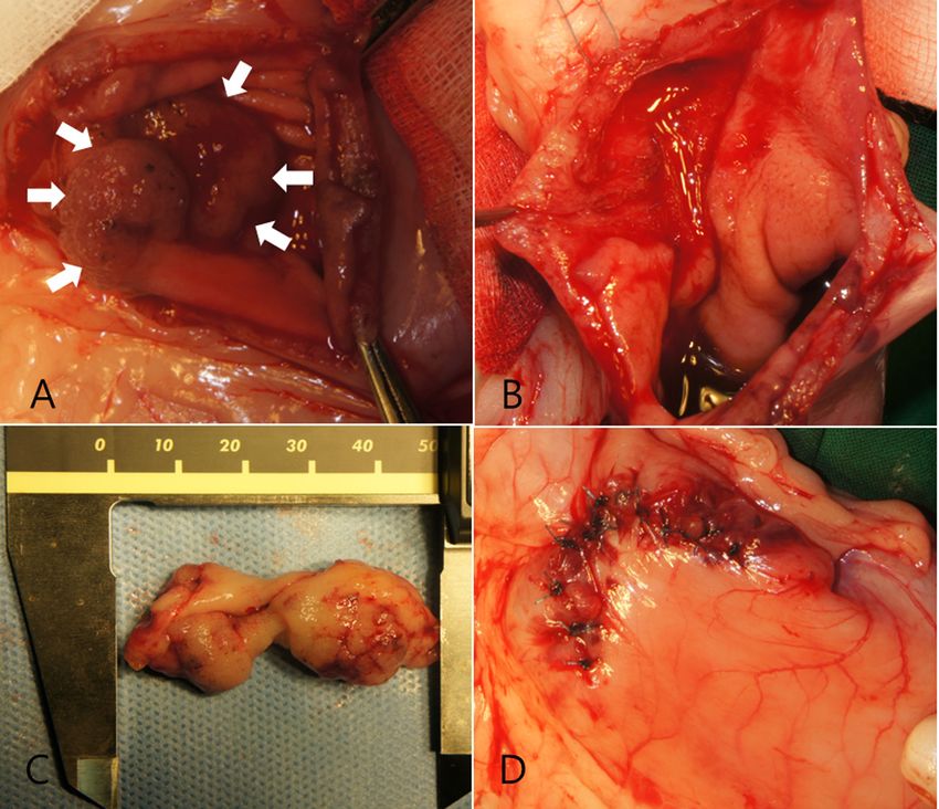
1034 H. LEE ET AL.
Fig. 1. Survey lateral abdominal radiography shows (A) the presence of a well-defined,
smooth, oval-shaped mass with soft tissue opacity overlapped with the stomach caudal to the
liver and (B) enhancement of the soft tissue opacity at the cranial and middle regions of the
abdomen and distension of the stomach with food, with the stomach deviating laterally and
being positioned vertically.
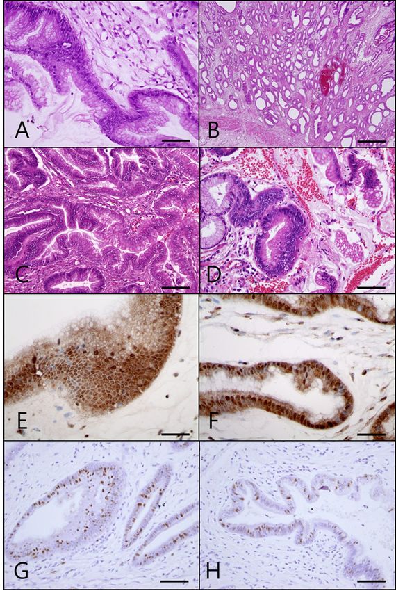
with grasping forceps (Fig. 3B), and the histopathological tions were placed in a 65°C oven for 20 min, dewaxed and
results for these specimens indicated hypertrophic gastritis rehydrated through xylene and graded ethanol solutions to
(Fig. 5A). Then, we performed marginal resection of the phosphate buffered saline (PBS; pH 7.4, 0.1 M). Endog-
tumor and a Y-U pyloroplasty (Fig. 4). After the surgery, the enous peroxidase was blocked by incubating the sections in
patient was given partial parenteral nutrition therapy for 10 3% H202 in PBS for 20 min at room temperature (RT). After
days, because of vomiting. Eleven days after the surgery, the three washes in PBS, antigens were retrieved by heating in
patient was allowed the liquid food, and clinical signs were citrate buffer (pH 6.0) for 20 min in a microwave oven (650
gradually alleviated including vomiting and diarrhea. W at high power). Each section was then overlaid with pri-
The histologic results for the surgically resected masses mary antibodies to CDX-2 (1:200) or Ki-67 (1:300) diluted
indicated intestinal type (tubular type) gastric adenocarci- in PBS. Sections were incubated with primary reagent at 4°C
noma. The gastric adenocarcinoma was formed by glands overnight and then washed in PBS three times. EnVision
with various degrees of differentiation (Fig. 5C and 5D). system-HRP (DakoCytomation, Carpinteria, CA, U.S.A.)
The gastric lesion was composed of well-formed tubules, was applied for detection of binding of primary reagents.
some of which were cystically dilated (Fig. 5B). Based on Sections were counterstained with Harris’s hematoxylin,
the histopathological findings, we strongly suspected gastric dehydrated and coverslipped under Permount™ (Fisher
adenocarcinoma. To confirm our tentative diagnosis, we Scientific, Fair Lawn, NJ, U.S.A.). Intense nuclear CDX-2
performed additional immunohistochemistry to evaluate the expression was noted in moderately differentiated tumor
expression of CDX-2 (Abcam, Cambridge, MA, U.S.A.) cells (Fig. 5E and 5F). Moderate expression of nuclear stain-
and Ki-67 (Clone MIB-1, Dako, Glostrup, Denmark). Sec- ing for Ki-67 was seen in tumor cells (Fig. 5G and 5H). The
Fig. 2. Abdominal ultrasonography shows (A) vascular flow of the pyloric region in the Doppler test, (B) the presence of thickening of the
gastric wall and (C) no pyloric obstruction in postoperative abdominal ultrasonography. (D) Fourteen months after surgery, a decrease in
gastric wall thickness can be seen.
Fig. 3. Serial endoscopic findings of the present patient. (A) Obstruction of the pyloric region by a proliferative mass was identified in the initial
examination. (B) Several endoscopic biopsy samples were obtained from the lesion. (C) Five months after surgery, occlusion of the pyloric
region was resolved. (D) Nineteen-months after surgery, the pylorus was open, and there were no ulcerative or hemorrhagic lesions.
Fig. 4. Resection of the mass and Y-U pyloroplasty. (A) Intraoperative photograph showing the mass at the pyloric region after gastrotomy
(arrows). (B) Resection of the mass is complete. The defect was closed by re-opposing the mucosa and submucosa with the Cushging pattern.
(C) The resected mass of the pyloric area. The margin of the mass, including the mucosa and part of the submucosa layer, was peeled off the
pylorus. (D) A completed Y-U antral advanced flap.
EARLY GASTRIC CANCER IN A DOG                                                     1035

Fig. 2.

                          Fig. 5. Histopathological results of endoscopic biopsy specimens
                             (A) and surgically resected specimens (B, C, D, E, F, G and
                             H). Panels A, B, C and D show hematoxylin and eosin staining
                             features. Panels E, F and G show CDX-2 immunohistochemical
                             features of surgically resected specimens. Panel H shows p53 im-
                             munohistochemical features of surgically resected specimens. (A)
                             A high power image shows the involved gastric pit (crypts), which
Fig. 3                       contains enlarged hyperchromatic nuclei that became crowded nu-
                             clei (scale bar: 32 µm). (B) The gastric lesion is composed of well-
                             formed tubules, some of which are cystically dilated (scale bar:
                             467 µm). (C and D) Intestinal type (tubular type) gastric adenocar-
                             cinoma formed by glands with various degrees of differentiation
                             (scale bar: 47 µm). (E and F) CDX-2 expression in a moderately
                             differentiated tubular type of canine gastric adenocarcinoma. High
                             power images show strong nuclear expressions of CDX-2 in mod-
                             erately differentiated tumor cells in the present patient (scale bar:
                             32 µm). (G and H) Moderate expression of nuclear staining for
                             Ki-67 seen in the present patient (scale bar: 47 µm).

Fig. 4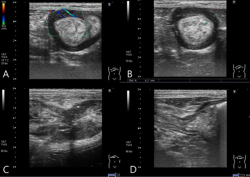
1036                                                      H. LEE ET AL.

expression of Ki-67 was partially positive, indicating that       present patient was restricted to the mucosal and submucosal
the tumor cells had progressed to a malignant form. Based         areas, and there was no metastasis to other organs or lymph
on the histopathological and immunohistochemical findings,        nodes. Furthermore, there were sessile nodules of mucosal
this case was definitively diagnosed as an early stage canine     proliferation instead of ulcerative, hemorrhage and infiltra-
gastric adenocarcinoma.                                           tive lesions which are seen in most cases of adenocarcinoma
   Unfortunately, marginal resection of the tumor and Y-U         on endoscopy.
pyloroplasty had been performed based on the endoscopic              CDX-2 is an intestine-specific homeobox product that
biopsy results (hypertrophic gastritis), and we could not be      plays a key role in the regulation, proliferation and differen-
confident that all cancer cells had been removed during the       tiation of intestinal epithelial cells during normal embryonic
surgery. Therefore, we decided to treat with carboplatin che-     and postnatal development. CDX-2 is known as an impor-
motherapy (Carbotinol®, Korea United Pharm., Seoul, South         tant nuclear transcription factor that regulates development
Korea; 250 mg/m2 for the first injection and then 200 mg/m2       of intestinal metaplasia and gastric carcinogenesis [5, 21,
3 times during 10 weeks). Then, we regularly performed            32, 35]. According to previous reports [5, 16, 22, 27, 35],
hematologic, radiographic and ultrasonographic examina-           CDX-2 is strongly associated with the intestinal phenotypic
tions to evaluate the dog for metastasis or recurrence of the     expression in gastric carcinomas.
cancer. We performed thoracic and abdominal computed                 One recent veterinary report [5] demonstrated that all ca-
tomographic (CT) examinations 2 times after surgery (5            nine gastric adenocarcinomas in the study expressed CDX-2
and 11 months after surgery), and the results were normal.        and that 86.4% of the colorectal adenocarcinomas expressed
Furthermore, gastrointestinal endoscopic examinations were        CDX-2. Interestingly, that report [5] indicated that CDX-2
performed again, and there were no remarkable findings            expression was detected in the normal canine colorectal mu-
(Fig. 3C and 3D). The status of the patient was well con-         cosa, but was not found in the normal canine gastric mucosa.
trolled until recently (more than 30 months after surgery).       Based on those study results [5], CDX-2 expression could be
   Canine carcinoma is the most common gastric neoplasm           useful for the diagnosis of canine gastric adenocarcinoma, as
in the dog [9, 24]. The WHO classifies this malignancy in         it was not detected in the normal canine gastric epithelium.
domestic animals based on the growth pattern of the car-          One other report suggested CDX-2 expression was increased
cinoma, but the relation between the histological type and        in gastric carcinomas with less invasiveness and an intestinal
other characteristics associated with the prognosis has not       phenotype, but CDX-2 expression was decreased in ad-
been previously reported [2, 13]. Carcinoma of the canine         vanced gastric carcinomas [16]. Strong nuclear expressions
stomach may arise in any part of the stomach. Canine gas-         of CDX-2 in moderately differentiated tumor cells were no-
tric carcinomas can assume a wide range of forms, but they        ticed in the present patient. However, Whether or not CDX-2
mostly have the features of tubular adenocarcinoma. This          is expressed in nonneoplastic conditions, e.g. hypertrophic
case was a mucosa form and was diagnosed as intestinal type       gastritis, is unknown, although the normal gastric mucosa is
(tubular type) canine gastric adenocarcinoma.                     almost negative.
   The diagnosis of gastric neoplasms is usually based on            Thus, we performed Ki-67 immunohistochemistry as pro-
clinical features and imaging findings and is confirmed by        liferative marker for evaluating malignancy in the present
histopathological and immunohistochemical examinations            patient. According to previous reports [18, 23], Ki-67 is a
[19]. Endoscopy is a useful definitive diagnostic investigation   nonhistone protein with expression that peaks during G2/M,
for observing the size, location and morphology of a tumor        so tumors with a higher proliferation rate demonstrate great-
and for obtaining biopsy samples [10, 20]. In humans, over        er Ki-67 expression. In the present patient, positive Ki-67
90% of tumor cases can be diagnosed by endoscopy [14].            expression was detected in tumor cells, and this suggested a
In the present case, the histologic result for the endoscopic     malignant tumor of the gastric mucosa.
forceps biopsy specimens was hypertrophic gastritis, but the         Therefore, the immunohistochemistry results for CDX-2
result for the surgically resected specimens was gastric tubu-    and Ki-67 could support the diagnosis of early stage gastric
lar adenocarcinoma. Interestingly, histologic discrepancies       adenocarcinoma (early gastric cancer) in the present patient.
have been reported for gastric cancer between endoscopic             Surgery is the only potentially effective treatment for
forceps biopsy and surgical endoscopic treatment sample in        localized canine gastric carcinoma [10, 24], and partial gas-
human medicine with the incidence ranging from 25 to 35%          trectomy is usually indicated for this malignancy [7]. Gener-
[11, 25]. According to one report of human gastric cancer,        ally, the prognosis after surgery is poor, and most affected
this discrepancy is found frequently in early gastric cancer      dogs expired within 6 months of diagnosis [3]. In several
(EGC). EGC is defined as adenocarcinoma confined to the           studies that described the prognosis of surgical treatment
mucosa or submucosa, irrespective of lymph node involve-          for canine adenocarcinoma, the median survival time was
ment, in human medicine [9]. Because endoscopy cannot             only 55 days for 29 dogs treated with surgery. [8, 10, 33].
detect diseases that primarily involve the submucosal, mus-       In human cases, endoscopic submucosal dissection or surgi-
cularis or serosal layers of the stomach and the size of the      cal resection is indicated for EGC [17]. In the present case,
biopsy sample is restricted by the cup size [20], EGC may         we performed surgical submucosal resection of the mass
not be identified with this procedure. Based on these stud-       and Y-U pyloroplasty for obstruction of the pyloric outflow
ies, the gastric tumor in the present case could be thought       tract based on the initial histopathological results. We then
of as equivalent to EGC in humans. The gastric tumor in the       initiated chemotherapy since identified malignant change
EARLY GASTRIC CANCER IN A DOG                                                            1037

of the mass. According to one study in humans, adjuvant                  9. Goldstein, N. S. and Lewin, K. J. 1997. Gastric epithelial dyspla-
chemotherapy was effective for prevention of postoperative                  sia and adenoma: historical review and histological criteria for
recurrence in patients with EGC [30]. In human stomach                      grading. Hum. Pathol. 28: 127–133. [Medline] [CrossRef]
tumor, chemotherapy is effective as an adjuvant therapy                 10. Gualtieri, M., Monzeglio, M. G. and Scanziani, E. 1999. Gastric
                                                                            Neoplasia. Vet. Clin. North Am. Small Anim. Pract. 29: 415–440.
followed by surgical resection, improving overall survival
                                                                            [Medline]
[26, 29]. Carboplatin chemotherapy for adenocarcinoma is                11. Hansson, L. E., Lindgren, A. and Nyrén, O. 1996. Can endo-
controversial these days, but carboplatin has been shown to                 scopic biopsy specimens be used for reliable Laurén classifica-
have an antitumor effect in human gastric tumor cells and                   tion of gastric cancer? Scand. J. Gastroenterol. 31: 711–715.
animal models of gastric tumor [1]. Furthermore, carbopla-                  [Medline] [CrossRef]
tin had marginal activity in patients with gastric cancer, with         12. Hayden, D. W. and Nielsen, S. W. 1973. Canine Alimen-
response rates of 6–10% as a single agent [29, 34]. In the                  tary Neoplasia. Zentralbl. Veterinarmed. A 20: 1–22. [Medline]
present case, we used 250 mg/m2 carboplatin for the first in-               [CrossRef]
jection and then used 200 mg/m2 for the 3 subsequent treat-             13. Head, K.W., Cullen, J.M., Dubielzig, R.R., Else, R.W., Misdorp,
ments. Despite the fact that this type of tumor is known to                 W., Patnaik, A.K., Tateyama, S. and Van Der. Gaag, I. 2003. His-
                                                                            tological Classification of Tumors of the Alimentary System of
have a guarded prognosis, the status of the patient has been
                                                                            Domestic Animals, 2nd Series, vol. IX. Armed Forces Institute
well controlled after treatment (30 months after surgery).                  of Pathology, Washington, D.C.
   In conclusion, this report describes the clinical features           14. McLoughlin, J. M. 2004. Adenocarcinoma of the stomach: a
and prognosis of a canine tubular type gastric adenocarci-                  review. Proc. (Bayl. Univ. Med. Cent.). 17: 391–399. [Medline]
noma treated with surgical resection and chemotherapy. We               15. Kapatkin, A. S., Mullen, H. S., Matthien, D. T. and Patnaik, A.
suggest the possibility that a gastric tumor similar to EGC in              K. 1992. Leiomyosarcoma in dogs: 44 cases (1983–1988). J.
humans could exist in dogs. This report is the first report of              Am. Vet. Med. Assoc. 201: 1077–1079. [Medline]
a human EGC-like gastric tumor in dogs. Therefore, more                 16. Kim, G. H., Song, G. A., Park, D. Y., Lee, S. H., Lee, D. H., Kim,
cases of suspected EGC in dogs should be studied in an at-                  T. O., Jo, H. J., Heo, J., Kang, D. H. and Cho, M. 2006. CDX2
tempt to investigate this type of tumor.                                    expression is increased in gastric cancers with less invasiveness
                                                                            and intestinal mucin phenotype. Scand. J. Gastroenterol. 41:
                                                                            880–886. [Medline] [CrossRef]
ACKNOWLEDGMENT. This research was supported by                          17. Kim, J. H., Kim, S. H., Park, W. H., Jang, J. S., Bang, J. S., Yang,
the Basic Science Research Program through the National                     S. H., Byun, J. H. and Kim, Y. J. 2012. Predictable factors of
Research Foundation of Korea (NRF) funded by the Ministry                   histologic discrepancy of gastric cancer between the endoscopic
of Education, Science and Technology (2011-0008358).                        forceps biopsy and endoscopic treatment specimen. Korean J.
                                                                            Gastroenterol. 59: 354–359. [Medline] [CrossRef]
REFERENCES                                                              18. Landberg, G., Tan, E. M. and Roos, G. 1990. Flow cytometric
                                                                            multiparameter analysis of proliferating cell nuclear antigen/
 1. Beer, M., Cavalli, F., Kaye, S. B., Lev, L. M., Clavel, M. and          cyclin and Ki-67 antigen: a new view of the cell cycle. Exp. Cell
    Smyth, J. 1987. A phase II study of carboplatin in advanced             Res. 187: 111–118. [Medline] [CrossRef]
    or metastatic stomach cancer. Eur. J. Cancer Clin. Oncol. 23:       19. Lee, H. C., Kwon, D. H., Moon, J. H., Kim, Y. K., Cho, K. W.,
    1565–1567. [Medline] [CrossRef]                                         Kang, B. T., Im, K. S., Sur, J. H. and Jung, D. I. 2012. Gastric
 2. Carrasco, V., Canfrán, S., Rodríguez-Franco, F., Benito, A.,            Adenoma in the Pyloric Outflow Tract of a Shih-tzu Dog. J. Vet.
    Sáinz, A. and Rodríguez-Bertos, A. 2011. Canine gastric carci-          Clin. 29: 169–172.
    noma; Immunohistochemical expression of cell cycle proteins         20. Moore, L. E. 2003. The advantages and disadvantages of en-
    (p53, p21, and p16) and heat shock proteins (Hsp27 and Hsp70).          doscopy. Clin. Tech. Small Anim. Pract. 18: 250–253. [Medline]
    Vet. Pathol. 48: 322–329. [Medline] [CrossRef]                          [CrossRef]
 3. Nielsen, C. and Anderson, G. M. 2005. Metastasis of gastric         21. Mallo, G. V., Soubeyran, P., Lissitzky, J. C., André, F., Farnarier,
    adenocarcinoma to the abdominal wall following placement of a           C., Marvaldi, J., Dagorn, J. C. and Iovanna, J. L. 1998. Expres-
    gastrostomy tube in a dog. Can. Vet. J. 46: 641–643. [Medline]          sion of the Cdx1 and Cdx2 homeotic genes leads to reduced
 4. Crow, S. E. 1985. Tumors of the alimentary tract. Vet. Clin.            malignancy in colon cancer-derived cells. J. Biol. Chem. 273:
    North Am. Small Anim. Pract. 15: 577. [Medline]                         14030–14036. [Medline] [CrossRef]
 5. Doster, A. R., Yhee, J. Y., Kim, J. H., Im, K. S. and Sur, J.       22. Mazziotta, R. M., Borczuk, A. C., Powell, C. A. and Man-
    H. 2011. CDX-2 and HER-3 expression in canine gastric and               sukhani, M. 2005. CDX2 immunostaining as a gastrointestinal
    colorectal adenocarcinomas. J. Comp. Pathol. 145: 12–19.                marker: expression in lung carcinomas is a potential pitfall.
    [Medline] [CrossRef]                                                    Appl. Immunohistochem. Mol. Morphol. 13: 55–60. [Medline]
 6. Elliott, G. S., Stoffregen, D. A., Richardson, D. C., Blevins, W.       [CrossRef]
    E. and Richardson, R. C. 1984. Surgical, medical, and nutritional   23. Morris, J. S., Nixon, C., King, O. J., Morgan, I. M. and Philbey,
    management of gastric adenocarcinoma in a dog. J. Am. Vet.              A. W. 2009. Expression of TopBP1 in canine mammary neopla-
    Med. Assoc. 185: 98–101. [Medline]                                      sia in relation to histological type, Ki67, ERα and p53. Vet. J.
 7. Fossum, T. W. and Hedlund, C. S. 2003. Gastric and intestinal           179: 422–429. [Medline] [CrossRef]
    surgery. Vet. Clin. North Am. Small Anim. Pract. 33: 1117–1145.     24. Ogilvie, G.K. and Moore, A.S. 1995. Managing the Veterinary
    [Medline] [CrossRef]                                                    Cancer Patient: A Practice Manual, Veterinary Learning System,
 8. Fonda, D., Gualtieri, M. and Scanziani, E. 1989. Gastric carci-         Trenton.
    noma in the dog; clinicopathological study of 11cases. J. Small     25. Palli, D., Bianchi, S. and Cipriani, F. 1991. Reproducibility
    Anim. Pract. 30: 353–360. [CrossRef]                                    of histologic classification of gastric cancer. Br. J. Cancer 63:
1038 H. LEE ET AL.
765–768. [Medline] [CrossRef] [Medline]
26. Paoletti, X., Oba, K., Burzykowski, T., Michiels, S., Ohashi, Y., 32. Silberg, D. G., Swain, G. P., Suh, E. R. and Traber, P. G. 2000.
Pignon, J. P., Rougier, P., Sakamoto, J., Sargent, D., Sasako, M., Cdx1 and cdx2 expression during intestinal development. Gas-
Van, C. E. and Buyse, M. 2010. Benefit of Adjuvant Chemo- troenterology 119: 961–971. [Medline] [CrossRef]
therapy for Resectable Gastric Cancer: A meta-analysis. JAMA 33. Swann, H. M. and Holt, D. E. 2002. Canine Gastric Adenocar-
303: 1729–1737. [Medline] [CrossRef] cinoma and leiomyosarcoma; a retrospective study of 21 cases
27. Park, Y., Srivastava, A., Kim, G. H., Mino-Kenudson, M., Desh- (1986–1999) and literature review. J. Am. Anim. Hosp. Assoc.
pande, V., Zukerberg, L. R., Song, G. A. and Lauwers, G. Y. 38: 157–164. [Medline]
2010. CDX2 expression in the intestinal-type gastric epithelial 34. Takahashi, H., Sasaki, Y., Saijo, N., Sakurai, M., Nakano, H.,
neoplasia: frequency and significance. Mod. Pathol. 23: 54–61. Nakagawa, K., Hoshi, A., Jett, J. R. and Hong, W. S. 1987. In
[Medline] [CrossRef] vitro colony inhibition of carboplatin against stomach and lung
28. Patnaik, A. K., Hurvitz, A. I. and Johnson, G. F. 1977. Canine cancer cell lines in comparison with cisplatin. Cancer Che-
gastrointestinal neoplasms. Vet. Pathol. 14: 547–555. [Medline] mother. Pharmacol. 19: 197–200. [Medline] [CrossRef]
29. Preusser, P., Wilke, H., Achterrath, W., Lenaz, L., Stahl, M. and 35. Werling, R. W., Yaziji, H., Bacchi, C. E. and Gown, A. M. 2003.
Casper, J. 1990. Phase II study of carboplatin in untreated inop- CDX2, a highly sensitive and specific marker of adenocarci-
erable advanced stomach cancer. Eur. J. Cancer 26: 1108–1109. nomas of intestinal origin: an immunohistochemical survey of
[Medline] [CrossRef] 476 primary and metastatic carcinomas. Am. J. Surg. Pathol. 27:
30. Sasaki, A. 1994. Effect of adjuvant chemotherapy for patients 303–310. [Medline] [CrossRef]
with early gastric cancer. Gan To Kagaku Ryoho 21: 37–45. 36. Withrow, S.J. 2007. Gastric cancer. pp. 480–483. In: Withrow &
[Medline] MacEwen’s Small Aminal clinical oncology, 4th ed. (Withrow,
31. Sautter, J. H. and Hanlon, G. F. 1975. Gastric Neoplasms in the S.J. and Vail, D.M., eds.), Saunders, Philadelphia.
Dog: a report of 20 cases. J. Am. Vet. Med. Assoc. 166: 691–696.You can also read