A Prognostic Nomogram of Colon Cancer With Liver Metastasis: A Study of the US SEER Database and a Chinese Cohort - Frontiers
←
→
Page content transcription
If your browser does not render page correctly, please read the page content below
ORIGINAL RESEARCH
                                                                                                                                            published: 26 February 2021
                                                                                                                                         doi: 10.3389/fonc.2021.591009

                                              A Prognostic Nomogram of Colon
                                              Cancer With Liver Metastasis: A
                                              Study of the US SEER Database
                                              and a Chinese Cohort
                                              Chuan Liu 1,2,3, Chuan Hu 4, Jiale Huang 1,2,3, Kanghui Xiang 1,2,3, Zhi Li 1,2,3, Jinglei Qu 1,2,3,
                                              Ying Chen 1,2,3, Bowen Yang 1,2,3, Xiujuan Qu 1,2,3, Yunpeng Liu 1,2,3, Guangwei Zhang 5*
                                              and Ti Wen 1,2,3*
                                              1 Department of Medical Oncology, The First Hospital of China Medical University, Shenyang, China, 2 Key Laboratory of

                                              Anticancer Drugs and Biotherapy of Liaoning Province, The First Hospital of China Medical University, Shenyang, China,
                                              3 Liaoning Province Clinical Research Center for Cancer, Shenyang, China, 4 Medical College, Qingdao University, Qingdao,

                           Edited by:         China, 5 Smart Hospital Management Department, The First Hospital of China Medical University, Shenyang, China
                   Alessandro Vitale,
    University Hospital of Padua, Italy
                                              Background: Among colon cancer patients, liver metastasis is a commonly deadly
                      Reviewed by:
                    Ziv Radisavljevic,
                                              phenomenon, but there are few prognostic models for these patients.
 Brigham and Women’s Hospital and             Methods: The clinicopathologic data of colon cancer with liver metastasis (CCLM)
Harvard Medical School, United States
                     Somaiah Aroori,          patients were downloaded from the Surveillance, Epidemiology and End Results (SEER)
      Plymouth Hospitals NHS Trust,           database. All patients were randomly divided into training and internal validation sets
                    United Kingdom
                                              based on the ratio of 7:3. A prognostic nomogram was established with Cox analysis in
                 *Correspondence:
                   Guangwei Zhang
                                              the training set, which was validated by two independent validation sets.
               gwzhang@cmu.edu.cn             Results: A total of 5,700 CCLM patients were included. Age, race, tumor size, tumor site,
                            Ti Wen
                  wenti@cmu.edu.cn            histological type, grade, AJCC N status, carcinoembryonic antigen (CEA), lung
                                              metastasis, bone metastasis, surgery, and chemotherapy were independently
                   Specialty section:         associated with the overall survival (OS) of CCLM in the training set, which were used
         This article was submitted to
                    Surgical Oncology,
                                              to establish a nomogram. The AUCs of 1-, 2- and 3-year were higher than or equal to
               a section of the journal       0.700 in the training, internal validation, and external validation sets, indicating the
                 Frontiers in Oncology
                                              favorable effects of our nomogram. Besides, whether in overall or subgroup analysis,
         Received: 07 August 2020
                                              the risk score calculated by this nomogram can divide CCLM patients into high-, middle-
        Accepted: 25 January 2021
       Published: 26 February 2021            and low-risk groups, which suggested that the nomogram can significantly determine
                            Citation:         patients with different prognosis and is suitable for different patients.
 Liu C, Hu C, Huang J, Xiang K, Li Z,
  Qu J, Chen Y, Yang B, Qu X, Liu Y,
                                              Conclusion: Higher age, the race of black, larger tumor size, higher grade, histological
        Zhang G and Wen T (2021) A            type of mucinous adenocarcinoma and signet ring cell carcinoma, higher N stage, RCC,
      Prognostic Nomogram of Colon            lung metastasis, bone metastasis, without surgery, without chemotherapy, and elevated
     Cancer With Liver Metastasis: A
     Study of the US SEER Database            CEA were independently associated with poor prognosis of CCLM patients. A nomogram
               and a Chinese Cohort.          incorporating the above variables could accurately predict the prognosis of CCLM.
            Front. Oncol. 11:591009.
     doi: 10.3389/fonc.2021.591009            Keywords: colon cancer with liver metastasis, overall survival, prognostic factors, nomogram, external validation

Frontiers in Oncology | www.frontiersin.org                                          1                                      February 2021 | Volume 11 | Article 591009
Liu et al.                                                                                                                    A Prognostic Nomogram for CCLM

INTRODUCTION                                                                           METHODS
Among all malignant tumors, the incidence and mortality of colon                       Population Selection
cancer (CC) ranked fourth and fifth worldwide in both genders,                          The Surveillance, Epidemiology and End Results (SEER) is a
respectively (1–3). In recent years, owing to the development of                       cancer database based on the US population, which collected
multiple therapeutic strategies [operation, chemotherapy,                              data on cancer patients from 18 registries and covered more than
neoadjuvant chemoradiotherapy, and radiotherapy (RT)], the                             30% of the population (16). The data of patients in the present
prognosis of CC has been improved. For example, Hu et al. (4)                          research were downloaded from the SEER∗Stat 8.3.6 software.
found that the duration of adjuvant chemotherapy may be related to                     Patients with histological diagnosis as CCLM from 2010–2015
improved DFS of CC patients. Besides, the adjuvant RT also benefits                     were included. According to the histology and site codes, patients
the cause-specific survival of CC patients (5). On these bases, the 5-                  with adenocarcinoma (8,140–8,147, 8,210–8,211, 8,220–8,221,
year survival rate of T1–T2 stage CC patients was up to 89.9%, while                   8,260–8,263), mucinous adenocarcinoma (MAC) (8,480–8,481),
71.3% in the T3–T4 stage (5). However, nearly 13% of CC patients                       and signet ring cell carcinoma (SRCC) (8,490) and the tumor site
have been found to have distant metastases at the time of diagnosis,                   of colon (site code: C18.0 and C18.2–18.9) were included.
of which the survival rate of five years is only 13.3% (6, 7). Among all                Meanwhile, patients were excluded if: (1) the information of
distant metastases patterns, liver metastases are the most common,                     race, histological grade, AJCC T stage, AJCC N stage, accurate
accounting for about one-third. In general, colorectal cancer (CRC)                    tumor size, tumor site, surgery, radiotherapy, chemotherapy,
is usually studied as a whole cohort. However, CC patients are more                    carcinoembryonic antigen (CEA), and metastatic status of liver,
likely to metastasize to the liver than rectal cancer (RC), which may                  lung, bone, and brain is unknown; (2) not the first tumor; (3)
be attributed to the different directions of blood metastasis of CC                    survival time < 1 month; (4) age at diagnosis < 18 years old. All
and RC and results in different metastasis patterns. Thus, patients of                 included CCLM patients were randomly divided into a training
CC with liver metastasis (CCLM) is a unique subset that deserves                       set (70%) and an internal validation set (30%). The training set
further study. Of all CCLM. only 10–25% are eligible for surgery and                   was used to determine the independent prognostic factors for
more than half of them will develop recurrence within three years, so                  CCLM patients and establish the prognostic nomogram, while
it is obvious that these patients have a worse prognosis than patients                 the internal validation sets were used to validate the nomogram.
without liver metastasis (8–10). Therefore, it is necessary to explore                     To further validate our nomogram responsibly, patients
the prognostic factors to accurately predict the prognosis of CCLM                     diagnosed as CCLM from August 1998 to May 2019 in The
patients for individual planning.                                                      First Hospital of China Medical University were used to form the
     In previous studies, some prognostic factors for CC patients                      external validation set. This validation set included 101 CCLM
were reported, including stage and metastatic status, which                            patients who were recruited according to inclusion and exclusion
revealed the association between clinicopathologic features and                        criteria the same as the training cohort. The time of the last
the prognosis of CC patients (11–14). Nevertheless, there is no                        follow-up was June 2020. This study was approved by the
large cohort-based study in exploring prognostic factors of                            institutional review board of The First Hospital of China
CCLM patients. Therefore, in the present study, we intended                            Medical University.
to identify overall survival (OS)-related variables of CCLM
patients and establish a nomogram as a more intuitive tool.                            Variable Collection
Besides, as for different advanced patients, the effect of                             The variables included in the present study were age at diagnosis,
treatments is significantly different, so we included treatments                        race, gender, tumor site, histological type, tumor size, histological
as prognostic factors to discover the benefit of treatment to                           grade, AJCC T status, AJCC N status, CEA, metastasis sites
patients and avoid over-medication.                                                    (lung, brain, and bone), and information of therapy (surgery,
     Additionally, the prognosis of different CC pathology is                          radiotherapy, and chemotherapy). The optimal cut-off values of
different. For example, in patients with stage III CC, proximal                        age and tumor size were determined by the X-tile software (17),
colon cancer was found to be worse than the distal (15). Hence,                        and the results showed that the best cut-off values of age were 61
we also conducted the subgroup analyses of the left-sided and                          and 76 years old, while the optimal cut-off values of the tumor
right-sided colon cancers and other subgroups to validate the                          size were 4.6 and 6.1 cm. In our research, the primary outcome
efficacy of our prognostic nomogram. Finally, we also included                          was OS, which was defined as the time interval between the day
an external validation to further verify the nomogram, which                           of diagnosis and death for all causes.
would provide treatment advice for patients with different risks
and help clinical decision-making.                                                     Statistical Analysis
                                                                                       The statistical analysis in our study was performed in SPSS 25.0
                                                                                       or R software (Version 3.6.1). A p value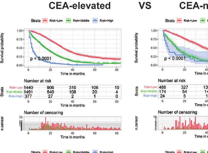
Liu et al.                                                                                                             A Prognostic Nomogram for CCLM

package in R software based on those independent prognostic              TABLE 1 | Baseline characteristics of CCLM patients in the training and internal
                                                                         validation groups.
factors. Meanwhile, the time-dependent receiver operating
characteristic (ROC) curves at 1-, 2-, and 3-years were plotted,         Variables                        Total     Training group      Validation group
and the corresponding time-dependent area under the curve                                                             (n = 3,992)          (n = 1,708)
(AUC) values were used to evaluate the discrimination of the
                                                                         Age(year)
nomogram. Besides, the corresponding calibration curves were               76                             865            613                  252
clinical benefit of the nomogram. Furthermore, based on the               Gender
                                                                           Male                           3,101          2,187                 914
risk score and X-tile software, the optimal cut-off values were
                                                                           Female                         2,599          1,805                 794
determined and all patients were stratified into low-, middle-,           Tumor size(cm)
and high-risk groups. The Kaplan-Meier survival curve was                  6.1                           1,732          1,221                 511
                                                                         Race
points of each patient in two validation sets were calculated
                                                                           White                          4,214          2,951                1,263
according to the nomogram developed in the training set, then              Black                           975            672                  303
Cox regression in this cohort was performed using the total                Other                           511            369                  142
points as a factor, and finally, the C-index, calibration curve and         Site
DCA were derived based on the regression analysis (18).                    LCC                            2,580          1,802                 778
                                                                           RCC                            3,120          2,190                 930
    Furthermore, to confirm that the effectiveness of the                 T stage
nomogram was better than a single factor, the ROC curves of                T1–2                            414            292                  122
all independent prognostic factors were generated. Subgroup                T3–4                           5,286          3,700                1,586
analysis was performed in left-side CC (LCC), right-side CC              N stage
                                                                           0                              1,095           765                  330
(RCC), liver-only metastasis, multiple metastases, CEA-elevated,
                                                                           1                              2,164          1,502                 662
CEA-normal, grade I–II, and grade III–IV. The Kaplan-Meier                 2                              2,441          1,725                 716
survival curves for each subgroup were generated.                        Grade
                                                                           I                               226            152                   74
                                                                           II                             3,999          2,793                1,206
                                                                           III                            1,230           877                  353
RESULTS                                                                    IV                              245            170                   75
                                                                         Type
Clinicopathologic Characteristics                                          Adenocarcinoma                 5,257          3,672                1,585
                                                                           Mucinous                        411            294                  117
According to the criteria of inclusion and exclusion, a total of
                                                                         adenocarcinoma                     32             26                   6
5,700 CCLM patients were included, which were divided into a               Signet ring cell carcinoma
training set (n=3,992) and an internal validation set (n=1,708).         Lung metastasis
The Chi-square test showed that there was no significant                    Yes                             906            649                  257
difference between the two sets (Table 1). The average age of              No                             4,794          3,343                1,451
                                                                         Bone metastasis
these patients was 62.05 ± 13.18 (range: 21–108) years old, and            Yes                             155            107                   48
54.4% of patients were male. Besides, the CEA was elevated in              No                             5,545          3,885                1,660
most patients. In comparison, the pathological type in most              Brain metastasis
CCLM patients is adenocarcinoma, accompanied by deep                       Yes                              30             19                   11
                                                                           No                             5,670          3,973                1,697
infiltration (T3–T4), grade II, and surgery-received, and the
                                                                         Surgery
distribution of which was similar to that of CC patients (19).             Yes                            5,242          3,655                1,587
Notably, we found that most CCLM patients have a relatively                No                              458            337                  121
higher proportion of lymph node metastasis (N1–N2) (80.8%)               Radiation
compared with CC patients (36.2%) (19).                                    Yes                             189            127                   62
                                                                           No                             5,511          3,865                1,646
                                                                         Chemotherapy
Identification of Prognostic Factors of                                     Yes                            4,368          3,040                1,328
CCLM Patients in the Training Set                                          No                             1,332           952                  380
To identify OS-related variables, sixteen variables were included        CEA
in the univariate Cox analysis. The result showed that age, tumor          Normal                          985            684                  301
                                                                           Elevated                       4,715          3,308                1,407
size, race, tumor site, histological type, grade, CEA, AJCC T
status, AJCC N status, extrahepatic metastasis (lung, brain, and
bone), and treatments (surgery and chemotherapy) were
identified as OS-related variables (Table 2). Then, the                   higher grade, histological type of mucinous adenocarcinoma and
multivariate Cox analysis was performed and the result                   signet ring cell carcinoma, higher AJCC N status, RCC, lung
indicated that higher age, the race of black, larger tumor size,         metastasis, bone metastasis, without surgery, without

Frontiers in Oncology | www.frontiersin.org                          3                                      February 2021 | Volume 11 | Article 591009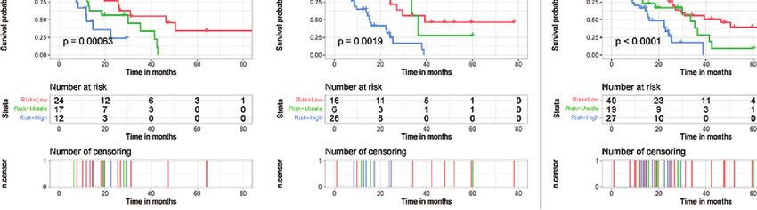
Liu et al.                                                                                                                           A Prognostic Nomogram for CCLM

TABLE 2 | Univariate and multivariate Cox regression analysis of CCLM patients based on clinicopathological characteristics in the training cohort.

                                                              Univariate Cox                                                       Multivariate Cox

                                          HR                      95% CI                        P               HR                      95%CI                    P

Age
   76                                   2.460                  2.220–2.726                  0.000             1.776                 1.590–1.984               0.000
Size
   6.1                                  1.353                  1.239–1.477                  0.000             1.266                 1.159–1.384               0.000
Race
   Black                                                                                     0.111                                                             0.006
   White                                 0.902                  0.818–0.994                  0.037             0.850                 0.770–0.937               0.001
   Other                                 0.908                  0.783–1.054                  0.206             0.863                 0.742–1.003               0.055
Sex
   Female
   Male                                  0.976                  0.906–1.050                  0.513
Site
   LCC
   RCC                                   1.545                  1.434–1.665                  0.000             1.339                 1.238–1.447               0.000
Histological type
   Adenocarcinoma                                                                            0.000                                                             0.001
   Mucinous adenocarcinoma               1.322                 1.157–1.510                   0.000             1.202                 1.050–1.377               0.008
   Signet ring cell carcinoma            2.756            1.827           4.158              0.000             1.932                 1.274–2.929               0.002
Grade
   I                                                                                         0.000                                                             0.000
   II                                    1.057                  0.869–1.286                  0.577             1.167                 0.959–1.420               0.123
   III                                   1.641                  1.338–2.013                  0.000             1.653                 1.346–2.030               0.000
   IV                                    1.781                  1.383–2.294                  0.000             1.834                 1.422–2.366               0.000
T
   T1–2
   T3–4                                  0.921                  0.799–1.061                  0.255
N
   0                                                                                         0.000                                                             0.000
   1                                     1.130                  1.014–1.259                  0.026             1.361                 1.218–1.521               0.000
   2                                     1.449                  1.305–1.608                  0.000             1.805                 1.611–2.022               0.000
CEA
   Normal
   Elevated                              1.538                  1.385–1.707                  0.000             1.612                 1.450–1.791               0.000
Surgery                                  0.520                  0.459–0.589                  0.000             0.399                 0.348–0.458               0.000
Radiation                                0.978                  0.792–1.208                  0.835
Chemotherapy                             0.343                  0.316–0.372                  0.000             0.356                 0.326–0.389               0.000
Bone                                     1.951                  1.593–2.390                  0.000             1.530                 1.244–1.882               0.000
Brain                                    2.160                  1.321–3.532                  0.002
Lung                                     1.606                  1.460–1.765                  0.000             1.404                 1.273–1.549               0.000

chemotherapy, and elevated CEA were independently associated                           Furthermore, DCA was performed and the results indicated that
with poor OS of CCLM patients (Table 2).                                               the nomogram can serve as an effective tool for clinical practice
                                                                                       (Supplementary Figure S1).
Development and Validation of the
Prognostic Nomogram                                                                    Risk Stratification for CCLM Patients
To predict the OS of CCLM, a nomogram was developed based                              Using our established prognostic nomogram, CCLM patients can
on all independent OS-related factors from the training set                            be divided into high-, middle- and low-risk groups. As shown in
(Figure 1). Meanwhile, the time-dependent ROC curves                                   Figure 2A, the results of Kaplan-Meier survival analysis with
showed that the AUC values in 1-, 2-, and 3-years were 0.792,                          log-rank test suggested that there existed different survival
0.769, and 0.763, respectively, which suggested the favorable                          patterns among patients in the three risk groups. Moreover,
discrimination of the nomogram (Supplementary Figure S1).                              patients in both validation sets were also divided into three risk
Then, the AUC values in 1-, 2- and 3-years were 0.754, 0.747, and                      groups with the result of X-tile. We can see that patients of the
0.751 in the internal validation set and 0.725, 0.738 and 0.700 in                     low-risk group had a better prognosis than patients in the high-
the external validation set, respectively. Besides, the calibration                    risk group (P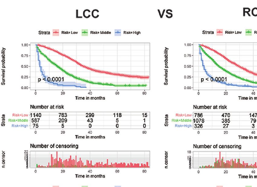
Liu et al.                                                                                                               A Prognostic Nomogram for CCLM

  FIGURE 1 | The nomogram for predicting the prognosis of CCLM patients.

three groups with different prognosis to provide a reference                     DISCUSSION
for treatment.
                                                                                 CC is a highly invasive cancer that is prone to distant metastases, and
Risk Stratification for Subgroup Analysis                                         the most common distant metastatic pattern is liver metastasis. Thus,
Although the ability of the nomogram has been confirmed in                        we included a range of clinicopathological variables to construct a
both training and validation sets, it remains unclear in subgroups.              clinical prognostic nomogram for OS of CCLM patients, which
Hence, to further verify the stability and performance of the                    achieved considerable discrimination ability and calibration accuracy
nomogram from different dimensions, we divided patients into                     when applied to the validation cohorts. According to the nomogram
different subgroups based on tumor site, CEA, the number of                      risk stratification model, patients in the training or verification group
distant metastasis sites, and grade. As shown in Figures 3, 4, no                could be effectively divided into three groups (high-, middle- and
matter in training or validation sets, risk stratification can divide             low-risk groups) with the significant OS. In addition, we included
patients with different OS into the subgroups of LCC, RCC, CEA-                  different treatments in the nomogram to clinicians for more facile
elevated, and CEA-normal, which indicated that the nomogram was                  individual survival prediction.
effective for the distinction of the prognosis in different CCLM                     Although some predictive models have been established in
patients subgroups. However, in the multiple metastases subgroup                 previous studies, we think our study improves upon the previous
of the external validation set, the survival of patients in the three risk       work. Compared with the study of Wu et al. (20), improvements in
groups was not significantly different (p=0.24), which may be                     ours are as follows. First, from the perspective of the subject, CC and
attributed to the relatively small sample size (n=15) (Figure 3L).               RC patients with liver-only metastasis were included in the study of
For the grade subgroups, because there are few patients (n=6, all of             Wu et al. Although the liver is the most common metastatic site of
them belong to the high-risk group) in grade III-IV, we only                     CC and RC, different molecular developmental mechanisms and
analyzed the survival status of patients in grade I-II (Figure 4K).              metastatic patterns require different staging methods and treatments
                                                                                 between CC and RC (21–23). Therefore, our study only included CC
Comparison of Predictive Accuracy                                                to provide a more accurate prediction of prognosis for CCLM.
AS shown in Figure 5, the AUC values of every independent                        Second, the study of Wu et al. focused on CRC patients with liver-
prognostic factor were higher than 0.5, including the training set               only metastasis, but it was discovered that multiple metastases occur
and the two validation sets. By comparing the predictive power                   in approximately 20% of CRC patients (24). Thus, this part of
between the nomogram and all independent factors, we found                       patients cannot be predicted through the nomogram established by
that the AUC value of the nomogram was higher than every                         Wu et al., while the nomogram we constructed can be used. More
single factor in 1-, 2- and 3-years, suggesting the effectiveness of             importantly, subgroup analyses of both liver-only patients and
the nomogram.                                                                    multiple metastases patients showed good performance of our

Frontiers in Oncology | www.frontiersin.org                                  5                                  February 2021 | Volume 11 | Article 591009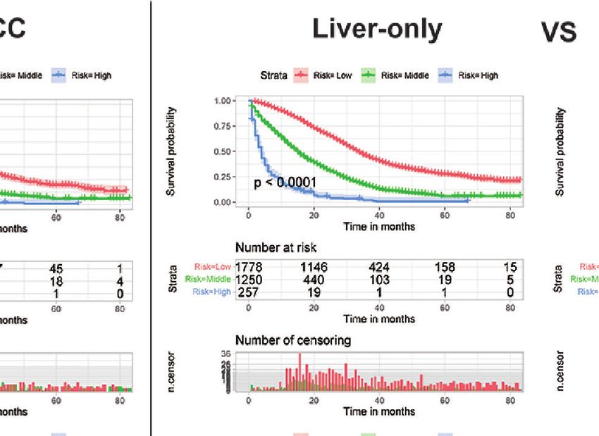
Liu et al.                                                                                                                              A Prognostic Nomogram for CCLM

             A

             B

             C

  FIGURE 2 | Survival curves showed the survival status classified by our nomogram of the training set (A), internal validation set (B), and external validation set (C) in
  CCLM patients.

Frontiers in Oncology | www.frontiersin.org                                          6                                       February 2021 | Volume 11 | Article 591009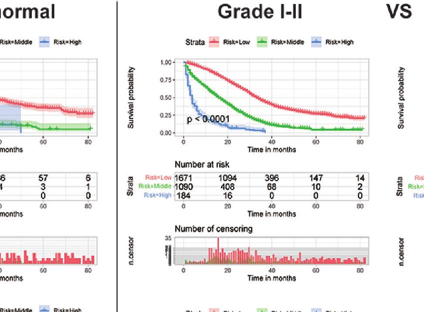
Frontiers in Oncology | www.frontiersin.org

                                                                                                                                                                                                                                                                        Liu et al.
                                                           A                                                  B                                                C                                                 D

                                                           E                                                  F                                                G                                                 H
7

                                                           I                                                  J                                                  K                                               L
February 2021 | Volume 11 | Article 591009

                                                                                                                                                                                                                                                                        A Prognostic Nomogram for CCLM
                                              FIGURE 3 | Subgroup analysis of LCC and RCC in the training set (A, B), internal validation set (E, F), and external validation set (I, J); Subgroup analysis of liver-only and multiple metastases in the training set
                                              (C, D), internal validation set (G, H), and external validation set (K, L).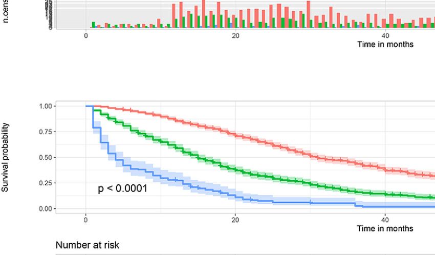
Frontiers in Oncology | www.frontiersin.org

                                                                                                                                                                                                                                                                             Liu et al.
                                                           A                                                    B                                              C                                                 D

                                                           E                                                    F                                              G                                                 H
8

                                                           I                                                    J                                              K
February 2021 | Volume 11 | Article 591009

                                                                                                                                                                                                                                                                             A Prognostic Nomogram for CCLM
                                              FIGURE 4 | Subgroup analysis of CEA-elevated and CEA-normal in the training set (A, B), internal validation set (E, F), and external validation set (I, J); Subgroup analysis of grade I–II and grade III–IV in the training
                                              set (C, D), internal validation set (G, H), and external validation set (K).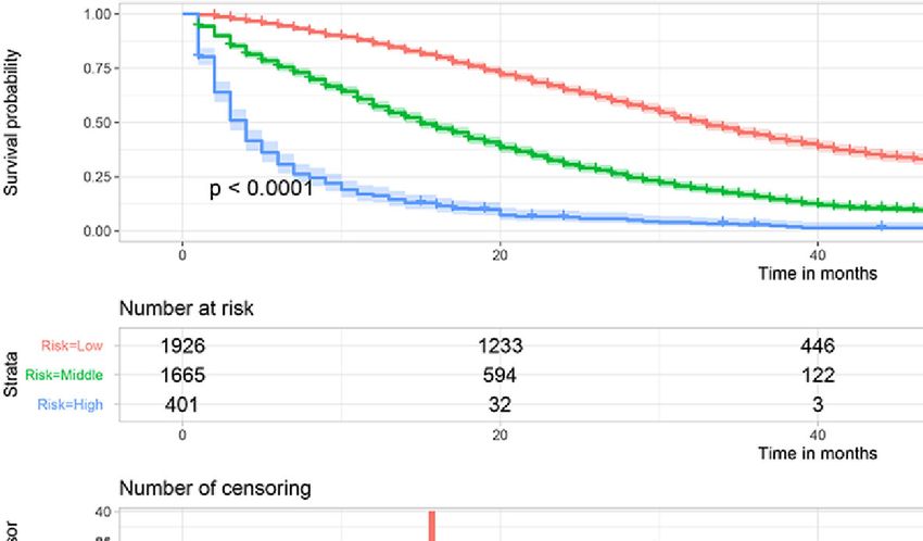
Liu et al.                                                                                                                       A Prognostic Nomogram for CCLM

    A                                                    B                                                     C

     D                                                   E                                                     F

  FIGURE 5 | Comparison of predictive accuracy between the nomogram and single independent factors in the training (A–C) and internal validation sets (D–F).

nomogram, which further confirm the improvement of our model.                             From the perspective of the tumor, tumor site, tumor size,
Then, among treatment factors, only surgery was included in the                      histological type, N stage, histological grade, and CEA level were
study of Wu et al. Whether it was used as a disease treatment method                 determined as independent prognostic factors of CCLM. Previous
or as an adjuvant treatment, chemotherapy was considered to be                       studies reported that RCC had lower OS and disease-free survival
beneficial for CCLM patient’s survival (25, 26). Thus, the factor of                  than LCC (32, 33), which may be associated with RCC usually
chemotherapy was also included in our study and was identified as a                   presents with a diagnosis of a more advanced stage (34). And
protective factor. Finally, we conducted external validation on the                  another reason may be that microsatellite instability and mutations
established nomogram, which is important and strong evidence.                        of KRAS and BRAF are more common in RCC patients (35).
    From the perspective of the patient’s condition, older age, the                  Lymph node metastasis is a common form of metastasis in CC, and
race of black, lung metastasis, and bone metastasis are independent                  high rates are also associated with a high risk of multiple metastatic
prognostic factors of CCLM patients’ prognosis. Elderly patients are                 sites and worse differentiation (36). Through the above indirect
often accompanied by dysfunction, malnutrition, and comorbidity,                     effects, the prognosis of patients is poor, which proves that the
which prompts the physicians to choose a less active treatment or                    prognosis is related to the N stage. And the conclusion of the higher
shorten the course of treatment and affect the outcome of treatment                  N stage, the worse the prognosis was consistent with our study (37).
(27–29). Meanwhile, it was reported that the prognosis of liver                      However, in the study of Wang et al, only the N1 stage was
metastasis alone was different from multiple metastases in the                       independently associated with the prognosis of stage IV CRC.
elderly group, but not in the middle-aged group in a previous                        While in our study, both N1 and N2 stages were the prognostic
study (30). And this study also found that CCLM patients with                        factors of CCLM, which may be contributed to the difference
extrahepatic metastasis had shorter survival times than patients with                between CC and RC and the difference in metastatic patterns.
liver-only metastases, including lung metastasis and bone metastasis                 Based on many studies, CEA was also closely related to the survival
(30). The results in our study suggested that the metastatic sites of                of advanced CRC patients with liver metastases (38). This
lung and bone are independently associated with the prognosis of                     conclusion coincided with the results shown in the present study.
CCLM patients, which was consistent with the conclusion of                               From the perspective of treatments, the traditional treatment for
previous studies (31).                                                               patients with stage I–III CC is surgery combined with adjuvant

Frontiers in Oncology | www.frontiersin.org                                      9                                     February 2021 | Volume 11 | Article 591009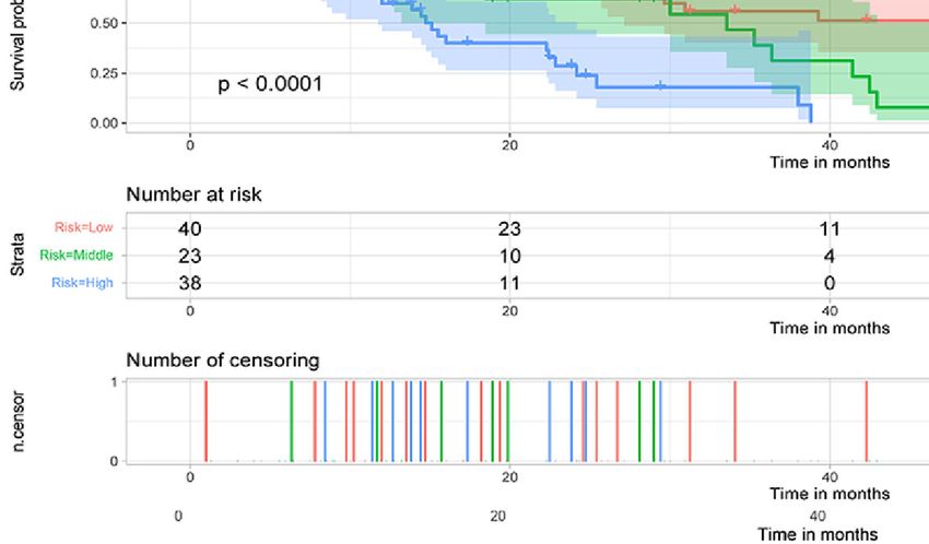
Liu et al.                                                                                                                          A Prognostic Nomogram for CCLM

chemotherapy. Partial or total colectomy is performed in 84% of                         patients. A nomogram incorporating the above 12 predictors could
patients with stage I and II CC, while 67% in stage III (39). And                       accurately predict the prognosis of CCLM patients.
adjuvant chemotherapy within 8 weeks after surgery significantly
improves the prognosis of patients. Besides, a recent study has
found that adjuvant radiotherapy may benefit CC patients,
implying that radiotherapy may also be a treatment option for                           DATA AVAILABILITY STATEMENT
CC patients. With the advancement of treatment, surgery has also
                                                                                        Publicly available datasets were analyzed in this study. This data
become the standard treatment option for CCLM patients, which
                                                                                        can be found here: Surveillance, Epidemiology, and End Results
can improve patients’ outcomes. In clinical practice, partial
                                                                                        (SEER) database (https://seer.cancer.gov/).
colectomy and total/subtotal colectomy are more effective for
CCLM patients than those without surgery. Additionally,
chemotherapy is also an important treatment approach for
CCLM patients to significantly prolong the survival time, such as                        ETHICS STATEMENT
5-fluorouracil/leucovorin (5-FU/lv), capecitabine, irinotecan, and
oxaliplatin (40). As neoadjuvant therapy, chemotherapy can also                         Written informed consent was obtained from the individual(s)
promote the likelihood of resectability and treat micro-metastases                      for the publication of any potentially identifiable images or data
(41, 42). Moreover, as the postoperative adjuvant therapy, the                          included in this article.
previous study reported that chemotherapy was related to OS and
DFS of CRC patients with liver metastasis (43). However, more
than 80% of CCLM patients are unresectable, and the prognosis of
these patients can also be improved with different chemotherapy                         AUTHOR CONTRIBUTIONS
regimens (6, 44). Thus, as with our results, surgery and
chemotherapy can improve the outcomes of CCLM patients.                                 CL and TW designed the research; CH performed the research
    In the present study, the nomogram could be used to                                 and analyzed results; CL and JH edited the manuscript; TW and
effectively predict the prognosis of CCLM patients. However,                            ZL provided critical comments and revised the manuscript; GZ,
some limitations should be stated. Firstly, this is a retrospective                     JQ, YC, XQ, and YL collected and organized data; GZ added the
study based on a publicly available database, which made it                             data, sorted them out and analyzed them in the revised
susceptible to the inherent weaknesses of retrospective data                            manuscript; KX wrote the revised manuscript; All authors
collection. Besides, specific information of liver metastases                            contributed to the article and approved the submitted version.
associated with the prognosis of CCLM, such as the large
size, more than three liver metastases, and presence of bi-lobar
metastases, is a lack in the SEER database. Secondly, most
patients in the external invalidation set were of other races                           FUNDING
(Asian) and have received chemotherapy, which may produce
                                                                                        This study was funded by the National Natural Science Foundation
selection bias. Thirdly, the sample size of the external
                                                                                        of China (No. 31770963); National Science and Technology Major
validation set was not very large, So, other validation cohorts
                                                                                        Project of the Ministry of Science and Technology of China (No.
with a larger sample size for the predictive nomogram
                                                                                        2017ZX09304025); Technological Special Project of Liaoning
are indispensable.
                                                                                        Province of China (2019020176-JH1/103); Science and
                                                                                        Technology Plan Project of Liaoning Province; NO.2013225585).

CONCLUSION
In summary, we found that higher age, the race of black, larger tumor                   SUPPLEMENTARY MATERIAL
size, higher grade, histological type of mucinous adenocarcinoma and
signet ring cell carcinoma, higher N stage, RCC, lung metastasis, bone                  The Supplementary Material for this article can be found online
metastasis, without surgery, without chemotherapy, and elevated                         at: https://www.frontiersin.org/articles/10.3389/fonc.2021.
CEA were independently associated with poor prognosis of CCLM                           591009/full#supplementary-material

REFERENCES                                                                               3. Dekker E, Tanis PJ, Vleugels JLA, Kasi PM, Wallace MB. Colorectal cancer.
                                                                                            Lancet (London England) (2019) 394(10207):1467–80. doi: 10.1016/s0140-
 1. Siegel RL, Miller KD, Jemal A. Cancer statistics, 2019. CA: Cancer J Clin               6736(19)32319-0
    (2019) 69(1):7–34. doi: 10.3322/caac.21551                                           4. Hu H, Wu Z, Wang C, Huang Y, Zhang J, Cai Y, et al. Duration of FOLFOX
 2. Arnold M, Sierra MS, Laversanne M, Soerjomataram I, Jemal A, Bray F.                    Adjuvant Chemotherapy in High-Risk Stage II and Stage III Colon Cancer
    Global patterns and trends in colorectal cancer incidence and mortality. Gut            With Deficient Mismatch Repair. Front Oncol (2020) 10:579478. doi: 10.3389/
    (2017) 66(4):683–91. doi: 10.1136/gutjnl-2015-310912                                    fonc.2020.579478

Frontiers in Oncology | www.frontiersin.org                                        10                                     February 2021 | Volume 11 | Article 591009
Liu et al. A Prognostic Nomogram for CCLM
5. McLaughlin C, Kim NK, Bandyopadhyay D, Deng X, Kaplan B, Matin K, et al. 24. Qiu M, Hu J, Yang D, Cosgrove DP, Xu R. Pattern of distant metastases in
Adjuvant radiation therapy for T4 non-rectal colon adenocarcinoma provides colorectal cancer: a SEER based study. Oncotarget (2015) 6(36):38658–66.
a cause-specific survival advantage: A SEER database analysis. Radiother doi: 10.18632/oncotarget.6130
Oncol J Eur Soc Ther Radiol Oncol (2019) 133:50–3. doi: 10.1016/ 25. Leone F, Artale S, Marino D, Cagnazzo C, Cascinu S, Pinto C, et al.
j.radonc.2018.11.026 Panitumumab in combination with infusional oxaliplatin and oral
6. Manfredi S, Lepage C, Hatem C, Coatmeur O, Faivre J, Bouvier AM. capecitabine for conversion therapy in patients with colon cancer and
Epidemiology and management of liver metastases from colorectal advanced liver metastases. MetaPan Study Cancer (2013) 119(19):3429–35.
cancer. Ann Surg (2006) 244(2):254–9. doi: 10.1097/01.sla.0000217629. doi: 10.1002/cncr.28223
94941.cf 26. Sereno Moyano M, Casado Sá enz E, de Castro-Carpeño J, Belda-Iniesta C.
7. Siegel RL, Miller KD, Fedewa SA, Ahnen DJ, Meester RGS, Barzi A, et al. The combination of FOLFOX4 and bevacizumab may enable salvage surgery
Colorectal cancer statistics, 2017. CA: Cancer J Clin (2017) 67(3):177–93. of unresectable liver metastases in colon cancer. Anti-Cancer Drugs (2009) 20
doi: 10.3322/caac.21395 Spec No 1:S4–6. doi: 10.1097/01.cad.0000349777.01991.25
8. Van Cutsem E, Cervantes A, Adam R, Sobrero A, Van Krieken JH, Aderka D, 27. Burdett N, Vincent AD, O’Callaghan M, Kichenadasse G. Competing
et al. ESMO consensus guidelines for the management of patients with Risks in Older Patients With Cancer: A Systematic Review of Geriatric
metastatic colorectal cancer. Ann Oncol (2016) 27(8):1386–422. Oncology Trials. J Natl Cancer Inst (2018) 110(8):825–30. doi: 10.1093/
doi: 10.1093/annonc/mdw235 jnci/djy111
9. Engstrand J, Nilsson H, Strömberg C, Jonas E, Freedman J. Colorectal cancer 28. Lee L, Cheung WY, Atkinson E, Krzyzanowska MK. Impact of comorbidity on
liver metastases - a population-based study on incidence, management and chemotherapy use and outcomes in solid tumors: a systematic review. J Clin
survival. BMC Cancer (2018) 18(1):78. doi: 10.1186/s12885-017-3925-x Oncol (2011) 29(1):106–17. doi: 10.1200/jco.2010.31.3049
10. Jones RP, Kokudo N, Folprecht G, Mise Y, Unno M, Malik HZ, et al. 29. Foster JA, Salinas GD, Mansell D, Williamson JC, Casebeer LL. How does
Colorectal Liver Metastases: A Critical Review of State of the Art. Liver older age influence oncologists’ cancer management? Oncol (2010) 15(6):584–
Cancer (2016) 6(1):66–71. doi: 10.1159/000449348 92. doi: 10.1634/theoncologist.2009-0198
11. Wang J, Li S, Liu Y, Zhang C, Li H, Lai B. Metastatic patterns and survival 30. Yang L, Yang X, He W, Liu S, Jiang C, Xie K, et al. Comparisons of
outcomes in patients with stage IV colon cancer: A population-based analysis. metastatic patterns of colorectal cancer among patients by age group: a
Cancer Med (2020) 9(1):361–73. doi: 10.1002/cam4.2673 population-based study. Aging (Albany NY) (2018) 10(12):4107–19.
12. Zheng P, Lai C, Yang W, Guo J, Xiao S, Chen Z. Nomogram predicting doi: 10.18632/aging.101700
cancer-specific survival in elderly patients with stages I-III colon cancer. Scand 31. Baek SJ, Hur H, Min BS, Baik SH, Lee KY, Kim NK. The Characteristics of
J Gastroenterol (2020) 55(2):202–8. doi: 10.1080/00365521.2020.1720280 Bone Metastasis in Patients with Colorectal Cancer: A Long-Term Report
13. Kawai K, Nozawa H, Hata K, Kiyomatsu T, Tanaka T, Nishikawa T, et al. from a Single Institution. World J Surg (2016) 40(4):982–6. doi: 10.1007/
Nomogram Predicting Survival After Recurrence in Patients With Stage I to s00268-015-3296-x
III Colon Cancer: A Nationwide Multicenter Study. Dis Colon Rectum (2018) 32. Signorelli C, Chilelli MG, Sperduti I, Giacinti S, Amodio PM, Palmieri RM,
61(9):1053–62. doi: 10.1097/dcr.0000000000001167 et al. Correlation of Tumor Location to Clinical Outcomes in Colorectal
14. Kazem MA, Khan AU, Selvasekar CR. Validation of nomogram for disease Cancer: A Single-institution Retrospective Analysis. Anticancer Res (2019) 39
free survival for colon cancer in UK population: A prospective cohort study. (9):4917–24. doi: 10.21873/anticanres.13679
Int J Surg (London England) (2016) 27:58–65. doi: 10.1016/j.ijsu.2015.12.069 33. Lim DR, Kuk JK, Kim T, Shin EJ. Comparison of oncological outcomes of
15. Zhang Y, Ma J, Zhang S, Deng G, Wu X, He J, et al. A prognostic analysis of right-sided colon cancer versus left-sided colon cancer after curative resection:
895 cases of stage III colon cancer in different colon subsites. Int J Colorectal Which side is better outcome? Medicine (2017) 96(42):e8241. doi: 10.1097/
Dis (2015) 30(9):1173–83. doi: 10.1007/s00384-015-2273-z md.0000000000008241
16. Park HS, Lloyd S, Decker RH, Wilson LD, Yu JB. Overview of the Surveillance, 34. Alexiusdottir KK, Möller PH, Snaebjornsson P, Jonasson L, Olafsdottir EJ,
Epidemiology, and End Results database: evolution, data variables, and quality Björnsson ES, et al. Association of symptoms of colon cancer patients with
assurance. Curr Probl Cancer (2012) 36(4):183–90. doi: 10.1016/ tumor location and TNM tumor stage. Scand J Gastroenterol (2012) 47
j.currproblcancer.2012.03.007 (7):795–801. doi: 10.3109/00365521.2012.672589
17. Camp RL, Dolled-Filhart M, Rimm DL. X-tile: a new bio-informatics tool for 35. Nitsche U, Stögbauer F, Späth C, Haller B, Wilhelm D, Friess H, et al.
biomarker assessment and outcome-based cut-point optimization. Clin Right Sided Colon Cancer as a Distinct Histopathological Subtype with
Cancer Res (2004) 10(21):7252–9. doi: 10.1158/1078-0432.ccr-04-0713 Reduced Prognosis. Dig Surg (2016) 33(2):157–63. doi: 10.1159/
18. Wang Y, Li J, Xia Y, Gong R, Wang K, Yan Z, et al. Prognostic nomogram for 000443644
intrahepatic cholangiocarcinoma after partial hepatectomy. J Clin Oncol 36. Derwinger K, Gustavsson B. A study of lymph node ratio in stage IV colorectal
(2013) 31(9):1188–95. doi: 10.1200/jco.2012.41.5984 cancer. World J Surg Oncol (2008) 6:127. doi: 10.1186/1477-7819-6-127
19. Stojadinovic A, Bilchik A, Smith D, Eberhardt JS, Ward EB, Nissan A, et al. 37. Wang X, Mao M, Xu G, Lin F, Sun P, Baklaushev VP, et al. The incidence,
Clinical decision support and individualized prediction of survival in colon associated factors, and predictive nomogram for early death in stage IV
cancer: bayesian belief network model. Ann Surg Oncol (2013) 20(1):161–74. colorectal cancer. Int J Colorectal Dis (2019) 34(7):1189–201. doi: 10.1007/
doi: 10.1245/s10434-012-2555-4 s00384-019-03306-1
20. Wu Q, Wang WJ, Huang YQ, Fang SY, Guan YJ. Nomograms for estimating 38. Thomas P, Toth CA, Saini KS, Jessup JM, Steele GJr. The structure, metabolism
survival in patients with liver-only colorectal metastases: A retrospective and function of the carcinoembryonic antigen gene family. Biochim Biophys Acta
study. Int J Surg (London England) (2018) 60:1–8. doi: 10.1016/ (1990) 1032(2-3):177–89. doi: 10.1016/0304-419x(90)90003-j
j.ijsu.2018.10.032 39. Miller KD, Siegel RL, Lin CC, Mariotto AB, Kramer JL, Rowland JH, et al.
21. Paschke S, Jafarov S, Staib L, Kreuser ED, Maulbecker-Armstrong C, Roitman Cancer treatment and survivorship statistics, 2016. CA: Cancer J Clin (2016)
M, et al. Are Colon and Rectal Cancer Two Different Tumor Entities? A 66(4):271–89. doi: 10.3322/caac.21349
Proposal to Abandon the Term Colorectal Cancer. Int J Mol Sci (2018) 19 40. Brandi G, De Lorenzo S, Nannini M, Curti S, Ottone M, Dall’Olio FG, et al.
(9):2577. doi: 10.3390/ijms19092577 Adjuvant chemotherapy for resected colorectal cancer metastases: Literature
22. Kalady MF, Sanchez JA, Manilich E, Hammel J, Casey G, Church JM. review and meta-analysis. World J Gastroenterol (2016) 22(2):519–33.
Divergent oncogenic changes influence survival differences between colon doi: 10.3748/wjg.v22.i2.519
and rectal adenocarcinomas. Dis Colon Rectum (2009) 52(6):1039–45. 41. Nordlinger B, Benoist S. Benefits and risks of neoadjuvant therapy for liver
doi: 10.1007/DCR.0b013e31819edbd4 metastases. J Clin Oncol (2006) 24(31):4954–5. doi: 10.1200/jco.2006.07.9244
23. Tamas K, Walenkamp AM, de Vries EG, van Vugt MA, Beets-Tan RG, van 42. Folprecht G, Gruenberger T, Bechstein WO, Raab HR, Lordick F,
Etten B, et al. Rectal and colon cancer: Not just a different anatomic site. Hartmann JT, et al. Tumour response and secondary resectability of
Cancer Treat Rev (2015) 41(8):671–9. doi: 10.1016/j.ctrv.2015.06.007 colorectal liver metastases following neoadjuvant chemotherapy with
Frontiers in Oncology | www.frontiersin.org 11 February 2021 | Volume 11 | Article 591009Liu et al. A Prognostic Nomogram for CCLM
cetuximab: the CELIM randomised phase 2 trial. Lancet Oncol (2010) 11 Conflict of Interest: The authors declare that the research was conducted in the
(1):38–47. doi: 10.1016/s1470-2045(09)70330-4 absence of any commercial or financial relationships that could be construed as a
43. Ychou M, Hohenberger W, Thezenas S, Navarro M, Maurel J, Bokemeyer C, et al. potential conflict of interest.
A randomized phase III study comparing adjuvant 5-fluorouracil/folinic acid with
FOLFIRI in patients following complete resection of liver metastases from Copyright © 2021 Liu, Hu, Huang, Xiang, Li, Qu, Chen, Yang, Qu, Liu, Zhang and
colorectal cancer. Ann Oncol (2009) 20(12):1964–70. doi: 10.1093/annonc/mdp236 Wen. This is an open-access article distributed under the terms of the Creative
44. Goldberg RM, Sargent DJ, Morton RF, Fuchs CS, Ramanathan RK, Commons Attribution License (CC BY). The use, distribution or reproduction in other
Williamson SK, et al. A randomized controlled trial of fluorouracil plus forums is permitted, provided the original author(s) and the copyright owner(s) are
leucovorin, irinotecan, and oxaliplatin combinations in patients with credited and that the original publication in this journal is cited, in accordance with
previously untreated metastatic colorectal cancer. J Clin Oncol (2004) 22 accepted academic practice. No use, distribution or reproduction is permitted which
(1):23–30. doi: 10.1200/jco.2004.09.046 does not comply with these terms.
Frontiers in Oncology | www.frontiersin.org 12 February 2021 | Volume 11 | Article 591009You can also read