Long non coding RNA SNHG20 promotes ovarian cancer development by targeting microRNA 338 3p to regulate MCL1 expression
←
→
Page content transcription
If your browser does not render page correctly, please read the page content below
ONCOLOGY LETTERS 21: 130, 2021

               Long non‑coding RNA SNHG20 promotes ovarian
              cancer development by targeting microRNA‑338‑3p
                         to regulate MCL1 expression
                                DING WANG, ZHIYING LI, HUI LI, JIAO LU and QI QIN

                 Department of Gynecology, The Affiliated Renhe Hospital of China Three Gorges University,
                                           Yichang, Hubei 443001, P.R. China

                                     Received May 13, 2020; Accepted November 4, 2020

                                                  DOI: 10.3892/ol.2020.12391

Abstract. Long non‑coding RNAs (lncRNAs) and microRNAs            Introduction
(miRNAs/miRs) were reported to be associated with the
development of ovarian cancer (OC). Increasing evidence           Ovarian cancer (OC), a common gynecological tumor, is the
demonstrated that lncRNA SNHG20 and miR‑338‑3p were               fifth leading cause of tumor‑associated death in women, and
involved in OC. However, the functional mechanism of              there were ~22,240 new cases and ~14,070 deaths associated
lncRNA SNHG20 and miR‑338‑3p in OC development                    with OC every year worldwide (1). Most patients with OC
remains unknown. The expression of SNHG20, miR‑338‑3p             were diagnosed at the advanced stage with tumor metastasis
and myeloid cell leukemia 1 (MCL1) was detected by reverse        and recurrence (2). Despite achieving great progress in the
transcription‑quantitative PCR. MTT assay, flow cytometry         technologies for the detection and therapy of OC, the overall
and transwell migration and invasion assays were used to          survival rate of patients with OC remains low. Therefore, it is
assess cell proliferation, apoptosis, migration and invasion,     urgent to find the effective therapeutic targets for OC.
respectively. The relative protein expression was detected by         Long non‑coding RNAs (lncRNAs), with over 200 nucleo‑
western blot analysis. The interaction between miR‑338‑3p         tides, are a group of conserved RNAs that play pivotal function
and SNHG20 or MCL1 was predicted by starBase v3.0, and            in cancer progression, such as cell proliferation, mobility,
subsequently confirmed by dual‑luciferase reporter assay.         apoptosis and autophagy (3‑6). In recent years, lncRNAs
Besides, mouse xenograft assay was carried out to explore         were reported to be involved in various human cancer types,
the effect of SNHG20 on tumor growth in vivo. The levels of       including OC (7,8). Small nucleolar RNA host gene 20
SNHG20 and MCL1 were upregulated, while miR‑338‑3p level          (SNHG20), 8,275 bases in length, was originally identified
was downregulated in OC tissues and cells. SNHG20 knock‑          as an oncogene in hepatocellular cancer (9). A previous study
down repressed OC cell proliferation, migration, invasion and     indicated that SNHG20 knockdown suppressed the growth
epithelial‑mesenchymal transition, and induced apoptosis.         of OC cells through modulating the levels of downstream
Interestingly, SNHG20 targeted miR‑338‑3p to regulate MCL1        genes (10). However, the functional mechanism of SNHG20 is
expression. miR‑338‑3p depletion or MCL1 overexpression           largely unknown in OC.
could reverse the effects of SNHG20 knockdown on OC                   MicroRNAs (miRNAs/miRs), ~22 nucleotides, exert
cells. Besides, SNHG20 knockdown impeded tumor growth             crucial roles via inhibiting translation or promoting degra‑
in vivo. In conclusion, the present study demonstrated that       dation (11,12). Multiple studies demonstrated that miRNAs
SNHG20 regulates OC development via modulation of the             participated in the progression of various cancer types,
miR‑338‑3p/MCL1 axis, providing the theoretical basis for the     including lung cancer (13), osteosarcoma (14), thyroid
treatment of OC.                                                  cancer (15) and OC (16). A recent study suggested that
                                                                  miR‑338‑3p level was decreased in OC, and upregulation of
                                                                  miR‑338‑3p repressed OC cell proliferation (17). Moreover,
                                                                  Liu et al (18) demonstrated that miR‑338‑3p acted as a tumor
                                                                  suppressor to regulate cell proliferation, mobility and apop‑
                                                                  tosis in OC. These data revealed that miR‑338‑3p played an
Correspondence to: Dr Ding Wang, Department of Gynecology,
                                                                  important role in OC. Thus, the present explored the underlying
The Affiliated Renhe Hospital of China Three Gorges University,
410 Yiling Avenue, Yichang, Hubei 443001, P.R. China              mechanism of miR‑338‑3p in OC.
E‑mail: opdsfl@163.com                                                Myeloid cell leukemia 1 (MCL1) belongs to B‑cell
                                                                  lymphoma 2 (BCL‑2) family, which is considered as a class of
Key words: ovarian cancer, SNHG20, microRNA‑338‑3p, MCL1          cell apoptosis regulators in human cancer (19). It was reported
                                                                  that many factors, including deubiquitinase and miRNAs,
                                                                  mediated the level of MCL1 to affect cell growth in OC. For
                                                                  instance, USP13 regulated MCL stabilization to modulate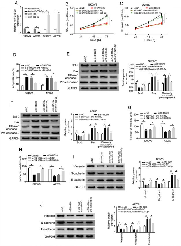
2                                     WANG et al: SNHG20 REGULATES OC DEVELOPMENT

tumor growth (20). Su et al (21) demonstrated that miR‑142‑5p    Table I. Reverse transcription‑quantitative PCR primer
mediated cisplatin‑induced cell apoptosis via regulating MCL     sequences.
expression. Thus, MCL pathway is associated with OC devel‑
opment. Therefore, it is important to examine the function of    Gene                               Sequence
MCL in OC cells.
    Here, we determined the levels of SNHG20, miR‑338‑3p         SNHG20          F: 5'‑ATGGCTATAAATAGATACACGC‑3'
and MCL1 in OC tissues and cells. Furthermore, the function of                   R: 5'‑GGTACAAACAGGGAGGGA‑3'
SNHG20 was investigated in OC in vitro and in vivo. Besides,     miR‑338‑3p      F: 5'‑AACCGGTCCAGCATCAGTGATT‑3'
the underlying mechanism of SNHG20 in OC progression was                         R: 5'‑CAGTGCAGGGTCCGAGGT‑3'
explored.                                                        MCL1            F: 5'‑GGGCAGGATTGTGACTCTCATT‑3'
                                                                                 R: 5'‑GATGCAGCTTTCTTGGTTTATGG‑3'
Materials and methods
                                                                 U6              F: 5'‑TGCGGGTGCTCGCTTCGGCAGC‑3'
Tissues and cell culture. 30 OC tissues and 30 adjacent                          R: 5'‑CCAGTGCAGGGTCCGAGGT‑3'
normal tissues were obtained from the patients at the hospital   GAPDH           F: 5'‑ATCACTGCCACCCAGAAGAC‑3'
of The Affiliated Renhe Hospital of China Three Gorges                           R: 5'‑TTTCTAGACGGCAGGTCAGG‑3'
University from April 2016 to January 2019. The average
age of OC patients was (52.02 ± 10.88) years, in which there     SNHG20, small nucleolar RNA host gene 20; MCL1, myeloid cell
were 18 patients ≥50 years and 12 patients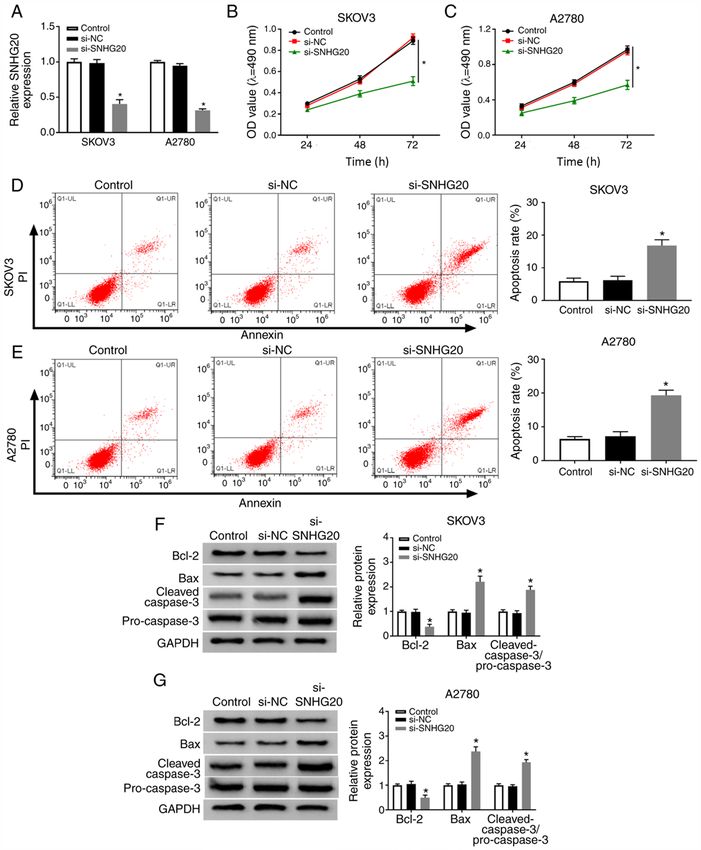
ONCOLOGY LETTERS 21: 130, 2021                                                                   3

Figure 1. SNHG20 expression level is higher in OC tissues and cells. Expression level of SNHG20 was detected by reverse transcription‑quantitative PCR assay
in OC tissues and paired adjacent normal tissues (A) as well as in OC cells and human ovarian surface epithelial cells (B) *P
4                                            WANG et al: SNHG20 REGULATES OC DEVELOPMENT

Figure 2. SNHG20 knockdown suppresses OC cell proliferation and promotes apoptosis. (A) Reverse transcription‑quantitative PCR was used to detect the
expression of SNHG20 in SKOV3 and A2780 cells transfected with si‑NC or si‑SNHG20. (B and C) MTT assay was performed to assess the proliferation
ability of SKOV3 and A2780 cells transfected with si‑NC or si‑SNHG20. (D and E) Flow cytometry was employed to measure the apoptotic rate of SKOV3
and A2780 cells transfected with si‑NC or si‑SNHG20. (F and G) Western blot assay was carried out to examine the levels of cell apoptosis‑associated
proteins. *P
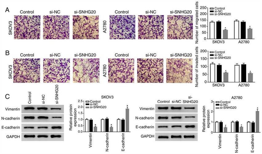
ONCOLOGY LETTERS 21: 130, 2021                                                                5

Figure 3. SNHG20 knockdown inhibits the migration, invasion and EMT of OC cells. SKOV3 and A2780 cells were transfected with si‑NC or si‑SNHG20.
(A and B) Cell migratory and invasive abilities were analyzed using transwell assay in SKOV3 and A2780 cells. (C) Relative protein levels of EMT markers
were determined by western blot assay. *P
6                                             WANG et al: SNHG20 REGULATES OC DEVELOPMENT

Figure 4. SNHG20 targets miR‑338‑3p. (A) The interaction between SNHG20 and miR‑338‑3p was predicted by starBase v2.0. Mutated sites were indicated
by the red color. (B and C) Dual‑luciferase reporter assay was used to detect the luciferase activity of SKOV3 and A2780 cells co‑transfected with miR‑NC
or miR‑338‑3p and WT‑SNHG20 or MUT‑SNHG20. (D) SNHG20 level in SKOV3 and A2780 cells transfected with pcDNA or SNHG20 was detected by
RT‑qPCR. (E) RT‑qPCR was used to measure miR‑338‑3p expression in SKOV3 and A2780 cells transfected with si‑NC, si‑SNHG20, pcDNA, or SNHG20,
respectively. (F and G) miR‑338‑3p level was detected by RT‑qPCR in OC tissues and the adjacent normal tissues (F) as well as OC cells and human ovarian
surface epithelial cells (G). (H) The correlation between SNHG20 and miR‑338‑3p was analyzed using linear regression. *P
ONCOLOGY LETTERS 21: 130, 2021                                                                        7

Figure 5. SNHG20 regulates OC progression through sponging miR‑338‑3p. (A) miR‑338‑3p level was investigated using reverse transcription‑quantitative
PCR in SKOV3 and A2780 cells transfected with miR‑338‑3p or anti‑miR‑338‑3p. (B‑J) SKOV3 and A2780 cells were transfected with si‑NC, si‑SNHG20,
si‑SNHG20 + anti‑miR‑NC, or si‑SNHG20 + anti‑miR‑338‑3p, respectively. (B and C) MTT assay was employed to measure cell proliferation ability.
(D) Cell apoptosis rate was analyzed using flow cytometry. (E and F) The levels of cell apoptosis‑associated proteins were determined by western blot assay.
(G and H) Cell migration and invasive abilities were assessed by transwell assay. (I and J) Western blot assay was used to detect the relative protein levels of EMT
markers. *P
8                                             WANG et al: SNHG20 REGULATES OC DEVELOPMENT

Figure 6. SNHG20 regulates miR‑338‑3p expression to modulate MCL1 level. (A) The interaction between miR‑338‑3p and MCL1 was predicted by starBase
v2.0. Mutated sites were indicated by the red color. (B and C) The luciferase activity of SKOV3 and A2780 cells co‑transfected with MCL1 3'UTR‑WT or MCL1
3'UTR‑MUT and miR‑NC or miR‑338‑3p was examined by dual‑luciferase reporter assay. (D and E) MCL1 expression was measured by western blot analysis
in SKOV3 and A2780 cells transfected with miR‑NC, miR‑338‑3p, miR‑338‑3p + pcDNA or miR‑338‑3p + SNHG20, respectively. (F and G) Relative mRNA
level and protein level of MCL1 were detected by RT‑qPCR and western blotting in OC tissues and the adjacent normal tissues. (H) The association between
MCL1 level and SNHG20 level was analyzed using linear regression. (I and J) Relative mRNA level and protein level of MCL1 were determined by RT‑qPCR
and western blotting in OC cells and human ovarian surface epithelial cells. *P
ONCOLOGY LETTERS 21: 130, 2021                                                                   9

Figure 7. SNHG20 depletion impeded OC progression by regulating MCL1 level. (A and B) Relative mRNA level and protein level of MCL1 were detected
by reverse transcription‑quantitative PCR and western blotting in SKOV3 and A2780 cells transfected with pcDNA or MCL1. (C‑L) SKOV3 and A2780
cells were transfected with si‑NC, si‑SNHG20, si‑SNHG20 + pcDNA or si‑SNHG20 + pcDNA‑MCL1, respectively. (C and D) MTT assay was used to
measure cell proliferation. (E and F) Cell apoptosis rate was detected using flow cytometry. (G and H) Relative levels of cell apoptosis‑associated proteins
were measured by western blot assay. (I and J) Transwell assay was used to determine cell migratory and invasive abilities. (K and L) Western blot assay
was carried out to determine the protein levels of EMT markers. *P
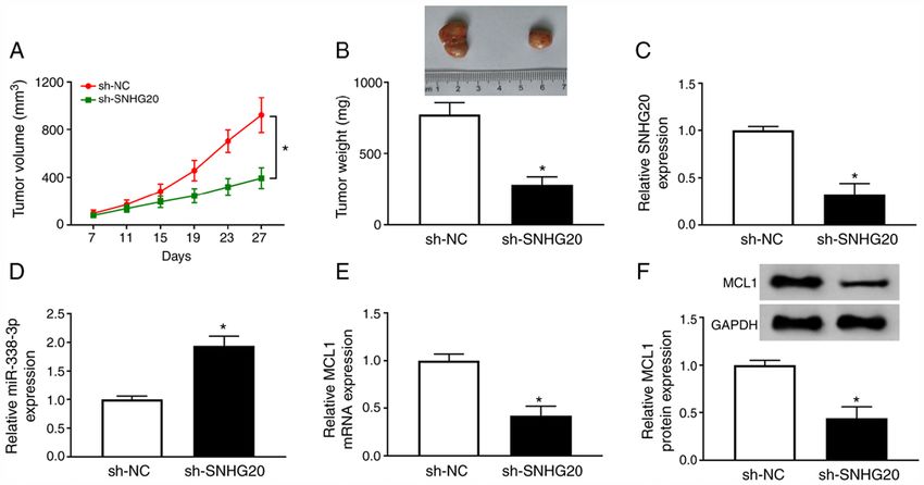
10                                           WANG et al: SNHG20 REGULATES OC DEVELOPMENT

Figure 8. SNHG20 depletion repressed tumor growth in vivo. (A) Tumor volume was calculated in sh‑SNHG20 group and sh‑NC group every 4 days. (B) Tumor
weight was analyzed. (C‑E) The levels of SNHG20, miR‑338‑3p, and MCL1 were detected by reverse transcription‑quantitative PCR assay. (F) Relative
protein level of MCL1 was determined by western blot assay. *P
ONCOLOGY LETTERS 21: 130, 2021                                                   11

found that SNHG20 level was remarkably elevated in OC               significantly suppressed by SNHG20 depletion. Therefore,
tissues and cells. This result corroborated with the previous       SNHG20 knockdown attenuated OC tumor growth in vivo.
data (30). Previous results indicated that SNHG20 positively            In conclusion, the present results demonstrate that SNHG20
regulated OC cell growth (10,31). SNHG20 promoted cell              knockdown represses the development of OC through modu‑
proliferation and migration via silencing the expression of         lating the miR‑338‑3p/MCL1 axis, providing a potential target
proliferation regulator p21 in non‑small cell lung cancer (32).     for the therapy of patients with OC.
Another study confirmed that SNHG20 promoted gastric
cancer progression by inhibiting p21 expression (33). In            Acknowledgements
glioma cells, downregulation of SNHG20 increased cell
apoptosis but inhibited cell proliferation by regulating            Not applicable.
PTEN/PI3K/AKT signaling pathway (34). Moreover,
SNHG20 could function as an oncogenic lncRNA by regu‑               Funding
lating miR‑140‑5p/ADAM10 axis and MEK/ERK signaling
pathway in cervical cancer (35). Furthermore, knockdown             No funding was received.
of SNHG20 remarkably inhibited cell proliferation, migra‑
tion and invasion via dysregulating the expression of p21,          Availability of data and materials
cyclin D1, E‑cadherin and vimentin in epithelial OC (31).
The aforementioned results demonstrated that SNHG20 was             The analyzed data sets generated during the present study are
closely associated with cancer cell progression. In the present     available from the corresponding author on reasonable request.
study, it was observed that SNHG20 depletion repressed
OC cell proliferation, mobility and promoted apoptosis,             Authors' contributions
suggesting that SNHG20 served as a positive regulator in
OC progression.                                                     DW, ZL, HL, JL and QQ made substantial contribution to
    Present evidence suggested that lncRNA exerted its              the concept and design, data analysis, and interpretation of
function through binding to target genes and modulating             the data; DW, ZL and HL performed the experiments and
the levels of targets in cancer (36). For instance, SNHG6           interpreted the data; DW and QQ drafted the manuscript; DW
elevated chemoresistance in colorectal cancer cell by targeting     and JL revised the manuscript critically for important intel‑
miR‑26a‑5p (37). Bioinformatics tool showed that miR‑338‑3p         lectual content. All authors have read and approved the final
is likely to interact with SNHG20. Moreover, SNHG20                 manuscript.
negatively regulated the expression of miR‑338‑3p in OC.
Besides, miR‑338‑3p level was reduced in OC tissues and             Ethics approval and consent to participate
cells, this result was consistent with the previous data (17). In
human cancer, many lncRNAs regulate cancer progression via          The present study was approved by the ethical review
regulating miR‑338‑3p expression. For instance, LINC00689           committee of the Affiliated Renhe Hospital of China Three
downregulated miR‑338‑3p level to accelerate glioma devel‑          Gorges University. Written informed consent was obtained
opment and metastasis (38). LncRNA‑SNHG15 repressed                 from all enrolled patients.
miR‑338‑3p expression to promote colorectal cancer develop‑
ment (39). Furthermore, miR‑338‑3p was reported to inhibit          Patient consent for publication
cell growth in OC (27,40). In the present study, the results
suggest that SNHG20 knockdown suppressed OC progression             Not applicable.
by inhibiting miR‑338‑3p expression.
    In the present study, the data demonstrated that MCL1 was       Competing interests
a potential target of miR‑338‑3p. Furthermore, miR‑338‑3p
was found to reduce MCL1 level in OC cells. Besides, MCL1           The authors declare that they have no competing interests.
was remarkably downregulated in OC tissues and cells. This
result was in agreement with the previous reporter (21).            References
MCL1, as an oncogene, positively regulated cell development
in many human cancer types, such as gastric cancer (41),             1. Siegel RL, Miller KD and Jemal A: Cancer statistics, 2018.
lung cancer (42), cervical cancer (43) and osteosarcoma (44).           CA Cancer J Clin 68: 7‑30, 2018.
                                                                     2. Doubeni CA, Doubeni AR and Myers AE: Diagnosis and
A recent study indicated that MCL1 depletion inhibited cell             management of ovarian cancer. Am Fam Physician 93: 937‑944,
proliferation and invasion in OC cells (21). The present data           2016.
indicate that SNHG20 repressed miR‑338‑3p expression to              3. Xue Y, Ma G, Gu D, Zhu L, Hua Q, Du M, Chu H, Tong N, Chen J,
                                                                        Zhang Z and Wang M: Genome‑wide analysis of long noncoding
increase MCL1 level, and MCL1 overexpression weakened                   RNA signature in human colorectal cancer. Gene 556: 227‑234,
the effect of SNHG20 depletion on OC progression. These                 2015.
results suggest that SNHG20 depletion suppressed OC                  4. Xiao J, Lai H, Wei SH, Ye ZS, Gong FS and Chen LC: lncRNA
                                                                        HOTAIR promotes gastric cancer proliferation and metastasis
progression via modulating the miR‑338‑3p/MCL1 axis.                    via targeting miR‑126 to active CXCR4 and RhoA signaling
    Previous results demonstrated that SNHG20 depletion                 pathway. Cancer Med 8: 6768‑6779, 2019.
remarkably repressed tumor growth of OC in vivo (10). The            5. Liang H, Su X, Wu Q, Shan H, Lv L, Yu T, Zhao X, Sun J,
                                                                        Yang R, Zhang L, et al: LncRNA promotes ischemic myocardial
effect of SNHG20 on tumor growth was also investigated in               injury by regulating autophagy through targeting. Autophagy
the present study. As expected, tumor volume and weight were            undefined: 1‑15, 2019.
12 WANG et al: SNHG20 REGULATES OC DEVELOPMENT
6. Li Q, Zhang J, Su DM, Guan LN, Mu WH, Yu M, Ma X and 28. Gao XF, He HQ, Zhu XB, Xie SL and Cao Y: LncRNA SNHG20
Yang RJ: lncRNA TUG1 modulates proliferation, apoptosis, promotes tumorigenesis and cancer stemness in glioblastoma via
invasion, and angiogenesis via targeting miR‑29b in trophoblast activating PI3K/Akt/mTOR signaling pathway. Neoplasma 66:
cells. Hum Genomics 13: 50, 2019. 532‑542, 2019.
7. Terashima M, Ishimura A, Wanna‑Udom S and Suzuki T: MEG8 29. Sun C, Sun Y and Zhang E: Long non‑coding RNA SNHG20
long noncoding RNA contributes to epigenetic progression of the promotes nasopharyngeal carcinoma cell migration and invasion
epithelial‑mesenchymal transition of lung and pancreatic cancer by upregulating TGF‑β1. Exp Ther Med 16: 4967‑4974, 2018.
cells. J Biol Chem 293: 18016‑18030, 2018. 30. Zhao W, Ma X, Liu L, Chen Q, Liu Z, Zhang Z, Ma S, Wang Z,
8. Lu YM, Wang Y, Liu SQ, Zhou MY and Guo YR: Profile and Li H, Wang Z and Wu J: SNHG20: A vital lncRNA in multiple
validation of dysregulated long noncoding RNAs and mRNAs in human cancers. J Cell Physiol: Jan 15, 2019 (Epub ahead of print).
ovarian cancer. Oncol Rep 40: 2964‑2976, 2018. 31. Wang D, Dai J, Hou S and Qian Y: LncRNA SNHG20 predicts
9. Zhang D, Cao C, Liu L and Wu D: Up‑regulation of LncRNA a poor prognosis and promotes cell progression in epithelial
SNHG20 predicts poor prognosis in hepatocellular carcinoma. ovarian cancer. Biosci Rep 39: BSR20182186, 2019.
J Cancer 7: 608‑617, 2016. 32. Chen Z, Chen X, Chen P, Yu S, Nie F, Lu B, Zhang T, Zhou Y,
10. He S, Zhao Y, Wang X, Deng Y, Wan Z, Yao S and Shen H: Chen Q, Wei C, et al: Long non‑coding RNA SNHG20 promotes
Up‑regulation of long non‑coding RNA SNHG20 promotes non‑small cell lung cancer cell proliferation and migration by
ovarian cancer progression via Wnt/β‑catenin signaling. Biosci epigenetically silencing of P21 expression. Cell Death Dis 8:
Rep 38: BSR20170681, 2018. e3092, 2017.
11. Trionfini P and Benigni A: MicroRNAs as master regulators of 33. Liu J, Liu L, Wan JX and Song Y: Long noncoding RNA
glomerular function in health and disease. J Am Soc Nephrol 28: SNHG20 promotes gastric cancer progression by inhibiting
1686‑1696, 2017. p21 expression and regulating the GSK‑3β/β ‑catenin signaling
12. Wang Y, Kim S and Kim IM: Regulation of metastasis by pathway. Oncotarget 8: 80700‑80708, 2017.
microRNAs in ovarian cancer. Front Oncol 4: 143, 2014. 34. Guo LP, Zhang ZJ, Li RT, Li HY and Cui YQ: Influences of
13. Sun J, Qiao Y, Song T and Wang H: MiR‑495 suppresses cell LncRNA SNHG20 on proliferation and apoptosis of glioma cells
proliferation by directly targeting HMGA2 in lung cancer. through regulating the PTEN/PI3K/AKT signaling pathway.
Mol Med Rep 19: 1463‑1470, 2019. Eur Rev Med Pharmacol Sci 23: 253‑261, 2019.
14. Liu C, Cai L and Li H: miR185 regulates the growth of osteo‑ 35. Guo H, Yang S, Li S, Yan M, Li L and Zhang H: LncRNA
sarcoma cells via targeting Hexokinase 2. Mol Med Rep 20: SNHG20 promotes cell proliferation and invasion via
2774‑2782, 2019. miR‑140‑5p‑ADAM10 axis in cervical cancer. Biomed
15. Luo Y, Hao T, Zhang J, Zhang M, Sun P and Wu L: MicroRNA‑592 Pharmacother 102: 749‑757, 2018.
suppresses the malignant phenotypes of thyroid cancer by regu‑ 36. Sun F, Liang W, Tang K, Hong M and Qian J: Profiling the
lating lncRNA NEAT1 and downregulating NOVA1. Int J Mol lncRNA‑miRNA‑mRNA ceRNA network to reveal potential
Med 44: 1172‑1182, 2019. crosstalk between inflammatory bowel disease and colorectal
16. Wang C, Zhang W, Xing S, Wang Z, Wang J and Qu J: MiR‑342‑3p cancer. PeerJ 7: e7451, 2019.
inhibits cell migration and invasion through suppressing fork‑ 37. Wang X, Lan Z, He J, Lai Q, Yao X, Li Q, Liu Y, Lai H, Gu C,
head box protein Q1 in ovarian carcinoma. Anticancer Drugs 30: Yan Q, et al: LncRNA SNHG6 promotes chemoresistance
917‑924, 2019. through ULK1‑induced autophagy by sponging miR‑26a‑5p in
17. Zhang Y, Shi B, Chen J, Hu L and Zhao C: MiR‑338‑3p targets colorectal cancer cells. Cancer Cell Int 19: 234, 2019.
pyruvate kinase M2 and affects cell proliferation and metabolism 38. Liu X, Zhu Q, Guo Y, Xiao Z, Hu L and Xu Q: LncRNA
of ovarian cancer. Am J Transl Res 8: 3266‑3273, 2016. LINC00689 promotes the growth, metastasis and glycolysis
18. Liu X, Wen J, Wang H and Wang Y: Long non‑coding RNA of glioma cells by targeting miR‑338‑3p/PKM2 axis. Biomed
LINC00460 promotes epithelial ovarian cancer progression Pharmacother 117: 109069, 2019.
by regulating microRNA‑338‑3p. Biomed Pharmacother 108: 39. Li M, Bian Z, Jin G, Zhang J, Yao S, Feng Y, Wang X, Yin Y,
1022‑1028, 2018. Fei B, You Q and Huang Z: LncRNA‑SNHG15 enhances cell
19. Yip KW and Reed JC: Bcl‑2 family proteins and cancer. proliferation in colorectal cancer by inhibiting miR‑338‑3p.
Oncogene 27: 6398‑6406, 2008. Cancer Med 8: 2404‑2413, 2019.
20. Zhang S, Zhang M, Jing Y, Yin X, Ma P, Zhang Z, Wang X, Di W 40. Zhang R, Shi H, Ren F, Liu Z, Ji P, Zhang W and Wang W:
and Zhuang G: Deubiquitinase USP13 dictates MCL1 stability Down‑regulation of miR‑338‑3p and Up‑regulation of MACC1
and sensitivity to BH3 mimetic inhibitors. Nat Commun 9: 215, indicated poor prognosis of epithelial ovarian cancer patients.
2018. J Cancer 10: 1385‑1392, 2019.
21. Su J, Ruan S, Dai S, Mi J, Chen W and Jiang S: NF1 regu‑ 41. Huang Y, Luo H, Li F, Yang Y, Ou G, Ye X and Li N: LINC00152
lates apoptosis in ovarian cancer cells by targeting MCL1 via down‑regulated miR‑193a‑3p to enhance MCL1 expression
miR‑142‑5p. Pharmacogenomics 20: 155‑165, 2019. and promote gastric cancer cells proliferation. Biosci Rep 38:
22. Livak KJ and Schmittgen TD: Analysis of relative gene expres‑ BSR20171607, 2018.
sion data using real‑time quantitative PCR and the 2(‑Delta Delta 42. Suzuki J, Nakajima W, Suzuki H, Asano Y and Tanaka N:
C(T)) method. Methods 25: 402‑408, 2001. Chaperone‑mediated autophagy promotes lung cancer cell
23. Sun Q, Li Q and Xie F: LncRNA‑MALAT1 regulates prolif‑ survival through selective stabilization of the pro‑survival
eration and apoptosis of ovarian cancer cells by targeting protein, MCL1. Biochem Biophys Res Commun 482: 1334‑1340,
miR‑503‑5p. Onco Targets Ther 12: 6297‑6307, 2019. 2017.
24. Wang Y, Huang Y, Liu H, Su D, Luo F and Zhou F: Long noncoding 43. Zhou C, Li G, Zhou J, Han N, Liu Z and Yin J: miR‑107 activates
RNA CDKN2B‑AS1 interacts with miR‑411‑3p to regulate ATR/Chk1 pathway and suppress cervical cancer invasion by
ovarian cancer in vitro and in vivo through HIF‑1a/VEGF/P38 targeting MCL1. PLoS One 9: e111860, 2014.
pathway. Biochem Biophys Res Commun 514: 44‑50, 2019. 44. Xu W, Li Z, Zhu X, Xu R and Xu Y: miR‑29 family inhibits resistance
25. You Q, Shi HY, Gong CF, Tian XY and Li S: Long non‑coding to methotrexate and promotes cell apoptosis by targeting COL3A1
RNA DLX6‑AS1 acts as an oncogene by targeting miR‑613 in and MCL1 in osteosarcoma. Med Sci Monit 24: 8812‑8821, 2018.
ovarian cancer. Eur Rev Med Pharmacol Sci 23: 6429‑6435,
2019. This work is licensed under a Creative Commons
26. Wu X, Xiao Y, Zhou Y, Zhou Z and Yan W: lncRNA SNHG20
promotes prostate cancer migration and invasion via targeting Attribution-NonCommercial-NoDerivatives 4.0
the miR‑6516‑5p/SCGB2A1 axis. Am J Transl Res 11: 5162‑5169, International (CC BY-NC-ND 4.0) License.
2019.
27. Liu J, Cheng LG and Li HG: LncRNA SNHG20 promoted the
proliferation of glioma cells via sponging miR‑4486 to regu‑
late the MDM2‑p53 pathway. Eur Rev Med Pharmacol Sci 23:
5323‑5331, 2019.You can also read