Spitz nevus with an uncertain malignant potential
←
→
Page content transcription
If your browser does not render page correctly, please read the page content below
Romanian Journal of Morphology and Embryology 2009, 50(2):275–282

                                                          CASE REPORT

                        Spitz nevus with an uncertain malignant
                                       potential
                       LIGIA STĂNESCU1), CARMEN FLORINA POPESCU2), IULIANA GEORGESCU3),
                              CLAUDIA VALENTINA GEORGESCU2), LILIANA ANGHELINA1),
                                          ILEANA PETRESCU1), G. CĂLIN1)
                                                                1)
                                                                  Pediatric Clinic,
                                                    “Filantropia” University Hospital, Craiova
                                              2)
                                                   Department of Pathology and Cytopathology,
                                                      Emergency County Hospital, Craiova
                                                     3)
                                                        Division of Dermatopathology,
                                              “Mediplus Diagnostica” Clinical Center, Craiova

   Abstract
   We present the case of 10-year-old girl who have had from birth a plane tumor, of tan color, 3–4 mm of diameter, localized on the face on
   the cutaneous part of the superior lip. This tumor has been stabile until 8-year-old. Then, after repeated sunlight exposures, the lesion has
   become more stark, hemispheric in shape, has increased in size becoming about 5–6 mm, with irregular borders, and after an accidental
   traumatism it began to bleed. We have performed the electroexcision of the lesion for diagnostic and therapeutic purpose.
   The histopathologic exam distinguished typical images of Spitz nevus on some of the histological sections but also of melanocytary tumor
   with uncertain malignant potential on the others where atypical mitoses localized in the deeper component of the tumor are being noticed.
   The immunohistochemical assessment of the tumoral cells showed positivity for the melanocytic markers HMB45 and Melan A, within
   junctional intraepidermic nevic cells and in the nevic cells from superficial dermis, and also for CD44 protein (belonging to the adhesion
   molecules family). However, cyclin D1 was positive in rare nevic cells, and the proliferation rate of the tumor was small, with a proliferation
   index for Ki67 lesser than 5%. The correlation between histopathological and immunohistochemical data conducive to final diagnosis of
   Spitz nevus with uncertain malignant potential. The clinical evolution confirmed the histopathological diagnosis by the fact that the patient
   did not presented clinical signs of local recurrences or metastasis at three years after the excision of the tumor.
   Keywords: Spitz nevus, malignant melanoma, child, neoplasia.

    Introduction                                                               Patient and Methods
    A Spitz nevus or a nevus with pipe stems is a                              We present the case of 10-year-old girl who has had
proliferation of melanocytes that usually appears                          from birth a plane tumor, of brownish color, 3–4 mm in
during childhood. Initially, Sophie Spitz has described                    diameter, with smooth margins, localized on the face on
these lesions under the name of “juvenile melanoma”                        the cutaneous part of the superior lip. After repeated
after the clinical behavior and the histopathological                      sunlight exposures for two years, it has been noticed
aspect [1].                                                                changes in shape, color and size, so the tumor has
    Soon, many authors that have previously accepted                       become more prominent, has increased in dimension
the name of benign “juvenile melanoma” have                                (5–6 mm diameter), with anfractuous borders, of a
confirmed the benignity of this kind of nevus.                             brown-blackish color. After an accidental traumatism,
Because this is not a melanoma and is not exclusively                      the tumoral formation was broken, abundantly bleeding,
the appanage of the child, being more frequently                           this being the reason for the electroexcision of the
met in children and adolescents, but being able to                         lesion. The clinical exam performed initially, in the
appear on adults too, it has finally been accepted                         moment of hospitalization, and periodically during the
the simple name of Spitz nevus. The melanocytar                            monitorizing have not been high-lightened ganglionar
nevi in children can be congenital or congenitally                         loco-regional or at a distance modifications and the
contracted, and the contracted ones are often Spitz                        performed paraclinic investigations could not sustain a
nevi. Their incidence decreases progressively with                         diagnosis of malignancy. The mentioned data have been
age [2].                                                                   sustained through a PET/CT exam (Budapesta –
    Spitz nevi are melanocytar neoplasias with                             Hungary, 2007; Oradea – Romania, 2008).
distinctive pathological aspects. However, sometimes                           The obtained resection piece has been worked in the
they can present some aspects that make the differential                   Pathology Department of the Emergency County
diagnosis with malignant melanoma very difficult.                          Hospital of Craiova, through the classical
Spitz nevus can be diagnosed wrong as malignant                            histopathological technique of inclusion in paraffin.
melanoma and vice versa [3].                                               There have been performed sections both for the usual
276 Ligia Stănescu et al.
Hematoxylin–Eosin stain, as well as for the immuno- 3rd 2007. The extension of the present disease evaluation
histochemical exam. The immunohistochemical investi- has not determined metastases at a distance.
gations have been realized both in the “Victor Babeş” In July 2007 was performed a second surgical
National Institute for Research and Development in intervention practicing the excision of a 1.5 cm scar,
Pathology and Biomedical Sciences, Bucharest and in that at the histopathological exam has highlighted a non-
the Research Center for Microscopic Morphology and specific inflammatory process, without tumoral lesions.
Immunology, University of Medicine and Pharmacy of The lympho-ganglionar evaluation through PET was
Craiova. We have used the tristadial method based on recommended.
the complex Streptavidine–Biotin (sABC Complex), After the PET investigations performed in
and was performed the evaluation of the immunohisto- September 2007, in Budapest, was noticed an
chemical expression of the following markers, using accumulation of 6 mm in the right parajugular region,
specific antibodies: anti-HMB45 (HMB45 clone, focal in a density of soft tissue, the accumulating
dilution 1:50, DAKO), anti-S100 protein (polyclonal, structures being probably reactive. There have not been
dilution 1:250, DAKO), anti-Vimentin (V9 clone, highlighted any pathological modifications in the head-
dilution 1:50, DAKO), anti-Melan A (A 103 clone, neck, thymus, mediastinum, lungs, liver, spleen,
dilution 1:50, DAKO), anti-CD44 (DF 1485 clone, suprarenal, kidneys region, and neither at the level of
dilution 1:50, DAKO), and anti-Cyclin D1 (DCS-6 the osseous system. After the PET/CT exam from
clone, dilution 1:40, DAKO). In order to evaluate the September 2008, in Oradea, there have not been
proliferation of the tumor, we have calculated the highlighted any detectable morphological modifications
proliferation index Ki67 based on the nuclear or of FDG assignation of a malignant type.
immunomarking with antibodies anti-protein Ki67 The histopathological exam of the lesion offers
(MIB 1 clone, dilution 1:50, DAKO). For each antibody typical images of Spitz nevus on some of the sections
have been used corresponding external check-ups. but also of melanocytary tumor with an uncertain
malignant potential on some other sections (Figure 1).
Results In the sickness of the epidermis, there have been
highlighted nests of fusiform/epithelioid cells grouped
The fragment has been extracted from June 2007 in a vertical position, perpendicular on the basal
and has been diagnosed histopathologically as a membrane, these being separated through gallants from
malignant Clark IV melanoma, Breslow 1.5, without the epidermic cells, aspect that does not appear in the
borders of oncological security, being treated through malignant melanoma (Figure 2).
chemotherapy within the period June 10th 2007–August
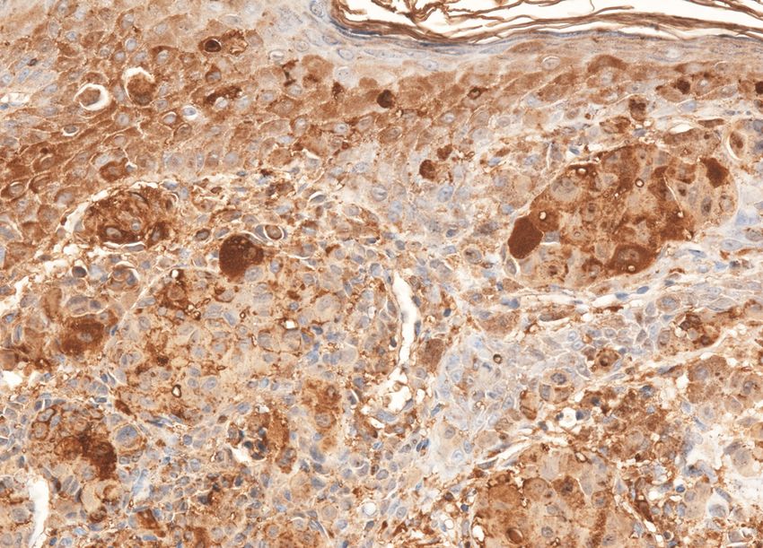
Figure 1 – Classical Spitz nevus, with features of Figure 2 – Compound nevus: nests of intraepidermal
tumor with uncertain malignant potential, rare atypical nevic cells separated through gallants from the
mitosis (H&E stain, 40×). epidermal cells, rare cells with melanic pigment (H&E
stain, 100×).
Also, in other regions of the tumor there have been the surface that become small at the base), aspect that is
highlighted also in the derma nests of epithelioid cells, not noticed in a malignant melanoma (Figure 4).
with an abundant cytoplasm, as well as multinucleated Even if, globally the tumoral proliferated cells
giant cells, but with nuclei of uniform shape and aspect have presented monomorphic nuclei, though there
– pleads for the Spitz nevus (Figure 3). have been areas in the tumor in which the cells were
Another feature evidentiated on the examined presenting polymorphic nuclei, with a heterogeneously
sections in our case has been presented to the big nevic condensed chromatin, and with macronucleoli
cells on the surface of the tumor (under-epithelial), with (Figure 5). The mitotic activity has been low, presenting
the progressive diminution of their dimensions towards rare typical mitoses – it pleads for the Spitz nevus.
the base of the tumor of the maturation of nevic cells However, in some regions of the tumor there have
from the epidermis to the profound dermis (big cells on been also highlighted very rare atypical mitoses and that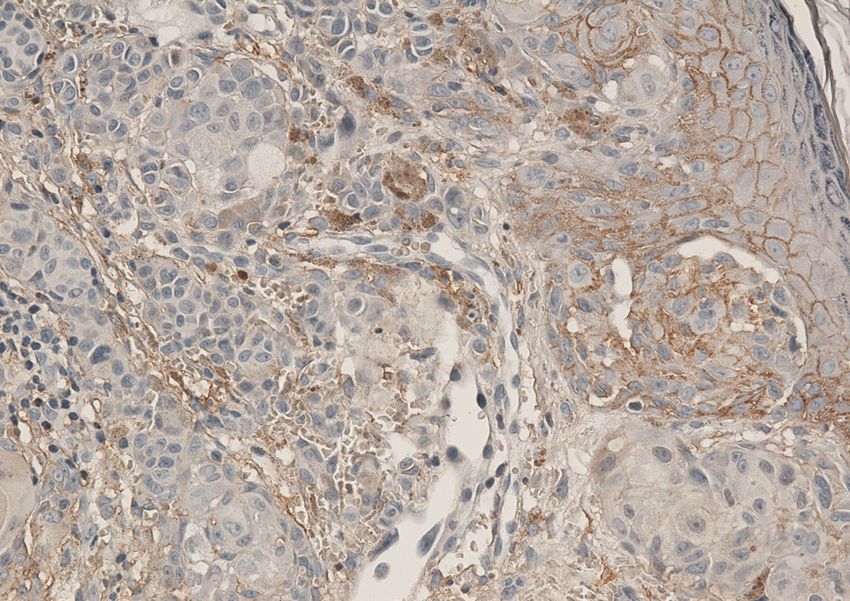
Spitz nevus with an uncertain malignant potential                                277
orient themselves towards a melanocytary tumor with            diagnosis with a malignant melanoma, we have
uncertain malignant potential (Figure 5); the melanic          performed the immunohistochemical marking of the
pigment has been reduced quantitatively, present in rare       sections from the tumor with antibodies for melano-
tumoral cells or has been completely absent in some            cytary markers (HMB45, Melan A), mesenchymal
zones of the tumor, that pleads for the Spitz nevus            markers (S100, Vimentin), for the molecules of
(Figure 2). Tumoral stroma has been poorly represented,        cellular adhesion (CD44), of tumoral growth (Cyclin
being constituted from thin fascicles of colagenous            D1), and of proliferation (assessing Ki67 proliferative
fibers that were surrounding the isles of nevic cells; the     index).
inflammatory lympho-plasmocytary infiltrate has been               The immunostaining for HMB45 has been focal,
reduced in quantity. The modifications of the epidermis        heterogeneous, at the level of tumoral cells. Thus, this
have consisted in atrophy, ulcerative zones, hyper-            has been intensely positive cytoplasmic both at the level
granulosis and hyperkeratosis.                                 of the nests of nevic intraepidermic cells and focally at
    In order to evidentiate the immunophenotype of             the level of some groups of tumoral cells from the
the proliferated tumoral cells and for the differential        dermis (Figure 6).

   Figure 3 – Spitz nevus: some epithelioid cells with           Figure 4 – Spitz nevus: progressive decreased of the
   abundant cytoplasm, with monomorphic nuclei with              maturation of the nevic cells from the surface of the
   similar shape and size (H&E stain, 100×).                     tumor (under-epithelial) towards the base of the tumor
                                                                 (H&E stain, 40×).

   Figure 5 – Spitz nevus with uncertain malignant               Figure 6 – Spitz nevus: intensely positive immuno-
   potential: areas with pleomorphic nuclei and very rare        staining for HMB45 in intraepidermal nevic cells and
   atypical mitoses (H&E stain, 400×).                           focally in the tumoral cells from the dermis (LSAB
                                                                 technique, 200×).

    As a completion of this staining, the expression of        through diffuse moderately positive immunoreaction for
the Melan A marker has been also positive at the level         vimetin at the level of tumoral cells and negative at the
of tumoral cells, being highlighted the same distribution      level of epithelial cells (Figure 9).
in the tumor as in the case of the HMB45 marker but of             The immunohistochemical expression of CD44
wicker intensity (Figure 7).                                   marker has been positive at the level of nevic cells both
    The mesenchymal origin of proliferated tumoral             in the intraepidermal nests and in those intradermal,
cells has been highlighted through nuclear and                 being also positive at the level of epidermal cells
cytoplasmic immunostaining intensely positive diffuse          (Figure 10).
for S100 in all tumoral cells (Figure 8), as well as               The immunostaining for Cyclin D1 has highlighted a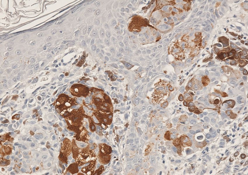
278 Ligia Stănescu et al.
positive nuclear reaction in rare tumoral cells, those differentiation of an atypical Spitz nevus by a
being disposed especially in the superficial dermis, malignant melanoma, we have evaluated the
under the covering epithelium (Figure 11). proliferative Ki67 index, this being lower than 5% in the
In order to evaluate the proliferative activity of the tumor (Figure 12), fact that leaded to the highlightening
tumoral cells, a very important factor in the of a much-reduced proliferation of nevic cells.
Figure 7 – Positive immunostaining for Melan A Figure 8 – Intensely positive nuclear and cytoplasmic
within tumoral nevic cells, with the same distribution immunostaining for S100, diffuse in all tumoral cells
as HMB45 marker but of wicker intensity (LSAB (LSAB technique, 200×).
technique, 200×).
Figure 9 – Diffuse moderately positive immuno- Figure 10 – Positive membrane immunohistochemical
reaction for vimetin at the level of tumoral cells (LSAB expression for CD44 in nevic cells, both in the
technique, 200×). intraepidermal nests and in those intradermal (LSAB
technique, 200×).
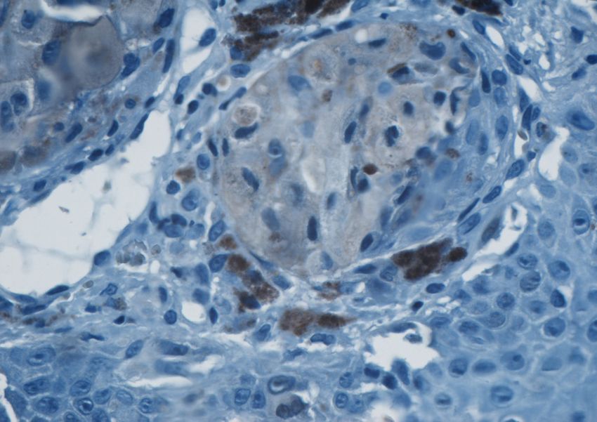
Figure 11 – Positive nuclear immunostaining for Figure 12 – Spitz nevus: the proliferative Ki67 index
Cyclin D1 in rare tumoral cells, disposed especially in lower than 5% within the tumor (LSAB technique,
the superficial dermis (LSAB technique, 400×). 200×).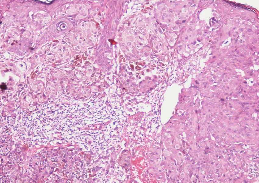
Spitz nevus with an uncertain malignant potential                              279

    Discussion                                                 reduced cellular maturation and more desmoplasia [9].
                                                                    The differential diagnosis of the Spitz nevus
    The Spitz nevus is a benign proliferation of                includes the juvenile xantogranuloma, the pyogenic
melanocytes that seems sometimes very difficult or              granuloma or the malignant melanoma. The most
impossible to differentiate from the malignant                  problematic lesion in the differential diagnosis of Spitz
melanoma through clinical and histopathological                 nevus is malignant melanoma – in such a way that
examination [4].                                                sometimes it can be clinically diagnosed wrong as a
    The tumor appears especially on children, but it can        melanoma. In our case was also raised the problem of
be found on adults as well; it has been communicated            differential diagnosis with a malignant melanoma, both
that it appeared before birth or frequently in the seventh      clinically and histopathologically, because the tumoral
decade of life. Both sexes are equally affected with a          formation has been present since birth but with a rapid
rare appearance in races Mongoloid and Negroid.                 increase in the last year, being influenced by repeated
It is impossible to estimate the prevalence of Spitz            sun exposures.
nevus, because the majority of the pathological surgical            The malignant melanoma is a tegumentary cancer
proofs have been in favor of an unusual lesion. Certain         highly aggressive that develops in melanocytes unlike
authors consider that it is likely to be less than 1% of        the Spitz nevus, which is a benign melanocytary lesion.
melanocytary nevi in a child [5].                               The malignant melanoma is a black tumor that increases
    Clinically, the Spitz nevus appears in early                rapidly. Any new black macula or papule that extends
childhood in the form of a head of a red-brown nodule;          should be biopsied in order to exclude the melanoma.
as compared to other gained nodules, this one increase          Besides, certain forms of melanoma are apigmentary
rapidly reaching a dimension of de 6–8 mm at six                and appear clinically as pink papules extended rapidly.
months.                                                         The melanoma on a child is very rare, being present on
    The lesions are highlighted, under the form of a            only 3% of the pediatric malignant tumors. Only 2% of
dome, without hair and often firm on palpation. When a          all melanoma appear during childhood [10].
light pressure is applied over the surface of the lesion            Clinically, it is impossible to make a distinction
with a glass slide, a maneuver called diascopy, the pink        between a Spitz tumor and an achrome melanoma.
coloration of the nevus will often disappear showing a          Moreover, the majority of the melanomas diagnosed on
tan-reddish pigmentation.                                       a baby and that do not develop in congenital nevus are
    The erythema of the Spitz nevus results from the            apygmentary. Early recognition of the melanoma is
vascular component of this tumor that can even have a           critical for survival because the prognosis depends
surface of telagiectasia. The surface may remain smooth         on the depth of the penetration of malignancy into the
and the epidermis is often thin and fragile. It is not          derma [11].
unusual; after major lesions may appear bleedings and               On the histopatological exam, in our case the
scalls.                                                         Spitz nevus has raised differential diagnostic problems
    The common areas for localization are the face,             with the malignant melanoma, because there have
especially chicks and legs, but other area can be affected      been highlighted some pleomorphic tumoral cells.
as well. After the initial increase, these can remain           The histopathological exam has revealed typical images
stationary for years [6]. Spitz nevus is usually solitary.      of Spitz nevus on some of the sections, but also of
The cases of Spitz nevi are rare, usually of a grouped          melanocytary tumor with an uncertain malignant
form [7] or less common as Spitz nevus eruptive                 potential on others, where there have been noticed
outspread [5, 8].                                               atypical mitosis localized in the dermal component.
    In the case described by us, this tumor has appeared            Many authors have highlightened the fact that
from birth, being localized at the level of the superior        there is no single factor that could differentiate the
lip as a plane formation, well delimited, of tan color that     Spitz nevus from the malignant melanoma, because
has then presented the above described modifications.           virtually any histopathological aspect from this
    From a histopathological point of view, general             nevus has been also described in the melanoma [12, 13].
architecture of the Spitz nevus usually is similar to that          The differentiation is even more complicated in a
of a composed nevus, being formed either from                   case of atypical Spitz nevus with: large dimension,
fusiform cells that penetrate into the derma in twined          ulceration, extension up to the adipose hypodermic
fascicles or from epithelioid cells arranged in nests, with     tissue and an increases mitotic activity. There is no
multinucleated giant cells among them.                          consensus between dermatologists and dermato-
    In the case described by us, the tumor has consisted        pathologists in what the two lesions are concerned.
of nests of nevic epithelioid cells situated both               Some consider that the Spitz nevus and the melanoma
intraepidermic, in the neighborhood of basal membrane           are part of a long sequence of lesions with the Spitz
and perpendicularly on this one, as well as within              nevus benign at an end, and the malignant melanoma on
superficial and profound dermis.                                the other end, between the two extremities existing
    After some authors, the histopathological                   different lesions with common aspects for them both
examination has shown the following differences                 [12, 14, 15]; others consider that there is no connection
between the Spitz nevus according to age [9]:                   between the two lesions [16].
achantosis, parakeratosis, pagetoidal infiltration, and             However, there are a few aspects that might help to
Kamino bodies (more frequent in children than in                the establishment of the differential diagnosis between
adults). In adults, the lesions are less pigmented, with a      the Spitz nevus and the malignant melanoma, that have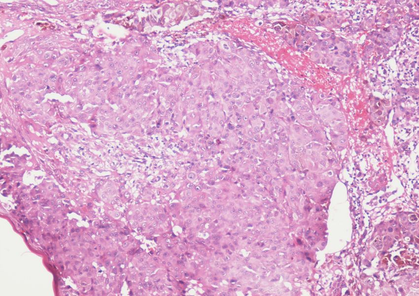
280                                                Ligia Stănescu et al.
to be taken into consideration [17]. Thus, the junctional          The immunostaining for markers S100 and Melan A
activity even though intense in the Spitz nevus, does not      has been used frequently in the clinical studies in
disorganize and do not wear down the epidermis, and            order to establish the diagnosis of malignant melanoma.
the individual segregation “in the rain” of melanocytes        In the case that we have presented, the immunoreaction
from the basal stratum is discrete or absent. Lever            for S100 has been intensely positive and diffuse at the
insists so on the importance for the diagnosis of the          level of all tumoral cells.
artifactual split that partially isolates the nevic nests;         This expression of S100 is not specified for the Spitz
these are present between the melanocytes in the               nevus because the same distribution of the
superficial dermis and thicken the collagenous fascicles       immunoreaction may be highlighted also in a malignant
that divide the small nests [10]. Sometimes, rare              melanoma. However, the interpretation of the immuno-
atypical mitoses may be highlighted, especially in the         reaction for S100 does not have to be made isolate,
cases of atypical Spitz nevus where a cellular                 but correlated to the immunostaining for the other
pleomorphism may be present, these aspects being met           melanocytary markers. From the data received from
in our case too.                                               the specialty literature it was noticed that virtually
    Due to the limits of the classical clinic and              all melanocytary benign lesions express S100 protein
histopathological exam in the differentiation of Spitz         that is also present in over 95% of the malignant
nevus (benign and atypical) from the malignant                 melanomas [21].
melanoma, many researchers have searched other                     Melan A, isolated as a specific antigen for the
methods in order to increase the accuracy of the               melanoma, is a transmembranar protein having an
diagnosis, namely the immunohistochemical exam and             uncertain function. In our case, the expression of the
the techniques of molecular biology in order to find out       marker has been similar to that of HMB45, being
the chromosomal abnormalities.                                 evidentiated only at the level of tumoral cells from the
    For the moment, the most accessible method is the          superficial dermis and in rare isolated cells from the
immunohistochemical exam. Even though up to                    deeper dermis.
the present moment, a specific marker that could                   The assessment of the vimetin expression is useful
clearly differentiate between the Spitz nevus and the          in order to show the mesenchymal origin of the tumoral
malignant melanoma, and as the used markers                    proliferated cells, but cannot differentiate between the
(melanocytary, mesenchymal, anti-apoptotic markers,            benignant and malignant melanocytary tumors and
molecules of cellular adhesion, markers for the tumoral        between the Spitz nevus and the malignant melanoma.
growth) are positive in both types of lesions,                 In our case, the expression of vimetin has been
however for the differential diagnosis is important to         positively diffuse at the level of the nevic cells.
evaluate the intensity and the distribution of the                 CD44 is a cellular adhesion protein expressed at a
immunostaining for these markers in melanocytary               membrane level and has a role both in the regulation of
cells (melanocytes), as well as the degree of the              intercellular interactions as well as in cellular migration
tumoral proliferation.                                         [24]. It is considered that it has an important role in the
    HMB45 (Human Melanosoma Black 45) is a                     tumoral invasion.
cytoplasmic antigen whose presence in the cells indicate           In our case, its expression has been homogenous in
the active formation of melanosomes and thus the               the tumor, CD44 being highlighted at the level of the
differentiation is melanocytary. It is also being              membrane of nevic cells, as well as at the level of the
expressed in normal fetal melanocytes [18], but not in         epidermal cells. The data from the specialty literature
normal non-active melanocytes [19, 20].                        showed that even in the malignant melanoma CD44 is
    The immunostaining for HMB45 in the                        present at the level of tumoral cells, but its expression is
melanocytary nevus depends on their localization at the        heterogenous in the tumor being correlated reversively
level of the skin. The dysplastic nevi usually express         proportional to the dimension of the tumor and the
HMB45 both in junctional cells as well as in nevic             depth of the invasion [24, 25].
dysplastic cells from the superficial dermis; more the             Cyclin D1 is involved in the regulation of cellular
severity of the dysplasia is bigger, the most the nevic        proliferation and in the cellular growth, being expressed
cells from the deeper dermis may focally express               also in the S phases of the cellular cycle. Its super-
HMB45 [20, 21], the same aspect being also                     extension in the dysplastic nevi and in the Spitz nevus
evidentiated in the case presented by us. This expression      present an areal pattern, is highlighted at the level of
is suggestive for the diagnosis of dysplastic or atypical      nevic cells from the dermo-epidemic junction and less at
Spitz nevus, in the disservice of the diagnosis of             the level of those from the papillary and reticular derma,
malignant melanoma. Rarely, the melanocytary benign            being correlated to cellular maturation, while in the
proliferations, such as nevi with fusiform cells, Spitz        malignant melanoma this marker is expressed in a
nevus and the atypical melanocytary hyperplasias               diffuse and intense manner at the level of cells in the
express HMB45 [20, 22].                                        tumor.
    In the malignant melanoma, the cytoplasmic                     In our case, Cyclin D1 has been expressed focally in
positivity for HMB45 is intense in a variable proportion       rare cells, disposed predominantly in the superficial
of cells, the tumors that can reach to over 100%,              derma. Thus, it can be concluded that an areal pattern of
exception making the desmoplasic malignant melanoma            the expression of Cyclin D1 may offer useful
in which the expression of this marker may be                  information in the differentiation of the Spitz nevus
completely negative [21, 23].                                  from the malignant melanoma [26].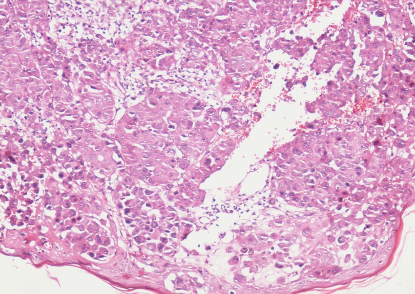
Spitz nevus with an uncertain malignant potential                                      281
    The analysis of the proliferative marker Ki67 has a           [4] BASTIAN B. C., WESSELMANN W., PINKEL D., LEBOIT P. E.,
                                                                       Molecular cytogenetic analysis of Spitz nevi shows
role in the differentiation of the malignant lesions from
                                                                       clear differences to melanoma, J Invest Dermatol, 1999,
the benign ones. Often, its expression in dysplastic                   113(6):1065–1069.
nevi and in the composed Spitz nevus is observed in               [5] FASS J., GRIMWOOD R. E., KRAUS E., HYMAN J., Adult onset
less than 6% of the cells, usually at the level of the                 of eruptive widespread Spitz nevi, J Am Acad Dermatol,
dermo-epidemic junction, being highlighted in very rare                2002, 46(5 Pt 2):S142–S143.
                                                                  [6] SANDERSON K. V., MACKIE RONA, Tumors of the skin.
cells from the profound derma (average proliferative                   In: ROOK A., WILKINSON D. S., EBLING F. J. G. (eds),
index of 3.2).                                                                                        rd
                                                                       Textbook of Dermatology, 3 edition, Blackwell Scientific
    In exchange, the malignant melanoma contains                       Publications, Oxford, 1982, 2197–2198.
multiple positive cells for protein Ki67, without a               [7] GLASGOW M. A., LAIN E. L., KINCANNON J. M., Agminated
particular distribution but diffusely disseminated in the              Spitz nevi: report of a child with a unique dermatomal
                                                                       distribution, Pediatr Dermatol, 2005, 22(6):546–549.
tumor (average proliferative index of 15.3%) [27, 28].            [8] SMITH S. A., DAY C. L. JR., VAN DER PLOEG D. E., Eruptive
    In our case, the Ki67-proliferative index has been                 widespread Spitz nevi, J Am Acad Dermatol, 1986,
less than 5% thing that was correlated to the slow                     15(5 Pt 2):1155–1159.
clinical evolution and to the absence of any secondary            [9] CESINARO A. M., FORONI M., SIGHINOLFI P., MIGALDI M.,
determinations up to the present moment.                               TRENTINI G. P., Spitz nevus is relatively frequent in adults:
                                                                       a clinico-pathologic study of 247 cases related to patient’s
    In what the evolution of the Spitz nevus is                        age, Am J Dermatopathol, 2005, 27(6):469–475.
concerned, from the beginning to maturation is of about           [10] LEVER F. W., SCHAUMBURG-LEVER G. (eds), Histopathology
six months, and then it remains stationary. Very seldom,               of the skin, J.B. Lippincott Co., Philadelphia, 1983.
it can ulcerate without constituting a malignant sign; its        [11] FERRARA G., ARGENZIANO G., SOYER H. P., CHIMENTI S.,
coloring when is observable being due to rarefaction of                DI BLASI A., PELLACANI G., PERIS K., PICCOLO D., RUBEGNI P.,
                                                                       SEIDENARI S., STAIBANO S., ZALAUDEK I., DE ROSA G.,
the epidermis.                                                         The spectrum of Spitz nevi: a clinicopathologic study of
    The lesion remains benign all over the evolution.                  83 cases, Arch Dermatol, 2005, 141(11):1381–1387.
There have been noticed, sometimes, some recidives                [12] PIEPKORN M., On the nature of histologic observations:
after its removal, leading to the confusion with a                     the case of the Spitz nevus, J Am Acad Dermatol, 1995,
                                                                       32(2 Pt 1):248–254.
melanoma.                                                         [13] SPATZ A., BARNHILL R. L., The Spitz tumor 50 years later:
    The approach of the Spitz nevus is controversial.                  revisiting a landmark contribution and unresolved contro-
The majority of the Spitz nevi act in a benign manner.                 versy, J Am Acad Dermatol, 1999, 40(2 Pt 1):223–228.
Sometimes, communications about Spitz tumors that are             [14] SPATZ A., CALONJE E., HANDFIELD-JONES S., BARNHILL R. L.,
acting aggressively just as the melanoma has led to the                Spitz tumors in children: a grading system for risk
                                                                       stratification, Arch Dermatol, 1999, 135(3):282–285.
uncertainty about the biological behavior. It has been            [15] BARNHILL R. L., FLOTTE T. J., FLEISCHLI M., PEREZ-ATAYDE A.,
described, also, the spitzoid melanoma (M). Many                       Cutaneous melanoma and atypical Spitz tumors in
dermatologists have recommended the excisional biopsy                  childhood, Cancer, 1995, 76(10):1833–1845.
of all Spitz nevi in order to allow the histological              [16] SHAPIRO P. E., Spitz nevi, J Am Acad Dermatol, 1993,
confirmation of the diagnosis.                                         29(4):667–668.
                                                                  [17] DIMITRESCU A., TRIFU P., Precancerele şi cancerele
                                                                       cutanate, vol. II, Ed. Medicală, Bucureşti, 1993, 72–74.
    Conclusions                                                  [18] KAPUR R. P., BIGLER S. A., SKELLY M., GOWN A. M.,
                                                                       Anti-melanoma monoclonal antibody HMB45 identifies an
    Although, the clinical aspect pleaded for malignant                oncofetal glycoconjugate associated with immature melano-
melanoma, and the first histopathological exam was in                  somes, J Histochem Cytochem, 1992, 40(2):207–212.
favor of a malignant melanocytary tumor, the immuno-              [19] SMOLLER B. R., HSU A., KRUEGER J., HMB-45 monoclonal
histochemical examination and clinical evolution                       antibody recognizes an inducible and reversible melanocyte
                                                                       cytoplasmic protein, J Cutan Pathol, 1991, 18(5):315–322.
disjoint the diagnosis of Spitz nevus.                            [20] BACCHI C. E., BONETTI F., PEA M., MARTIGNONI G.,
    Regarding the evolutive potential to malignancy of                 GOWN A. M., HMB45: a review, Appl Immunohistochem,
the Spitz nevus, we consider that must become                          1996, 4:73–85.
established a careful supervision of the patient                  [21] LEONG A. S.-Y., COOPER K., JOEL F., LEONG W.-M., HMB 45.
presenting this type of a nevus, its excision being                    In: LEONG A. S.-Y., COOPER K., JOEL F., LEONG W.-M. (eds),
                                                                       Manual of diagnostic antibodies for immunohistology,
imposed with safety oncological margins.                               Oxford University Press, London, 1999, 175–177.
    The classical histopathological examination must be           [22] DABBS D. J., Immunohistochemistry of soft tissue
completed with an immunohistochemical exam, often                      neoplasms and immunohistochemistry of the skin tumors.
capable to evidentiate differences to the malignant                    In: DABBS D. J. (ed), Diagnostic immunohistochemistry,
                                                                         st
                                                                       1 edition, Churchill Livingstone, 2002, 59–113, 536–559.
melanoma.
                                                                  [23] LEONG A. S.-Y., MILIOS J., An assessment of a melanoma-
                                                                       specific antibody (HMB45) and other immunohistochemical
References                                                             markers of malignant melanoma in paraffin-embedded
[1] SPITZ S., Classics in oncology: Melanomas in childhood,            tissues, Surg Pathol, 1989, 2:137–145.
    CA Cancer J Clin, 1991, 41(1):40–51.                          [24] LEIGH C. J., PALECHEK P. L., KNUTSON J. R., MCCARTHY J. B.,
[2] SULIT D. J., Spitz nevus, DermatologyReview.com Journal,           COHEN M. B., ARGENYI Z. B., CD44 expression in benign
    http://dermatologyreview.org/journal/spitz.pdf, June 2005,         and malignant nevomelanocytic lesions, Hum Pathol, 1996,
    1–6.                                                               27(12):1288–1294.
[3] WETTENGEL G. V., DRAEGER J., KIESEWETTER F., SCHELL H.,       [25] FERNÁNDEZ-FIGUERAS M. T., ARIZA A., CALATRAVA A., PUIG L.,
    NEUBAUER S., GEBHART E., Differentiation between                   FERNÁNDEZ-VASALO A., FERRÁNDIZ C., CD44 and melano-
    Spitz nevi and malignant melanomas by interphase                   cytic tumors: a possible role for standard CD44 in the
    fluorescence in situ hybridization, Int J Oncol, 1999,             epidermotropic spread of melanoma, J Cutan Pathol, 1996,
    14(6):1177–1183.                                                   23(2):133–139.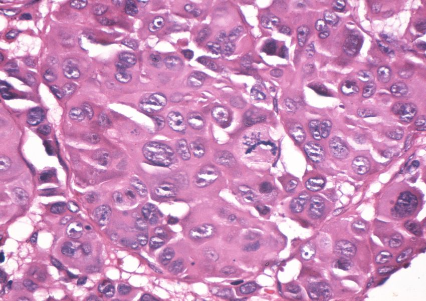
282                                                     Ligia Stănescu et al.
[26] EWANOWICH C., BRYNES R. K., MEDEIROS L. J., MCCOURTY A.,       [28] NAGASAKA T., LAI R., MEIDEIROS L. J., BRYNES R. K.,
     LAI R., Cyclin D1 expression in dysplastic nevi: an immuno-         MCCOURTY A., HARADA T., SADDIK M., Cyclin D1 over-
     histochemical study, Arch Pathol Lab Med, 2000,                     expression in Spitz nevi: an immunohistochemical study,
     125(2):208–210.                                                     Am J Dermatopathol, 1999, 21(2):115–120.
[27] PRIETO V. G., Immunohistochemistry and molecular biology
     in the management of melanocytic lesions, Rev Esp Pathol,
     1999, 32(3):452–453.

    Corresponding author
    Ligia Stănescu, Associate Professor, MD, PhD, Department of Pediatry, “Filantropia” University Hospital
    of Craiova, University of Medicine and Pharmacy, 2–4 Petru Rareş Street, 200349, Craiova, Romania;
    Phone +40748–182 406, Fax +40251–420 896, e-mail: ligstanescu@yahoo.com, cpopescu67ro@yahoo.com

    Received: February 6th, 2009

    Accepted: April 15th, 2009
You can also read