BRAIN TUMOR SEGMENTATION USING COVOLUTIONAL NEURAL NETWORK IN MRI IMAGES
←
→
Page content transcription
If your browser does not render page correctly, please read the page content below
International Journal of Pure and Applied Mathematics
Volume 119 No. 17 2018, 1585-1592
ISSN: 1314-3395 (on-line version)
url: http://www.acadpubl.eu/hub/
Special Issue
                                                                                                      http://www.acadpubl.eu/hub/

               BRAIN TUMOR SEGMENTATION USING
          COVOLUTIONAL NEURAL NETWORK IN MRI IMAGES
                                         Manda SSSNMSRL Pavan1, P. Jagadeesh2
                                    Department of Electronics and Communication Engg,
                                              Saveetha School of Engineering,
                                    Saveetha Institute of Medical and Technical Sciences,
                                                           Chennai.
          Abstract

          Brain tumor segmentation methodology is based on Convolutional Neural Networks (CNN), by
          exploring into small 3x3 kernels. The employment of small kernels permits coming up with a
          deeper architecture, besides having a positive impact against over fitting, given the less variety
          of masses within the network and also investigating on the utilization of intensity normalization
          as a pre-processing step, which is not common in Convolution Neural Network based
          segmentation methods, and well-tried in conjunction with information augmentation to be
          intolerably in effect for neoplasm segmentation in magnetic resonance imaging pictures.

          Keywords: Convolutional Neural Networks (CNN)

          Introduction                                            of the tumor to decide         Highlights, for
                                                                  example, volume, spread, and the area are
          Brain tumors have a normal occurrence rate of           basic to conclusion and shaping a course of
          26.55 for every 100,000 for ladies and 22.37            treatment. As of now, tumor areas are
          for every 100,000 for men . Gliomas are the             sectioned physically by radiologists, however,
          most ordinarily happening kind of Brain tumor           progresses in PC vision have made
          and are conceivably risk, with around 90% of            conceivable the capacity to robotize the
          gliomas having a place with an exceedingly              division procedure. In particular, tumor
          forceful class of dangerous tumor known as              division calculations in view of convolutional
          Glioblastomas[2].Glioblas-toma is the most              neural net-works (CNNs) have been appeared
          common type of Brain malignancy and is                  to be at any rate as successful as other
          profoundly forceful, with a 5 year survival rate        mechanized tumor division strategies[1].
          of 5.3 % for patients matured 40 to 64 and              Here, they showed a novel way to deal with
          middle survival time of 331 to 529 days. In             glioma division in light of profound neural
          additionto    high     mortalityrates     [1-7].        systems. They displayed two fix shrewd CNN
          Glioblastoma is very costly to treat, with a            models for fix shrewd twofold grouping of
          meaningful use of over $100,000 in the half             tumor and non-tumor locales and a full-picture
          year post-surgery. Subsequently, there exists a         CNN design. They prepared and tested the two
          significant need to precisely analyse gliomas           models on the BRATS Challenge data set, and
          and glioblastoma in their beginning times.              investigate exchange figuring out how to the
          Multimodality attractive reverberation imaging          Rembrandt dataset [12]. Because of the
          is the essential strategy for screening and             moderately little size of the informational
          determination for gliomas [13]. Exact division          collections included, they additionally

                                                              1

                                                       1585
International Journal of Pure and Applied Mathematics Special Issue
investigate a few techniques to avert show savvy nonlinearity connected toward the finish
over fitting and enhance heartiness. In the of or after each layer. A convolution operation
accompanying, they present a concise review on little locales of information is acquainted
of previous work for biomedical picture with diminish the quantity of free parameters
division and exchange learning. At that point and enhance speculation. One noteworthy
propose and assess our model architectures for preferred standpoint of convolutional systems
tumor division [18]. At last, they introduce is the use of shared height in convolutional
comes about for exchange learning between layers, which implies that a similar channel is
neuroimaging datasets. utilized for every pixel in the layer; this both
lessens memory and enhances execution [2].
Brain Tumor
Brain tumor is the tumor form when the Image Segmentation
abnormal cell forms in the brain. The brain There exist two principle ways to deal with
tumor is of two types namely, malignant tumor semantic division: pixel-wise division, where a
which consists of cancerous cells and benign little fix of a picture is utilized to order the
tumor it does not have any cancerous cells [4]. middle pixel, and completely convolutional
The most common primary brain tumors are designs as first proposed by, where the system
Gliomas, Meningioma’s, Pituitary adenomas,
input is the full picture and yield is a semantic
and Nerve sheath tumors. A brain tumor starts division volume. And have investigated the
with the brain tissue and spreads the cancerous last utilizing VGG-motivated models and
cells to entire body, which grow in the brain. indicated completely convolutional systems to
These tumors are known as metastatic brain
have exactness practically identical to pixel-
tumors. They may occur at any age. Even wise methodologies with an altogether bring
researchers and doctors do not know the exact down computational cost.
reason for the occurrence of brain tumor. Risk
factors include exposure to ionization radiation A few CNN-based strategies have been
from high dose X-rays and family history of proposed for Brain tumor division from
brain tumors [3,4]. multimodal MRI, including those in view of
dividing singular MRI cuts, volumetric
division, and CNN joined with other factual
Image Recognition techniques. Almost all present designs for
Convolutional neural networks (CNNs) Brain tumor division utilize a pixel-wise U-net
comprise of numerous layers of responsive approaches as in, which have been promising
fields. These are little neuron accumulations yet at the same time demonstrate a restricted
which process bits of the information picture achievement. Besides, while has connected
[19]. The yields of these accumulations are completely convolutional systems to other
then tiled with the goal that their info areas biomedical issues, no investigation up to this
cover, to acquire a superior portrayal of the point has utilized a completely convolutional
first picture; this is rehashed for each such approach for the particular issue of Brain
layer. Tilling enables CNNs to endure tumor division [5].
interpretation of the info picture. In the field of Brain tumor division, late
Convolutional systems may incorporate nearby recommendations additionally research the use
or worldwide pooling layers which join the
of CNNs. utilized a shallow CNN with two
yields of neuron bunch. They likewise convolutional layers isolated by max-pooling
comprise of different mixes of convolutional with walk 3, trailed by one fully associated
and completely associated layers, with point
2
1586International Journal of Pure and Applied Mathematics Special Issue
(FC) layer and a delicate max layer assessed chances for success of the other
the use of 3D channels, in spite of the fact that processes.
the dominant part of creators settled on 2D Image Segmentation: To screens an
channels. 3D channels can exploit the 3D idea input image into its essential parts or
of the pictures; however, it builds the objects.
computational load[2]. A few proposition Image Representation: To translate
assessed two-pathway systems to permit one the input data to obtain
of the branches to get greater patches than suitableimage for processing.
alternate, in this manner having a bigger Image Description: To extract feature
setting view over the picture. Notwithstanding s that result in some quantitative
their two-pathway organize, manufactured a information
course of two systems and played out a two- Image Recognition: To assign a tag
arrange preparing, via preparing with adjusted to an object based on the information
classes and after that refining it with extents provided by its descriptors
close to the firsts twofold CNN to distinguish Image Interpretation: To assign
the total tumor. At that point, a cell automata meaning to collect recognized objects.
smooth the division, before a multilayer CNN
separates the sub-areas of tumor removed fixes Deep learning in Medical Imaging
in each plane of each voxel and prepared a
CNN in every MRI succession [5]; the yields The prominent investigation to apply profound
of the last FC layer with delicate max of each neural systems to biomedical picture handling
CNN are connected and used to prepare a RF was which utilized a CNN design to perform a
classifier the ' Brain tumor locales division pixel-wise arrangement of electron microscopy
assignments into parallel sub-undertakings and neuron pictures into film and non-layer pixels.
proposed organized forecasts utilizing a CNN Because of the early accomplishments of and
as learning technique [3]. Patches of marks are others, enthusiasm for applying CNN
bunched into a word reference of name structures to Medical pictures has prospered as
patches, and the CNN must foresee the of late[6].
participation of the contribution to each of the
groups. Thedesigns with little convolutional Medical Image Analysis and division issues
bits for division of gliomas in MRI pictures exhibit a few remarkable difficulties. To start
proposed the use of little 3 × 3 pieces to with, persistent information in Medical
acquire further CNNs [10]. With littler pieces imaging issues has a tendency to be
they can stack more convolutional layers, exceedingly heterogeneous, where a similar
while having the same responsive field of pathology can show in altogether different
greater portions. For example, two 3×3 fell routes crosswise over patients. Additionally,
convolutional layers have the same viable confusing the test of restorative picture
open field of one 5×5 layer. division is the moderately little size of the
informational collections accessible, and the
Steps in image processing access information being deficient or
conflicting [7]. While most PC vision
Image Processing consists of number of steps. informational collections, for example, contain
Namely, thousands or even a large number of cases, in
Image Acquisition: To obtain a medicinal imaging issues there are once in a
digital image. while more than a couple of hundred cases in
Image Pre-Processing: To improve an informational index; thusly, CNN prepared
the image in ways that increases the on these information collections are
exceedingly inclined to over fitting. By and
3
1587International Journal of Pure and Applied Mathematics Special Issue
by, CNN-based methods have been appeared recognition or classification of tasks since the
to perform at any rate and also different tiling of neuron yields should be possible in
strategies (e.g. bolster vector machine, coordinated stages, in a way valuable for
generative models), and are extremely investigation of pictures. Contrasted with other
encouraging for applications in Medical picture characterization calculations, [16]
picture division [8]. convolutional neural systems utilize
moderately little pre-preparing. This implies
Convolutional Neural Network the system is in charge of taking in the
channels that in customary calculations are
Convolutional Neural Network is also called hand-built. The lack of better knowledge and
as ConvNet. It is a deep machine learning human effort is the main benefit for
algorithm which is used in analysing the Convolution Neural Network[12].
Image. CNN uses many multilayer perceptions
designed to get a less pre processing time [17]. In the same time, it has the benefits of
These are also called as Space invariant or applying more non-linearity and being less
Shift invariant artificial neural network. inclined to over fitting since little bits have
Convolutional networks they are enlivened by greater pieces. The utilization of maximum
natural procedures and are varieties of pooling with stride proposed as a pre-'
multilayer perceptron's intended to utilize preparing step that means to address
negligible measures of pre-processing. They information heterogeneity caused by multi-site
have wide applications in picture and video multi-scanner acquisitions of MRI pictures.
acknowledgement, recommender frameworks The vast spatial and auxiliary inconstancy in
and preparing. The convolutional neural mind tumors is likewise an essential worry that
system is otherwise called move invariant or they ponder utilizing two sorts of information
space invariant fake neural system (SIANN), enlargement [6].
which is named in view of its mutual weights
design and interpretation invariance Pre-Processing: MRI images are altered by the
qualities.CNN uses a less time consuming bias field distortion. This makes the influence
algorithms when compared to other of similar tissues to shift over the picture. To
segmentation techniques. The human effort is revise it, and connected the N4ITK strategy.
more in this segmentation algorithm. Notwithstanding, this isn't sufficient to
Convolutional neural network has numerable guarantee that the force circulation of a tissue
applications such as Image and Video sort is in a comparative power scale crosswise
recognition, Natural language processing, over various subjects for a similar MRI
Recommender systems [9]. succession, which is an express or certain
suspicion in most division techniques. Truth
Model Architecture be told, it can change regardless of the
possibility that the picture of a similar patient
Based on the study, Convolutional Neural is gained in a similar scanner in various time
Network consists of three architectures such as focuses, or within the sight of pathology. [14].
Baseline Convolution Network, Fully Along these lines, to make the complexity and
Convolutional Network, and Fully Image Fully force runs more comparable crosswise over
Convolutional Network [5]. patients and acquisitions, and applied the
power standardization technique. Along these
Time Delay in Neural Network
lines, the histogram of each grouping is more
In some cases, the delay neural network and comparative crosswise over subjects. In the
convolutional neural network may use same wake of normalizing the MRI pictures, they
type of architectures, mainly for Image register the mean power esteem and standard
4
1588International Journal of Pure and Applied Mathematics Special Issue
deviation over all preparation patches
separated for each arrangement. At that point,
they standardize the patches on each grouping
to have zero mean and unit variance [11]
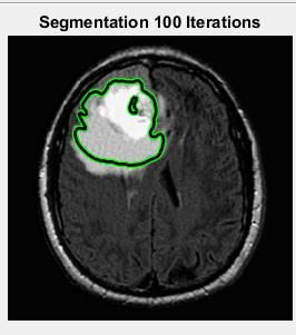
Result
References
[1] V. Badrinarayanan, A. Kendall, and R.
(a) Input Image Cipolla. SegNet: A Deep Convolutional
Encoder-Decoder Architecture for Im-age
Segmentation. pages 1–14, 2015.
[2] www.medicinenet.com/braintumor/article
[3] https://en.wikipedia.org/wiki/Braintumor
[4] M.Dhivya, R.Prema, Brain tumor
segmentation using convolution neural
network in MRI image, IJSIET.
[5] G. Tabatabai et al., “Molecular diagnostics
of gliomas: The clinical perspective,” Acta
Neuropathological, vol. 120, no. 5, pp.
(b) Estimated Bias Field 585–592, 2010.
[6] B. Menze et al., “The multimodal brain
tumor image segmentation benchmark
(BRATS),” IEEE Trans. Med. Imag., vol.
34, no. 10, pp. 1993–2024, Oct. 2015.
[7] N. J. Tustison et al., “N4ITK: Improved
n3 bias correction,” IEEETrans. Med.
Imag., vol. 29, no. 6, pp. 1310–1320, Jun.
2010.
[8] L. G. Nyúl, J. K. Udupa, and X. Zhang,
“New variants of a method of MRI scale
standardization,” IEEE Trans. Med. Imag.,
vol. 19, no. 2, pp. 143–150, Feb. 2000.
[9] M. Prastawa et al., “A brain tumor
segmentation framework based on outlier
detection,” Med. Image Anal., vol. 8, no.
3, pp. 275–283, 2004.
[10] B. H. Menze et al., “A generative
model for brain tumor segmentation in
multi-modal images,” in Medical Image
Computing and Comput.-Assisted
5
1589International Journal of Pure and Applied Mathematics Special Issue
Intervention-MICCAI 2010. New York: with high-level features. In: 2015 37th
XujiongYe,Mohammadreza, Annual Interna-tional Conference of the
Multimodal MRI brain tumor IEEE Engineering in Medicine and
segmentation using random forests with Biology Society (EMBC). pp. 3037–
features learned from fully 3040 (2015).
convolutional neural network. [19] H. Chen, Q. Dou, L. Yu, and P.-A.
[11] Patel, M.R., Tse, V.: Diagnosis and Heng. VoxResNet: Deep Voxelwise
staging of brain tumors. Semin Residual Networks for Volumetric
Roentgenol. 39, 347–360 (2004). Brain Segmentation. pages 1–9, 2016.
[12] Gordillo, N., Montseny, E., [20] D. Ciresan, A. Giusti, L.
Sobrevilla, P.: State of the art survey on Gambardella, and J. Schmidhu-ber.
MRI brain tumor segmentation. Magn Deep Neural Networks Segment
Reson Imaging. 31, 1426–1438 (2013). Neuronal Membranes in Electron
[13] Menze, B.H., Jakab, A., et.al.: The Microscopy Images. Nips, pages 1–9,
Multimodal Brain Tumor Image 2012.
Segmentation Benchmark (BRATS). [21] J. Deng, W. Dong, R. Socher, L.-J.
IEEE Transactions on Medical Imaging. Li, K. Li, and L. Fei-Fei. ImageNet: a
34, 1993–2024 (2015). large-scale hierarchical image database.
[14] Bauer, S., Wiest, R., Nolte, L.-P., In CVPR, 2009.
Reyes, M.: A survey of MRI-based [22] M. Everingham, L. Van Gool, C. K.
medical image analysis for brain tumor I. Williams, J. Winn, and A. Zisserman.
studies. Phys Med Biol. 58, R97-129 The PASCAL Visual Object Classes
(2013). Challenge 2012 (VOC2012) Results.
[15] Hamamci, A., Kucuk, N., Karaman, http://www.pascal-
K., Engin, K., Unal, G.: Tumor-Cut: network.org/challenges/VOC/voc2012/
Segmentation of Brain Tu-mors on workshop/index.html.
Contrast Enhanced MR Images for [23] R. Girshick. Fast r-cnn. In
Radiosurgery Applications. IEEE International Conference on Com-puter
Transactions on Medical Imaging. 31, Vision (ICCV), 2015.
790–804 (2012). [24] M. Havaei, A. Davy, D. Warde-
[16] Kleesiek, J., Biller, A., Urban, G., Farley, A. Biard, A. Courville, Y.
Köthe, U., Bendszus, M., Hamprecht, Bengio, C. Pal, P.-M. Jodoin, and H.
F.: ilastik for Multi-modal Brain Tumor Larochelle. Brain Tumor Segmentation
Segmentation. with Deep Neural Networks. 2015.
[17] Subbanna, N.K., Precup, D., [25] E. C. Holland. Progenitor cells and
Collins, D.L., Arbel, T.: Hierarchical glioma formation. Cur-rent Opinion in
Probabilistic Gabor and MRF Seg- Neurology, 14(6):683–688, 2001.
mentation of Brain Tumours in MRI [26] K. Kamnitsas, C. Ledig, V. F.
Volumes. In: Mori, K., Sakuma, I., Newcombe, J. P. Simpson, A. D. Kane,
Sato, Y., Barillot, C., and Navab, N. D. K. Menon, D. Rueckert, and B.
(eds.) Medical Image Computing and Glocker. Efficient multi-scale 3D CNN
Computer-Assisted Intervention – with fully connected CRF for accurate
MICCAI 2013. pp. 751–758. Springer brain lesion segmentation. Medical
Berlin Heidelberg (2013). Image Analysis, 36:61–78, 2017.
[18] Pinto, A., Pereira, S., Correia, H., [27] B. Kayalibay, G. Jensen, and P. van
Oliveira, J., Rasteiro, D.M.L.D., Silva, der Smagt. CNN-based Segmentation of
C.A.: Brain Tumour Segmen-tation Medical Imaging Data. 2017.
based on Extremely Randomized Forest
6
1590International Journal of Pure and Applied Mathematics Special Issue
[28] B. A. Kohler, E. Ward, B. J.
McCarthy, M. J. Schymura, L. A. G.
Ries, C. Eheman, A. Jemal, R. N.
Anderson, U. A. Ajani, and B. K.
Edwards. Annual report to the nation on
the status of cancer, 1975-2007,
featuring tumors of the brain and other
nervous system. Journal of the National
Cancer Institute, 103(9):714–736, 2011.
[29] A. Krizhevsky, I. Sutskever, and G.
E. Hinton. Imagenet classification with
deep convolutional neural networks. In
F. Pereira, C. J. C. Burges, L. Bottou,
and K. Q. Weinberger, editors,
Advances in Neural Information
Processing Systems 25, pages 1097–
1105. Curran Associates, Inc., 2012.
[30] S.H.S.A. Ubaidillah, R.
Sallehuddinand N.A. Ali, “Cancer
detection using artificial neural network
and support vector machine: A
Comparative study”.
[31] O.P. Verma, M. Hammandlu, S.
Susan, M. Kulkami and P.K. Jain, "A
simple single seeded region growing
algorithm for color image segmentation
using adaptive thresholding," 2011
International Conference on
Communication Systems and Network
Technologies, ©2011 IEEE.
7
15911592
You can also read