Original Article Embryonal tumor with multilayered rosettes, C19MC-altered (ETMR): a newly defined pediatric brain tumor
←
→
Page content transcription
If your browser does not render page correctly, please read the page content below
Int J Clin Exp Pathol 2019;12(8):3156-3163
www.ijcep.com /ISSN:1936-2625/IJCEP0094128

Original Article
Embryonal tumor with multilayered rosettes,
C19MC-altered (ETMR): a newly defined
pediatric brain tumor
Yu-Chun Pei1*, Guo-Hao Huang1*, Xiao-Hong Yao2, Xiu-Wu Bian2, Fei Li3, Yan Xiang1, Lin Yang1, Sheng-Qing
Lv1, Jun Liu1
1
 Department of Neurosurgery, Xinqiao Hospital, Third Military Medical University, Chongqing 400037, China;
Departments of 2Pathology, 3Neurosurgery, Southwest Hospital, Third Military Medical University, Chongqing
400038, China. *Equal contributors and co-first authors.
Received March 20, 2019; Accepted April 18, 2019; Epub August 1, 2019; Published August 15, 2019

Abstract: Embryonal tumor with multilayered rosettes (ETMR), C19MC-altered, is a newly defined and rare pediatric
malignant tumor of the central nervous system (CNS) in the 2016 WHO Classification of Tumors of the Central Ner-
vous System. Here we present two cases of ETMR with amplification of the C19MC locus at chromosome 19q13.42.
Case 1 is a fifteen-year-old boy, who underwent gamma knife surgery two times three years ago, after presenting
with seizures. Magnetic resonance imaging (MRI) identified a large mass in the left frontotemporal lobe. Case 2 is
a three-year-old boy who underwent surgery for a right frontal lobe tumor followed by chemotherapy. Eight months
later, MRI identified a recurrent tumor in the bilateral frontal lobe. Histologically, cases 1 and 2 exhibited a typical
papillary/trabecular and a multilayered rosette pattern resembling medulloepithelioma (ME) and ependymoblas-
toma (EBL), respectively. Immunohistochemically, CD99, synaptophysin, vimentin, and LIN28A were positive in both
cases. Most importantly, both cases displayed amplification in the C19MC locus at 19q13.42 in a fluorescence in
situ hybridization (FISH) analysis.

Keywords: Embryonal tumor with multilayered rosettes (ETMR), C19MC locus, LIN28A, chromosome 19q13.42,
amplification

Introduction                                                    which ME and EBL are also included irrespec-
                                                                tive of the C19MC locus amplification status.
Embryonal tumor with multilayered rosettes
C19MC-altered (ETMR) is a WHO grade IV                          Amplification of the C19MC locus at 19q13.42
aggressive embryonal tumor, which is newly                      was observed in 37/40 (93%) of the tumors
defined in the 2016 WHO Classification of                       morphologically diagnosed as EBL or ETANTR
Tumors of the Central Nervous System (CNS)                      [2]. Nobusawa et al. [3] found 19q13.42 ampli-
[1]. ETMR mainly affects children aged < 4                      fication in ETANTR, EBL, and ME, but not in AT/
years old and demonstrates a rapid growth and                   RT. Korshunov et al. [4] showed that LIN28A, an
an aggressive clinical course (the mean surviv-                 RNA-binding protein that inhibits the process-
                                                                ing of pre-let-7 miRNAs, is a highly specific
al is 12 months after combination therapies)
                                                                immunohistochemical diagnostic marker of
[1]. Most pediatric CNS embryonal neoplasms
                                                                ETMR [4].
were previously diagnosed as embryonal tumor
with abundant neuropil and true rosettes                        To date, ETMR in the literature remains rare.
(ETANTR), ependymoblastoma (EBL) and me-                        Here, we present the clinicopathological char-
dulloepithelioma (ME), and any CNS embryonal                    acteristics of two cases of ETMR (medical his-
tumor with C19MC amplification or fusion were                   tory, imaging data, H&E staining, FISH analysis
included in this entity [1]. This definition distin-            of the C19MC locus, and IHC detection of
guished ETMR from the previously defined CNS                    LIN28A are included) to help provide a better
primitive neuroectodermal tumors (PNETs), in                    understanding of this new entity.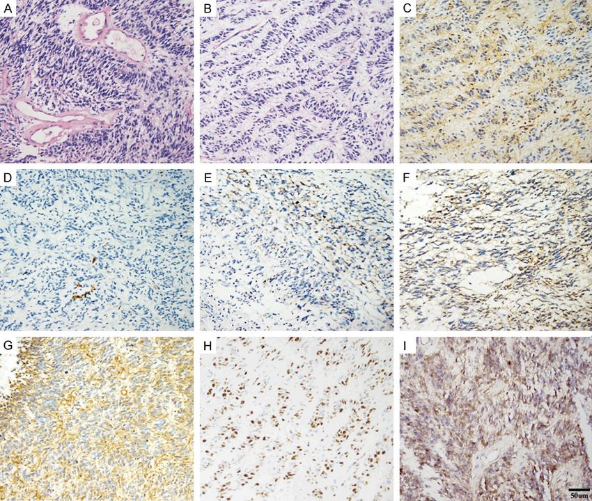
ETMR

Figure 1. Magnetic resonance (MR) of Case 1: (A) Initial axial T1-weighted image shows an irregular mass in the left
frontotemporal lobe; (B) Postoperative Axial Gadolinium (Gd)-enhanced T1-weighted image shows no enhancement,
confirming that this mass was gross totally resected; Axial (C) and coronal (D) Gd-enhanced T1-weighted images
show a heterogenous enhanced recurred mass in the primary lesion.

Materials and methods                                         ethanol. Ten microliters of probe were applied
                                                              on the sections for hybridization at 78°C for 10
We reviewed 29 cases of pediatric CNS embry-                  min and 37°C for 16 h. Following hybridization,
onal tumors diagnosed in three affiliated ho-                 the sections were washed with 2x saline sodi-
spitals of Third Military Medical University                  um citrate (SSC) and stained with 4’6’-diamino-
(Southwest Hospital, Xinqiao Hospital and                     2-phenylindole (DAPI). The sections were ana-
Daping Hospital), Chongqing, China, and in the                lyzed with a fluorescence microscope with
Children’s Hospital of Chongqing Medical                      appropriate filters. Cell nuclei with similar sizes,
University, China. We finally identified 2 cases              intact borders with no overlap, and with clear
which were initially diagnosed as CNS primitive               dual probe signals were considered effective
neuroectodermal tumor (PNETs) with 19q13.42                   cell nuclei. Dual color signals in two hundred
amplification according to the 2007 WHO clas-                 cell nuclei were randomly counted for each sec-
sification of CNS tumors. Case 1 was from                     tion. The sections were analyzed with a fluores-
Xinqiao Hospital, and case 2 was from                         cence microscope with appropriate filters. Cell
Children’s Hospital of Chongqing Medical                      nuclei with similar sizes, intact borders with no
University. Written informed consents were                    overlap, and with clear dual probe signals were
obtained from their parents. Formalin-fixed par-              considered effective cell nuclei. Dual color sig-
affin-embedded tissue sections were first sub-                nals in two hundred cell nuclei were randomly
jected to hematoxylin and eosin (H&E) staining.               counted for each section. The specimens were
Then, IHC staining was performed using the                    considered to have 19q13.42 amplification
standard two-step EnVision method. The anti-                  when the Target (Green)/Reference (Red) ratio
bodies used in this study were vimentin, S-100,               was > 1.5.
GFAP, EMA, CD99, LIN28A, and Ki-67. The anti-
bodies were obtained from Abcam (Abcam,                       Clinical summary, pathological findings, and
UK), Santa Cruz Biotechnology (Santa Cruz, CA,                FISH analysis
USA), and ZSGB-Bio (ZhongShan Golden Bridge,
Beijing, China).                                              Case 1: In 2012, a 15-year-old boy presented
                                                              with focal seizures with his limbs twitching,
Dual color fluorescence in situ hybridization                 accompanied by urinary incontinence, an
(FISH) was performed on formalin-fixed paraf-                 upward gaze, and vomiting. Other physical find-
fin-embedded tissue sections using ZytoLight                  ings and family histories were negative. An
SPEC C19MC/19q13 Dual Color Probe                             unenhanced MRI identified a mass in the left
(ZytoVision GmbH, Germany), which contains                    frontotemporal lobe (Figure 1A). The patient
ZyGreen labeled polynucleotides (target se-                   underwent gamma knife radiosurgery two
quences mapping in 19q13.42) and ZyOrange                     times, in June and July 2009, but failed to con-
labeled polynucleotides (target sequences                     trol the seizures. In August 2012, the patient
mapping in 19q13.3). The sections were                        started to complain of headaches and dizzi-
dewaxed in xylene, dehydrated in ethanol, and                 ness. Computed tomography (CT) indicated an
pretreated in a 30% sodium bisulfite solution,                enlarged mass measuring 90 mm × 60 mm in
digested with pepsin, and then dehydrated in                  size, which was removed via total gross resec-

3157                                                              Int J Clin Exp Pathol 2019;12(8):3156-3163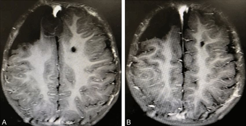
ETMR

                                                                              200 mg/m2/day, 146 mg for 3
                                                                              days; etoposide, 150 mg/m2/
                                                                              day, 109.6 mg for 3 days. After
                                                                              combined chemotherapy, the
                                                                              recurrent masses were gross
                                                                              totally resected (Figure 2B).
                                                                              The patient is currently being
                                                                              followed-up.

                                                                                  Histologically, the tumor was
                                                                                  presented with sheets and
                                                                                  clusters of round/oval-shaped
                                                                                  cells incorporating numerous
Figure 2. Gadolinium (Gd)-enhanced magnetic resonance (MR) images at
                                                                                  multilayered rosettes lacking
preoperative and postoperative presentation for Case 2, who underwent             a neuropil-like matrix (Figure
tumor resection followed by combined chemotherapy: (A) Axial Gadolinium           4A, 4B). In these cells, deep-
(Gd)-enhanced T1-weighted image shows a recurrent solid and mixed cystic          ly stained nuclei, dispersed
lesion in the bilateral frontal lobe, with heterogenous ring-enhancement; (B)     chromatin, and mitoses fig-
Axial Gadolinium (Gd)-enhanced T1-weighted image shows that the recur-
rent lesion was gross totally resected and no enhancement was observed.
                                                                                  ures were observed (Figure
                                                                                  4A, 4B). Immunohistochemi-
                                                                                  stry showed the expressions
tion (Figure 1B). One year later, MRI revealed a                of CD99 (Figure 4C), Syn (Figure 4F) and
large gadolinium-enhanced mass at the prima-                    LIN28A (Figure 4I). The immunoreactivity to
ry lesion, indicating a recurrence (Figure 1C,                  vimentin was intense (Figure 4G), but the
1D). The patient’s parents did not allow further                expressions of CK (Figure 4D) and EMA (Figure
treatment, and the patient was lost for                         4E) were negative. The Ki-67 labeling index was
follow-up.                                                      approximately 70% (Figure 4H). The FISH analy-
                                                                sis showed amplification of the 19q13.42
Histologically, the tumor consisted of spindle                  locus, with an amplification index (Green/Red)
shaped cells arranged in clusters tending to                    of 2.8 (Figure 5B).
form a typical papillary or trabecular structure
(Figure 3A, 3B). Occasionally, deeply stained                   Discussion
nuclei and mitotic figures can be found.
Abundant blood vessels, necrosis and calcifica-                 CNS embryonal tumors include several groups
tion were observed (Figure 3A, 3B). Immu-                       of pediatric brain tumors with typical multilay-
nohistochemically, the tumor cells were posi-                   ered rosettes. These are supratentorial primi-
tive for CD99 (Figure 3C), EMA (Figure 3E), Syn                 tive neuroectodermal tumors (PNET), medullo-
(Figure 3F) and LIN28A (Figure 3I). Vimentin                    blastoma (infratentorial location), medulloepi-
was strongly expressed (Figure 3G), but CK                      thelioma, neuroblastoma, ependymoblastoma,
expression was absent (Figure 3D). The Ki-67                    and atypical teratoid/rhabdoid tumors (AT/RT)
labeling index was approximately 60% (Figure                    [7]. The 2016 WHO Classification proposed a
3H). The FISH analysis showed amplification at                  new integrated diagnostic criterion for C19MC-
the 19q13.42 locus, with an amplification index                 altered ETMR [1]. If ETMR diagnosis is suggest-
(Green/Red) of 1.55 (Figure 5A).                                ed histologically, then 19q13.42 amplification
                                                                should be assessed using FISH. If ETMR is diag-
Case 2: A three-year-old boy was admitted to                    nosed on the basis of histology alone, the
Children’s Hospital of Chongqing Medical                        tumors may be diagnosed as ETMR, NOS (not
University to undergo chemotherapy following a                  otherwise specified) [1].
tumor resection 8 months earlier. The physical
findings were negative. However, MRI identified                 Most C19MC-altered ETMRs have been report-
a recurrent tumor mass (27 mm × 22 mm ×                         ed as case reports (Table 1) [8-15]. ETMRs may
28.5 mm in size) in the bilateral frontal lobe                  develop in both the supratentorial and infraten-
with mild enhancement (Figure 2A). The patient                  torial compartments. The most common site is
first received combined chemotherapy with the                   the cerebral hemisphere, with a frequent
following regimen: methotrexate, intraventricu-                 involvement of the frontal and parietotemporal
lar injection, 2 mg/day for 2 days; carboplatin,                regions [16]. In addition to the supratentorial

3158                                                            Int J Clin Exp Pathol 2019;12(8):3156-3163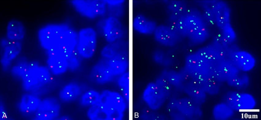
ETMR
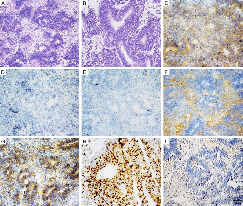
Figure 3. Microscopic appearance of Case 1: (A, B) Spindle shaped cells are arranged in clusters tending to form
a typical papillary or trabecular structure (H&E); (C-I) Immunohistochemistry shows the expression of CD99 (C),
epithelial membrane antigen (EMA, E), synaptophysin (Syn, F), vimentin (G) and LIN28A (I). The immunoreactivity
of cytokeratin was negative (CK, D); the Ki-67 labeling index was approximately 60% (H). Scale bar 50 mm: (A-I).
compartment, they can also originate in the and is associated with an aggressive clinical
cerebellum, brainstem, and spinal cord [17, course after combination therapies. Tumor
18]. For the radiological features, the head recurrence, wide-spread leptomeningeal dis-
computed tomographic (CT) image shows a semination, and extraneural metastases are
hyper attenuating mass in the cerebral hemi- frequent in the terminal stages of the disease
sphere. MRI shows well-defined margins, mini- [1]. So, it is important to elucidate the tumori-
mal vasogenic edema, and subtle enhance- genesis well and to seek an effective therapy
ment lesions [18]. There are no specific for this aggressive disease.
radiological features distinguishing ETMR and
other brain tumors [19]. In our clinic, we also treated a 12-year-old girl
with ETMR in the right frontal lobe. The tumor
In the present report, the two cases were locat- was total resected, and the histopathological
ed in the frontal lobes (Figures 1, 2). The features included multilayered and mitotically
patients had a smooth recovery after tumors active structures consisting of pseudostratified
were gross totally resected. Like other brain neuroepithelium with a central, round, or slit-
tumors, gross total resection and radiation may like lumen. IHC staining showed expressions of
provide some benefit for ETMR cases [1]. synaptophysin, vimentin and LIN28A. Unfor-
However, ETMR demonstrates rapid growth tunately, since it was not possible to assess the
3159 Int J Clin Exp Pathol 2019;12(8):3156-3163ETMR
Figure 4. The microscopic appearance of Case 2: (A, B) Sheets and clusters of round/oval-shaped cells incorporate
numerous multilayered rosettes lacking a neuropil-like matrix; (C-I) Immunohistochemistry shows the expression of
CD99 (C), synaptophysin (F), vimentin (G) and LIN28A (I), but an absence of the expression of cytokeratin (D) and
epithelial membrane antigen (EMA, E); the Ki-67 labeling index was approximately 70% (H). Scale bar 50 mm: (A-I).
status of 19q13.42, the tumor
was diagnosed as ETMR, NOS.
Two years later, the tumor
recurred, and she underwent
a second surgery. One year
later, she died of a recurrent
tumor with widespread lepto-
meningeal dissemination (un-
published data).
In summary, C19MC-altered
ETMR is a new entity that has
Figure 5. Fluorescence in situ hybridization (FISH) analysis detects the am- a poor outcome in children.
plification of the 19q13.42 locus of Case 1 and Case 2: the amplification The incidence of ETMR re-
index (Green/Red) for Case 1 is 1.55 (A) and for Case 2 is 2.8 (B). The green
lights represent polynucleotides targeting sequences mapping in 19q13.42; mains unclear, because only
the red lights represent polynucleotides targeting sequences mapping in single cases reports have
19q13.3 as an internal reference. become available so far.
3160 Int J Clin Exp Pathol 2019;12(8):3156-3163ETMR
Table 1. Summary of cases of the C19MC-altered intracranial embryonal tumor with multilayered rosettes in recent years
Age Sex Initial symptoms Location Resection Chemotherapy Immunohistochemistry Prognosis Ref.
2 ys M Personality changes and ataxia for 1 Cerebellum Sub-total Yes Positive for CD99, GFAP, INI1, NeuN, NF, Syn, > 17 months of PFS 8
month, morning headache, nausea and vermis Vimentin; Negative for Actin, CK, EMA; Ki67 index:
vomiting for 2 weeks 47%
33 ms F Episodic headaches, increased head Left parieto- Near-total Yes Positive for EMA, INI1, NF and Syn 10 months of PFS 9
circumference and mild gait disturbance occipital lobes
for 6 months
29 ms M Progressive visual disturbance for 1 Bilateral parieto- Sub-total No Positive for CD99, CgA, EMA, INI1, MAP2, Nestin, NA 10
months; headache, nausea and vomiting occipital lobes NeuN, NF, p53, S-100, Syn and Vimentin; Negative
for 1 weeks for Actin, CK, Desmin, Germ cell markers and Olig2;
4 ys M Headache, nausea, vomiting, gait and Right mid-pons, Sub-total No Ki67 index: 70% and 58% NA
balance disturbances for 2 months, more mesencephalon
recently strabismus and left hemiplegia
2 ys M Ataxic gait, dysarthria, and dysphagia Basilar part of the Sub-total Yes Positive for GFAP, INI1, LIN28A, Syn Died 7 months after surgery 11
pons
33 ms M Vomiting and gait disturbance for 3 Intramedullary Sub-total Yes Positive for EMA, INI1, LIN28A, NF, NeuN, Syn; Died 6 months after surgery 12
months mass, right dorsal Negative for GFAP; Ki67 index: 70%
part of the pons
8 ms F Complaints of vomiting and drooping of Left cerebellar Sub-total No Positive for CD99, CgA, EMA, INI1, NF, p53, Syn Expired 1 week after diagnosis 13
left eyelid for 3 weeks hemisphere and Vimentin; Negative for CK, Desmin, GFAP and because of widespread CNS drop
NSE; Ki67 index: 70%-80% metastases
2 ys F Two episodes of seizure, multiple epi- Right parietooc- Total Yes Positive for Syn, vimentin; Negative for CK > 6 months of no evidence for 14
sodes of vomiting, and weakness of the cipital lobes recurrence
left side of the body for 7 days
3161 Int J Clin Exp Pathol 2019;12(8):3156-3163ETMR
However, epidemiological data may be obtained Shimoyama Y, Nakazawa A, Nishizawa S, Kishi-
in the future, as a new ICD-O code (9478/3) has moto H, Matsuoka K, Nakayama M, Okura N,
been assigned to this new entity (2016 WHO Nakazato Y. Analysis of chromosome 19q13.42
Classification). The most common clinical mani- amplification in embryonal brain tumors with
ependymoblastic multilayered rosettes. Brain
festations are symptoms and signs of increased
Pathol 2012; 22: 689-697.
intracranial pressure and focal neurological
[4] Korshunov A, Ryzhova M, Jones DT, Northcott
signs. The radiological features are similar to PA, van Sluis P, Volckmann R, Koster J, Ver-
other brain tumors. The integrated diagnosis steeg R, Cowdrey C, Perry A, Picard D, Rosen-
should be based on histology (CNS embryonal blum M, Giangaspero F, Aronica E, Schüller U,
tumor with multilayered rosettes), immunoreac- Hasselblatt M, Collins VP, von Deimling A, Lich-
tivity (synaptophysin and vimentin, and the spe- ter P, Huang A, Pfister SM, Kool M. LIN28A im-
cific biomarker LIN28A), and genetics (amplifi- munoreactivity is a potent diagnostic marker
cation of C19MC locus at 19q13.42 by FISH) to of embryonal tumor with multilayered rosettes
reliably diagnose this novel aggressive pediat- (ETMR). Acta Neuropathol 2012; 124: 875-
ric brain tumor. 881.
[5] Spence T, Sin-Chan P, Picard D, Barszczyk M,
Acknowledgements Hoss K, Lu M, Kim SK, Ra YS, Nakamura H,
Fangusaro J, Hwang E, Kiehna E, Toledano H,
We thank Dr Hiroko Ohgaki, Institute of Neuro- Wang Y, Shi Q, Johnston D, Michaud J, La Spi-
pathology, Charite Medical University, Berlin, na M, Buccoliero AM, Adamek D, Camelo-Pira-
gua S, Peter Collins V, Jones C, Kabbara N,
for critically reading the manuscript, Prof. Hua
Jurdi N, Varlet P, Perry A, Scharnhorst D, Fan X,
Feng from the Department of Neurosurgery,
Muraszko KM, Eberhart CG, Ng HK, Gururan-
Southwest Hospital, Third Military Medical Uni- gan S, Van Meter T, Remke M, Lafay-Cousin L,
versity for his helpful comments, and Mrs. Rong Chan JA, Sirachainan N, Pomeroy SL, Clifford
Xin from Central Laboratory, Xinqiao Hospital, SC, Gajjar A, Shago M, Halliday W, Taylor MD,
Third Military Medical University for her techni- Grundy R, Lau CC, Phillips J, Bouffet E, Dirks
cal assistance. PB, Hawkins CE, Huang A. CNS-PNETs with
C19MC amplification and/or LIN28 expression
Disclosure of conflict of interest comprise a distinct histogenetic diagnostic
and therapeutic entity. Acta Neuropathol
None. 2014; 128: 291-303.
[6] Scheithauer BW. Development of the WHO
Address correspondence to: Sheng-Qing Lv and Jun classification of tumors of the central nervous
Liu, Department of Neurosurgery, Xinqiao Hospital, system: a historical perspective. Brain Pathol
Third Military Medical University, Chongqing 400- 2009; 19: 551-564.
037, China. Tel: +86-23-68774910; E-mail: lvsq- [7] Wang J, Liu Z, Fang J, Du J, Cui Y, Xu L, Li G.
0518@hotmail.com (SQL); Tel: +86-23-68774541; Atypical teratoid/rhabdoid tumors with multi-
E-mail: liujdoctor@163.com (JL) layered rosettes in the pineal region. Brain Tu-
mor Pathol 2016; 33: 261-266.
References [8] Pfister S, Remke M, Castoldi M, Bai AH, Muck-
enthaler MU, Kulozik A, von Deimling A,
[1] Louis DN, Ohgaki H, Wiestler OD, Cavenee WK, Pscherer A, Lichter P, Korshunov A. Novel ge-
editors. WHO classificaiton of tumours of the nomic amplification targeting the microRNA
central nervous system (Revised 4th edition). cluster at 19q13.42 in a pediatric embryonal
Lyon: IARC; 2016. tumor with abundant neuropil and true ro-
[2] Korshunov A, Remke M, Gessi M, Ryzhova M, settes. Acta Neuropathol 2009; 117: 457-464.
Hielscher T, Witt H, Tobias V, Buccoliero AM, [9] Woehrer A, Slavc I, Peyrl A, Czech T, Dorfer C,
Sardi I, Gardiman MP, Bonnin J, Scheithauer B, Prayer D, Stary S, Streubel B, Ryzhova M, Kor-
Kulozik AE, Witt O, Mork S, von Deimling A, Wi- shunov A, Pfister SM, Haberler C. Embryonal
estler OD, Giangaspero F, Rosenblum M, Pi- tumor with abundant neuropil and true ro-
etsch T, Lichter P, Pfister SM. Focal genomic settes (ETANTR) with loss of morphological but
amplification at 19q13.42 comprises a power- retained genetic key features during progres-
ful diagnostic marker for embryonal tumors sion. Acta Neuropathol 2011; 122: 787-790.
with ependymoblastic rosettes. Acta Neuro- [10] Wang Y, Chu SG, Xiong J, Cheng HX, Chen H,
pathol 2010; 120: 253-260. Yao XH. Embryonal tumor with abundant neu-
[3] Nobusawa S, Yokoo H, Hirato J, Kakita A, Taka- ropil and true rosettes (ETANTR) with a focal
hashi H, Sugino T, Tasaki K, Itoh H, Hatori T, amplification at chromosome 19q13.42 locus:
3162 Int J Clin Exp Pathol 2019;12(8):3156-3163ETMR
further evidence of two new instances in Chi- [16] Zagzag D, Miller DC, Knopp E, Farmer JP, Lee
na. Neuropathology 2011; 31: 639-647. M, Biria S, Pellicer A, Epstein FJ, Allen JC. Prim-
[11] Nobusawa S, Orimo K, Horiguchi K, Ikota H, Yo- itive neuroectodermal tumors of the brain-
koo H, Hirato J, Nakazato Y. Embryonal tumor stem: investigation of seven cases. Pediatrics
with abundant neuropil and true rosettes with 2000; 106: 1045-1053.
only one structure suggestive of an ependymo- [17] Benesch M, Sperl D, von Bueren AO, Schmid I,
blastic rosette. Pathol Int 2014; 64: 472-477. von Hoff K, Warmuth-Metz M, Ferrari R, Lassay
[12] Sato H, Terakawa Y, Tsuyuguchi N, Kuwae Y, L, Kortmann RD, Pietsch T, Rutkowski S. Pri-
Ohsawa M, Ohata K. Embryonal tumor with mary central nervous system primitive neuro-
abundant neuropil and true rosettes in the ectodermal tumors (CNS-PNETs) of the spinal
brainstem: case report. J Neurosurg Pediatr cord in children: four cases from the German
2015; 16: 291-295. HIT database with a critical review of the litera-
[13] Tariq MU, Ahmad Z, Minhas MK, Memon A, ture. J Neurooncol 2011; 104: 279-286.
Mushtaq N, Hawkins C. Embryonal tumor with [18] Wang B, Gogia B, Fuller GN, Ketonen LM. Em-
multilayered rosettes, C19MC-altered: report bryonal tumor with multilayered rosettes,
of an extremely rare malignant pediatric cen- C19MC-altered: clinical, pathological, and neu-
tral nervous system neoplasm. SAGE Open roimaging findings. J Neuroimaging 2018; 28:
Med Case Rep 2017; 5: 2050313X17745208. 483-489.
[14] Bouali S, Zehani A, Mahmoud M, Said IB, Kal- [19] Shih RY, Koeller KK. Embryonal tumors of the
lel J, Jemel H. Embryonal tumor with multilay- central nervous system: from the radiologic pa-
ered rosettes: illustrative case and review of thology archives. Radiographics 2018; 38:
the literature. Childs Nerv Syst 2018; 34: 525-541.
2361-2369.
[15] Wesseling P. Embryonal tumor with multilay-
ered rosettes (ETMR): signed, sealed, deliv-
ered. Acta Neuropathol 2014; 128: 305-308.
3163 Int J Clin Exp Pathol 2019;12(8):3156-3163You can also read