Molecular clarification of brainstem astroblastoma with EWSR1 BEND2 fusion in a 38 year old man
←
→
Page content transcription
If your browser does not render page correctly, please read the page content below
Free Neuropathology 2:16 (2021)                                                                                                                                                   Smith‐Cohn et al
doi: https://doi.org/10.17879/freeneuropathology‐2021‐3334                                                                                                                             page 1 of 8

                                                                                                                                                                           Case Report

Molecular clarification of brainstem astroblastoma with EWSR1‐
BEND2 fusion in a 38‐year‐old man
Matthew A. Smith‐Cohn,1,2 Zied Abdullaev,3 Kenneth D. Aldape,3 Martha Quezado,3 Marc K. Rosenblum,4
Chad M. Vanderbilt,4 Fausto J. Rodriguez,5 John Laterra,**2 Charles G. Eberhart**5
1
    Neuro‐Oncology Branch, National Cancer Institute, National Institutes of Health, Bethesda, MD, USA
2
    Department of Neurology, The Johns Hopkins University School of Medicine, Baltimore, MD, USA
3
    Laboratory of Pathology, Center for Cancer Research, National Cancer Institute, Bethesda, MD, USA
4
    Department of Pathology, Memorial Sloan‐Kettering Cancer Center, New York, NY, USA
5
    Department of Pathology, The Johns Hopkins University School of Medicine, Baltimore, MD, USA

** These authors contributed equally to this manuscript

Corresponding author:
Charles Eberhart, MD, Ph.D. ∙ Department of Pathology ∙ The Johns Hopkins University School of Medicine ∙ 1800 Orleans St. ∙ Sheikh
Zayed Tower ∙ Baltimore, MD 21287 ∙ USA
ceberha@jhmi.edu

Submitted: 21 April 2021                     ∙     Accepted: 17 June 2021                     ∙     Copyedited by: Calixto‐Hope Lucas                           ∙     Published: 21. June 2021

Abstract
The majority of astroblastoma occur in a cerebral location in children and young adults. Here we describe the
unusual case of a 38‐year‐old man found to have a rapidly growing cystic enhancing circumscribed brainstem
tumor with high grade histopathology classified as astroblastoma, MN1‐altered by methylome profiling. He was
treated with chemoradiation and temozolomide followed by adjuvant temozolomide without progression to
date over one year from treatment initiation. Astroblastoma most frequently contain a MN1‐BEND2 fusion, while
in this case a rare EWSR1‐BEND2 fusion was identified. Only a few such fusions have been reported, mostly in
the brainstem and spinal cord, and they suggest that BEND2, rather than MN1, may have a more critical func‐
tional role, at least in these regions. This unusual clinical scenario exemplifies the utility of methylome profiling
and assessment of gene fusions in tumors of the central nervous system.

Keywords: Astroblastoma, Brainstem, Fusion, Methylation, Neoplasms, Neuro‐oncology

Copyright: © 2021 The author(s). This is an open access article distributed under the terms of the Creative Commons Attribution 4.0 International License (https://creativecommons.org/licenses/by/4.0/),
which permits unrestricted use, distribution, and reproduction in any medium, provided the original author and source are credited, a link to the Creative Commons license is provided, and any changes are
indicated. The Creative Commons Public Domain Dedication waiver (https://creativecommons.org/publicdomain/zero/1.0/) applies to the data made available in this article, unless otherwise stated.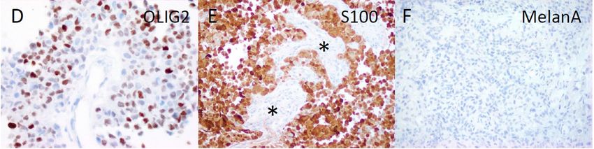
Free Neuropathology 2:16 (2021)                                                                                            Smith‐Cohn et al
doi: https://doi.org/10.17879/freeneuropathology‐2021‐3334                                                                      page 2 of 8

Introduction                                                               gressing to affect his right chest and leg over two
                                                                           weeks. MRI of the brain and cervical spine demon‐
      Astroblastomas are rare central nervous sys‐                         strated a well‐localized cystic enhancing anterior
tem (CNS) neoplasms that most frequently occur in                          medullary‐cervical lesion measuring 1.3 x 1.1 x 1.7
cerebral locations in children and young adults. Here                      cm, with T2 hyperintensity of the lesion and medul‐
we describe the unusual case of a 38‐year‐old man                          lary pyramids (Fig.1A, 1B, 1C). He was admitted to
with a brainstem tumor with an integrated diagnosis                        the neurology service for an expedited evaluation of
of malignant neoplasm consistent with astroblas‐                           infectious, rheumatologic, neoplastic, and inflam‐
toma with MN1 alteration following methylome                               matory etiologies. MRI of the spine and whole‐body
profiling, and found to have a non‐canonical EWSR1‐                        PET/CT revealed no other lesions. Serum studies
BEND2 fusion. This unusual clinical scenario exem‐                         were unremarkable, and lumbar CSF contained 1
plifies the utility of methylome profiling and assess‐                     WBC, 3 RBC, glucose 55, protein 57 (ref 15‐45), no
ment of gene fusions in tumors of the CNS.                                 oligoclonal bands and negative infectious studies,
                                                                           flow cytometry, and cytopathology. He was treated
Case Report                                                                with high dose IV methylprednisolone for five days
                                                                           and discharged home on a steroid taper. He had pro‐
      A 38‐year‐old male with a past medical history                       gression of symptoms, and a repeat MRI two months
of melanoma in situ of the trunk, status post exci‐                        later showed growth of the lesion to 3.3 x 1.7 x 1.5
sion, and no family history of cancer, presented with                      cm (Fig.1D, 1E, 1F). Subsequently, he underwent a
subacute onset of progressively worsening dyses‐                           suboccipital C1 laminectomy and subtotal surgical
thesia first involving his upper extremities and pro‐                      resection of the mass.

Figure 1. Radiographic findings. Coronal (A, D), sagittal (B, E), axial (C, F) post‐contrast T1 MRI of the brain. The top row (A‐C) shows MRI
imaging of the brain at presentation, and the bottom row (D‐F) is two months later before tumor resection.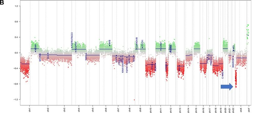
Free Neuropathology 2:16 (2021) Smith‐Cohn et al
doi: https://doi.org/10.17879/freeneuropathology‐2021‐3334 page 3 of 8
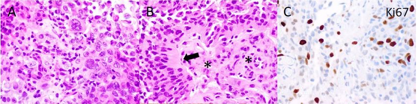
Figure 2. Histopathology. The tumor included more pleomorphic regions (A), as well as neoplastic cells with thick, short processes (arrow,
B) arrayed around proliferating blood vessels (asterisks, B). The tumor was quite proliferative on Ki67 immunostain (C), and diffusely
positive for OLIG2 (D) and S100 (E), while blood vessels did not express these glial markers (asterisks, E). MelanA was not expressed in the
tumor (F). (Original magnifications: A‐E 400X, F 200X).
Pathology no known pathologic variants, and several variants
of unknown significance, including ADGRA2
Microscopic evaluation showed a cellular tu‐ (p.G407S), AR (p.A646D), BLM (p.R643H), EPHA5
mor with compact growth. Large, pleomorphic to (p.L907V), and PRKDC (p.I1013V), all with allele fre‐
epithelioid cells predominated in some regions quencies of 46% or higher. No alterations in TP53,
(Fig.2A), while in other areas prominent perivascu‐ ATRX, BRAF, H3F3A or HIST1H3B were detected on
lar growth was noted. This included scattered cells NGS. DNA methylation profiling was consistent with
with a somewhat astroblastic phenotype, exhibiting a “high‐grade neuroepithelial tumor with MN1 alter‐
stout processes extending to the surface of blood ation”, with a calibrated score of 0.994 (Fig.3A).
vessels (Fig.2B). Necrotic foci without pseudopali‐ Copy number evaluation using data from the meth‐
sading were present. Cellular regions of tumor had 1 ylation array demonstrated alterations in chromo‐
to 5 mitotic figures per high power field, and the some 22 and X (Fig.3B). Subsequent gene fusion
Ki67 proliferation index was moderate to high, up to testing found a Ewing Sarcoma breakpoint region
20‐30% (Fig.2C). S100, OLIG2 and EMA were 1/EWS RNA binding protein 1 (EWSR1) ‐ BEN Domain
strongly positive on immunohistochemical analysis Containing 2 (BEND2) fusion between loci on chro‐
(Fig.2D, E and data not shown), and GFAP was fo‐ mosomes 22 and X (Fig.3C). The integrated diagno‐
cally positive, supporting glial differentiation. In con‐ sis was malignant neoplasm consistent with astro‐
trast, markers of melanocytic (MelanA, SOX10, blastoma with MN1 alteration.
HMB45), epithelial (cytokeratin AE1/AE3), and neu‐
ronal (synaptophysin) differentiation were all nega‐
tive (Fig.2F and data not shown). Subsequent Clinical Course
The patient was treated with fractionated radi‐
Molecular Diagnostics
ation (5040 cGy in 28 fractions) with concurrent
Next‐generation sequencing (NGS) found a tu‐ daily temozolomide (75 mg/m2) followed by adju‐
mor mutation rate of 0.88 mutations per megabase, vant temozolomide (150‐200 mg/m2, 5 days‐on/23
Free Neuropathology 2:16 (2021)                              Smith‐Cohn et al
doi: https://doi.org/10.17879/freeneuropathology‐2021‐3334        page 4 of 8
Free Neuropathology 2:16 (2021) Smith‐Cohn et al
doi: https://doi.org/10.17879/freeneuropathology‐2021‐3334 page 5 of 8
Figure 3. Methylation and copy number profiling of astroblastoma with an EWSR1‐BEND2 fusion. (A) The lesion (blue box) was plotted
on an X–Y coordinate graph (red dot to the lower left of the x–y intersection), where closer proximity to other dots indicates greater
similarity of the index tumor’s genomic CpG methylation pattern to existing cases in the library. (B) Copy number profiling demonstrates
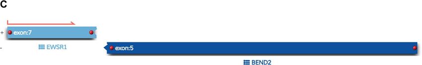
the location for EWSR1‐BEND2 fusion at chromosome 22 and X (blue arrow). (C) Fusion between exon 7 of EWSR1 and exon 5 of BEND2
genes with breakpoints at genomics positions chr22:29683123 and chrX:18234853 respectively. The red arrow represents the direction
of the gene specific primary utilized by the Archer assay to enrich the amplicons for EWSR1 fusion events. Legend: MB,G3, medulloblas‐
toma, subclass group 3; EPN, PF A, ependymoma, posterior fossa group A; EPN, PF B, ependymoma, posterior fossa group B; SUBEPN, PF,
subependymoma, posterior fossa; MB, SHH CHL AD, medulloblastoma, subclass SHH A (children and adult); MB, SHH INF, medulloblas‐
toma, subclass SHH B (infant); CONTR, CEBM, control tissue, cerebellar hemisphere; LIPN, cerebellar liponeurocytoma; CNS NB, FOXR2,
CNS neuroblastoma with FOXR2 activation; SP‐EPN‐MYCN, MYCN amplified spinal cord ependymoma; EPN, YAP, ependymoma, YAP fu‐
sion; HGNET, MN1; high grade neuroepithelial tumor with MN1 alteration; GBM, G34, glioblastoma, IDH wildtype, H3.3 G34 mutant; GBM,
MES, glioblastoma, IDH wildtype, subclass mesenchymal; GBM, MID, glioblastoma, IDH wildtype, subclass midline; GBM, MYCN, glioblas‐
toma, IDH wildtype, subclass MYCN; GBM, RTK I, glioblastoma, IDH wildtype, subclass RTK I; GBM, RTK II, glioblastoma, IDH wildtype,
subclass RTK II; GBM, RTK III, glioblastoma, IDH wildtype, subclass RTK III; HGNET, BCOR, CNS high grade neuroepithelial tumor with BCOR
alteration; PTPR, A, papillary tumor of the pineal region group A; PTPR, B, papillary tumor of the pineal region group B; CONTR, PONS,
control tissue, pons; CONTR, WM, control tissue, white matter; CONTR, HYPTHAL, control tissue, hypothalamus; CONTR, HEMI, control
tissue, hemispheric cortex; CN, central neurocytoma; SUBEPN, SPINE, subependymoma, spinal; LGG, MYB, low grade glioma, MYB/MYBL1;
LGG, DNT, low grade glioma, dysembryoplastic neuroepithelial tumor; LGG, GG, low grade glioma, ganglioglioma; IHG, infantile hemi‐
spheric glioma; LGG, PA/GG ST, low grade glioma, rosette forming glioneuronal tumor; LGG, RGNT, rosette forming glioneuronal tumor;
CONTR, REACT, reactive tumor microenvironment; DLGNT, diffuse leptomeningeal glioneuronal tumor; ANA PA, anaplastic pilocytic as‐
trocytoma; PXA, (anaplastic) pleomorphic xanthoastrocytoma; LYMPHO, lymphoma; DMG, K27, diffuse midline glioma H3 K27M mutant;
LGG, PA PF, subclass posterior fossa pilocytic astrocytoma; LGG, PA MID, midline pilocytic astrocytoma; CONTR, INFLAM, control tissue,
inflammatory tumor microenvironment; EPN, RELA, ependymoma, RELA fusion; ATRT, TYR, atypical teratoid/rhabdoid tumor, subclass
TYR; CHGL, chordoid glioma of the third ventricle; LGG, SEGA, subependymal giant cell astrocytoma.
days‐off) with a partial response. At the time of writ‐ CNS tumors with a EWSR1‐BEND2 fusion (Tab. 1).3–5
ing, the patient has not progressed since initiation of Aside from one case that did not provide additional
treatment over one year ago. Clinically the patient information, these tumors occurred in males and
was able to return to work full time with continued were infratentorial, involving the spinal cord and
central neuropathic pain of the left face and right‐ sometimes the brainstem.3–5
side extremities and trunk managed with gabapen‐
tin. Although non‐specific, MRI of astroblastomas
have been described to enhance, as well as appear
Discussion well‐demarcated, cystic, and lobulated, which is
Astroblastoma has historically been a contro‐ consistent with our case and similar to other cases
versial entity since its introduction in the 1924 clas‐ of EWSR1‐BEND2 fused astroblastoma tumors
sification of CNS brain neoplasms by Cushing and (Fig.1).3,4,6 Comparable to conventional MN1‐al‐
Bailey.1 MN1‐altered astroblastomas arise primarily tered astroblastomas and other cases of EWSR1‐
in cerebral locations in pediatric patients and young BEND2 fused tumors, our case showed perivascular
adults.2 In addition to the presence of a EWSR1‐ growth and immunohistochemistry was positive for
BEND2 fusion, our case is unusual compared to S100, OLIG2, EMA and GFAP.2–4 Ultrastructural ob‐
other described MN1‐altered astroblastoma in that servations in astroblastomas suggest a relationship
the patient is near his fourth decade of life and with to tanycytes, an ependymal cell subtype, but it is un‐
tumor located in the brainstem and upper cervical clear if this applies also to MN1‐altered and EWSR1‐
spine, rather than the cerebrum. Including the case BEND2 fused astroblastomas.1,2
presented, there are three other described cases ofFree Neuropathology 2:16 (2021) Smith‐Cohn et al
doi: https://doi.org/10.17879/freeneuropathology‐2021‐3334 page 6 of 8
Table 1: Known Cases of Astroblastoma with an EWSR1‐BEND2 fusion in the Literature
Molecular profiling has had a huge impact on The MN1 alteration is usually a fusion between
the diagnosis of “astroblastic” tumors and has MN1 and BEND2.2 While half or more of microscop‐
helped clarify subtypes of astroblastoma. In 2016, ically defined astroblastomas harbor an MN1 altera‐
Sturm et al. identified a group of 41 tumors with a tion, a significant number do not.3,10 Our patient is
common methylation profile and MN1 fusions, des‐ unique due to the presence of a rare non‐canonical
ignating them ‘CNS high‐grade neuroepithelial tu‐ EWSR1‐BEND2 fusion between chromosomes 22
mor with MN1 alteration’ (CNS‐HGNET‐MN1).7 No‐ and X. Interestingly, early molecular analyses of as‐
tably, the majority of these contained astroblastic or troblastomas identified deletions in chromosomes
ependymal perivascular pseudorosettes, although a 22q and X.1,2 Including our case, there are at least
number of other histopathological appearances four known cases of CNS tumor with EWSR1‐BEND2
were also represented. Subsequent studies of MN1‐ fusions in the literature (Table 1).3–5 One case was a
altered brain tumors have confirmed that many, but 3‐month ‐old male with tumor spanning the lower
not all, have ependymal or astroblastic features.8 medulla to the C4 spinal cord level,3 with a second in
Based on these and other studies, the Consortium to a 36‐year‐old male patient with a thoracic spinal
Inform Molecular and Practical Approaches to CNS “ependymoma” with EWSR1‐BEND2 fusion.4 A third
Tumor Taxonomy (cIMPACT‐NOW) has proposed case was found in a series of molecularly character‐
designating these tumors “astroblastoma, MN1‐al‐ ized pediatric tumors which did not provide clinical
tered”.2 Early case series of astroblastoma reported information.5 Remarkably, in at least two of these
prior to these newer molecular tools likely con‐ cases, as in ours, methylation clustering led to the
tained other tumor subtypes, as exemplified by a re‐ tumor falling into the category of astroblas‐
cent study in which molecular profiling of 14 adult toma/HGNET with MN1 alteration.3,4 These reports
tumors with histologic feature of astroblastoma highlight the possibility of astroblastic or ependymal
found they were pleomorphic xanthoastrocytomas tumors with a methylation profile consistent with
or high grade gliomas with alterations activating the “astroblastoma, MN1‐altered” being driven by fu‐
mitogen‐activated protein kinase pathway, and sions in genes other than MN1, and also suggest that
none had clear evidence of a MN1‐BEND2 fusion, or it may be BEND2, rather than MN1, which has a
clustered with that group on methylation profiling more critical functional role. Supporting this notion,
analysis.9Free Neuropathology 2:16 (2021) Smith‐Cohn et al
doi: https://doi.org/10.17879/freeneuropathology‐2021‐3334 page 7 of 8
there is one reported case of an neuro‐epithelial tu‐ Given the rarity of these tumors, there is no de‐
mor with an MN1‐PATZ1 fusion found with RNA se‐ fined standard established treatment. Based on
quencing a supratentorial mass in a 1‐year‐old girl studies of non‐molecularly characterized astroblas‐
that suggest a similar pathogenic role as a chimeric toma, resection remains the cornerstone of therapy
protein.11 However, this case lacked astroblastoma‐ to improve outcomes, as supported by an analysis of
tous rosettes and did not have epigenetic similarities 116 patients that found that gross total resection
with CNS HGNET‐MN1, but did have similarities to improved outcome with a 5‐year progression‐free
PATZ1‐sarcomas.11 survival of 83% versus 55% in those with subtotal re‐
section.1 Regarding radiation, a systemic review of
The grading and clinical behavior of astroblas‐ 95 histologic astroblastoma patients did not show a
tomas is not well understood. As noted by the 6th survival benefit with radiation therapy; however,
cIMPACT‐NOW update and other case series, a sig‐ the series had a broad age range from 1 to over 61
nificant proportion of tumors in the methylation years of age and did not compare survival differ‐
class Astroblastoma/CNS‐HGNET‐MN1 do not con‐ ences in astroblastoma with and without anaplastic
form to astroblastoma histologically, and it is un‐ features.14 Review of the literature found that a 20‐
clear if disparate histologic patterns have biologic year‐old woman with a spinal cord MN1‐altered as‐
relevance aside from high grade features.1,2 The troblastoma, and a 36‐year‐old man with a spinal as‐
2016 WHO, completed before identification of the troblastoma with EWSR1‐BEND2 both had a reduc‐
MN1‐altered molecular group, does not assign tion in tumor size after radiation, temozolomide,
grades, but observed that astroblastic neoplasms and bevacizumab.4,15 In another case, a 3‐month old
generally fall into two general categories: well‐dif‐ boy with EWSR1‐BEND2 astroblastoma had progres‐
ferentiated or anaplastic/malignant.1,12 Case series sive disease after five days of temozolomide and
that predated molecular evaluation of astroblasto‐ etoposide.3 Given the significant growth of the pre‐
mas likely included other tumor subtypes, but found sented patient's tumor in two months (Fig.1) and
that in histologically defined astroblastomas, an ele‐ dramatically elevated Ki67 proliferation index, he
vated proliferation rate is associated with worse was treated with concurrent radiation with te‐
outcomes.1,12 A case series of 14 neuroepithelial tu‐ mozolomide followed by adjuvant temozolomide
mors with MN1 alterations, including three spinal based on the “Stupp” protocol and this appears to
cases, two of which were the oldest (14.6 and 36 be a viable option, as he has had a partial response
years old) and only males in the series, describes control of his tumor over one year later at the time
heterogeneous treatments consisting of focal or cra‐ of writing.16
nial spinal radiation without chemotherapy.8 The
event‐free survival ranged considerably from 6 to This case exemplifies the use of advanced mo‐
100 months, with some cases resulting in metastasis lecular testing, including the evaluation of chromo‐
in the CNS, arguing that intermittent monitoring some fusions and methylation profiling, to accu‐
with completed neuroaxis imaging is warranted.8 A rately diagnose rare neoplasms of the CNS. Through
meta‐analysis of 73 patients with MN1‐altered neu‐ these technologies, improved diagnosis accuracy
roepithelial tumors (astroblastomas) found a 5‐ and provides clinically impactful insights that enhance
10‐year progression‐free survival of 38% and 0%, the understanding of tumor biology.
and 5‐ and 10‐year overall survival of 89% and 55%,
respectively.13 The natural history of astroblastoma Funding
with EWSR1‐BEND2 fusion has not been well de‐
scribed in the literature, and it is unclear if the clini‐ This work was supported by the National Insti‐
cal behavior is significantly different from astroblas‐ tutes of Health T32 research training grant.
tomas with a MN1 alteration. The case we present
showed anaplastic microscopic features, as well as
Authors’ Contributions
aggressive clinical behavior with rapid growth over
two months ‐ histologic features and clinical behav‐
M.A.S case report concept, design, and critical
ior was similar to other described cases of astroblas‐
revision of content. Z.A., K.D.A, M.Q., M.K.R., F.J.R.,
toma with a EWSR1‐BEND2 fusion.3,4
C.G.E and L.J.L. provided critical revision of content.Free Neuropathology 2:16 (2021) Smith‐Cohn et al
doi: https://doi.org/10.17879/freeneuropathology‐2021‐3334 page 8 of 8
Acknowledgments for help with formatting the methylation plot for
publication. The authors also thank the patient and
The authors thank Rust Turakulov at the NIH his caregivers.
References
1. Brat, D. J., Hirose, Y., Cohen, K. J., Feuerstein, B. G. & Burger, P. C. 9. Boisseau, W. et al. Molecular Profiling Reclassifies Adult
Astroblastoma: Clinicopathologic Features and Chromosomal Astroblastoma into Known and Clinically Distinct Tumor Entities with
Abnormalities Defined by Comparative Genomic Hybridization. Brain Frequent Mitogen‐Activated Protein Kinase Pathway Alterations.
Pathology 10, 342–352 (2006). Oncologist 24, 1584–1592 (2019).
2. Louis, D. N. et al. cIMPACT‐NOW update 6: new entity and diagnostic 10. Mhatre, R. et al. MN1 rearrangement in astroblastoma: study of
principle recommendations of the cIMPACT‐Utrecht meeting on future eight cases and review of literature. Brain Tumor Pathol 36, 112–120
CNS tumor classification and grading. Brain Pathol bpa.12832 (2020) (2019).
doi:10.1111/bpa.12832.
11. Burel‐Vandenbos, F. et al. A polyphenotypic malignant paediatric
3. Yamasaki, K. et al. Spinal cord astroblastoma with an EWSR1‐BEND2 brain tumour presenting a MN1‐PATZ1 fusion, no epigenetic similarities
fusion classified as a high‐grade neuroepithelial tumour with MN1 with CNS High‐Grade Neuroepithelial Tumour with MN1 Alteration (CNS
alteration. Neuropathol Appl Neurobiol 46, 190–193 (2020). HGNET‐MN1) and related to PATZ1 ‐fused sarcomas. Neuropathol Appl
Neurobiol 46, 506–509 (2020).
4. Tsutsui, T. et al. PATH‐23. ADULT SPINAL CORD ASTROBLASTOMA
WITH EWSR1‐BEND2 FUSION. Neuro‐Oncology 22, iii429–iii429 (2020). 12. Bonnin, J. M. & Rubinstein, L. J. Astroblastomas: a pathological study
of 23 tumors, with a postoperative follow‐up in 13 patients.
5. Ramkissoon, S. H. et al. Clinical targeted exome‐based sequencing in Neurosurgery 25, 6–13 (1989).
combination with genome‐wide copy number profiling: precision
medicine analysis of 203 pediatric brain tumors. Neuro Oncol 19, 986– 13. Chen, W. et al. Central nervous system neuroepithelial tumors with
996 (2017). MN1‐alteration: an individual patient data meta‐analysis of 73 cases.
Brain Tumor Pathol 37, 145–153 (2020).
6. Sener, R. N. Astroblastoma: diffusion MRI, and proton MR
spectroscopy. Comput Med Imaging Graph 26, 187–191 (2002). 14. Sughrue, M. E. et al. Clinical features and post‐surgical outcome of
patients with astroblastoma. J Clin Neurosci 18, 750–754 (2011).
7. Sturm, D. et al. New Brain Tumor Entities Emerge from Molecular
Classification of CNS‐PNETs. Cell 164, 1060–1072 (2016). 15. Yamada, S. M. et al. Primary spinal cord astroblastoma: case report.
Journal of Neurosurgery: Spine 28, 642–646 (2018).
8. Baroni, L. V. et al. Treatment response of CNS high‐grade
neuroepithelial tumors with MN1 alteration. Pediatr Blood Cancer 67, 16. Stupp, R. et al. Radiotherapy plus Concomitant and Adjuvant
(2020). Temozolomide for Glioblastoma. N Engl J Med 352, 987–996 (2005).You can also read