Clamshell thoracotomy for en bloc resection of a 3-level thoracic chordoma: technical note and operative video
←
→
Page content transcription
If your browser does not render page correctly, please read the page content below
NEUROSURGICAL
           FOCUS                                                                                                           Neurosurg Focus 49 (3):E16, 2020

                         Clamshell thoracotomy for en bloc resection of a 3-level
                         thoracic chordoma: technical note and operative video
                         John F. Burke, MD, PhD,1 Andrew K. Chan, MD,1 Rory R. Mayer, MD,1 Joseph H. Garcia, BS,2
                         Brenton Pennicooke, MD,1 Michael Mann, MD,3 Sigurd H. Berven, MD,4 Dean Chou, MD,1 and
                         Praveen V. Mummaneni, MD1
                         1
                             Department of Neurological Surgery, 2School of Medicine, 3Department of Surgery, Division of Adult Cardiothoracic Surgery, and
                         4
                             Department of Orthopedic Surgery, University of California, San Francisco, California

                         The clamshell thoracotomy is often used to access both hemithoraxes and the mediastinum simultaneously for cardio-
                         thoracic pathology, but this technique is rarely used for the excision of spinal tumors. We describe the use of a clamshell
                         thoracotomy for en bloc excision of a 3-level upper thoracic chordoma in a 20-year-old patient. The lesion involved T2,
                         T3, and T4, and it invaded both chest cavities and indented the mediastinum. After 2 biopsies to confirm the diagnosis,
                         the patient underwent a posterior spinal fusion followed by bilateral clamshell thoracotomy for 3-level en bloc resection
                         with simultaneous access to both chest cavities and the mediastinum. To demonstrate how the clamshell thoracotomy
                         was used to facilitate the tumor resection, an operative video and illustrations are provided, which show in detail how the
                         clamshell thoracotomy can be used to access both hemithoraxes and the mediastinum.
                         https://thejns.org/doi/abs/10.3171/2020.6.FOCUS20382
                         KEYWORDS chordoma; en bloc resection; clamshell thoracotomy; operative video; surgical technique

C
         hordomas   are malignant spinal neoplasms arising                        tient with a 3-level thoracic chordoma that invaded both
         from notochordal remnants and most frequently                            chest cavities and indented the mediastinum. Because of
         occur in the clivus and sacral spine.1–3 Within the                      the upper thoracic spine location and because of the inva-
mobile spine (C1–L5), chordomas most commonly occur                               sion into both chest cavities by the tumor, a formal clam-
in the cervical and lumbar levels; it is rare for a chordoma                      shell thoracotomy was performed to achieve an en bloc
to occur in the thoracic spine, especially the upper tho-                         excision. The description of the approach presented here
racic spine.4 Boriani et al. presented 52 consecutive cases                       includes illustrations and a video.
of chordomas in the mobile spine over a 50-year period,
and only 1 tumor arose from the upper thoracic spine (T4)                         Methods
in an adult patient.5 In 2012, Fontes and O’Toole reviewed
the literature and found only 22 reported cases of thoracic                       History, Workup, and Surgical Planning
chordomas.4 Moreover, chordomas usually appear after                                 A 20-year-old otherwise healthy woman presented with
age 40 years and are rare in young adults and pediatric                           shortness of breath and wheezing. Chest radiograph re-
patients.1 When chordomas do occur in pediatric patients,                         vealed an 8-cm left eccentric mediastinal mass. Standing
they occur almost exclusively along the clivus.6                                  scoliosis radiographs, cervicothoracic spine CT, and total
   En bloc excision of thoracic chordoma can require an                           spine MRI with and without contrast were obtained. An
anterior approach, but this strategy usually involves either                      MRI neurogram was also obtained to evaluate possible
a standard thoracotomy or a sternotomy. When the tumor                            involvement of the left brachial plexus, but the evaluation
invades both chest cavities and the mediastinum, a clam-                          results were negative. Imaging revealed an enhancing le-
shell thoracotomy can be employed to access all 3 anterior                        sion involving the T2–4 vertebral bodies and expanding
compartments simultaneously. We describe the use of the                           predominantly into the left hemithorax, right hemithorax,
clamshell thoracotomy to excise a 3-level upper thoracic                          and mediastinum (Fig. 1). The tumor was indenting the
chordoma. We present the case of a 20-year-old female pa-                         anterior upper mediastinal structures, including the great

SUBMITTED May 4, 2020. ACCEPTED June 16, 2020.
INCLUDE WHEN CITING DOI: 10.3171/2020.6.FOCUS20382.

©AANS 2020, except where prohibited by US copyright law                                               Neurosurg Focus Volume 49 • September 2020         1

                                                                                                             Unauthenticated | Downloaded 10/13/20 11:23 AM UTC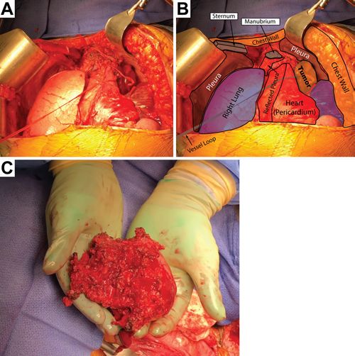
Burke et al.

                                                                                FIG. 2. Left-sided thoracotomy and tumor exposure. A: The first left-
                                                                                sided thoracotomy is shown with exposure of the pericardium, medias-
                                                                                tinum, tumor, pleura, chest wall, and sternum. B: These structures are
                                                                                labeled for orientation.

                                                                                discectomies were completed. The left-sided T2, T3, and
FIG. 1. Preoperative MRI of the tumor. Coronal (A), sagittal (B), and           T4 nerve roots were sacrificed to enable en bloc resec-
axial (C) contrast MR images of the tumor. D: The axial MRI is shown            tion during the second stage. In order to release the dorsal
with vital structures highlighted, including the trachea, the junction of the
aorta with the left common carotid artery (L. CCA) and right brachioce-
                                                                                spine from the ventral tumor, the entire posterior elements
phalic artery (R BCA), and the left subclavian artery (L. SCA).                 were removed at T2, T3, and T4, including the pedicles,
                                                                                the transverse processes, and the facets. Posterior element
                                                                                resection was correlated with tumor extension by MRI;
                                                                                posterior element excision was carried out until a definite
vessels, esophagus, and trachea, but there was no radio-                        margin between normal bone and tumor was ensured by
graphic evidence of invasion of these structures (Fig. 1D).                     continual cross referencing the MRI intraoperatively. In
The radiographic findings were consistent with a thoracic                       addition, the biopsy tract was included and excised during
chordoma. The patient underwent a CT-guided biopsy to                           the posterior stage.
establish the diagnosis, which was determined to be chor-                          Although the patient had consented to a complete clam-
doma. Because of the atypical presentation in a 20-year-                        shell thoracotomy, the second stage was begun with a left-
old patient, a repeat CT-guided biopsy was performed,                           sided hemiclamshell thoracotomy by thoracic surgery to
again confirming the diagnosis of chordoma. Once the                            expose the tumor as well as the pericardium and pleural
diagnosis of chordoma was confirmed by pathological                             space. Specifically, the surgical saw was used to vertically
analysis, en bloc resection was planned. The invasion into                      divide the sternum in the midline, and then to extend the
both chest cavities and the mediastinum made removing                           sternotomy laterally at the level of the fifth interspace.
the chordoma entirely from a posterior-only approach too                        Chest wall retractors were placed to expose the upper left
risky for possible spinal cord damage and intralesional vi-                     hemithorax, and the tumor was identified (Fig. 2). The left
olation. Moreover, the upper thoracic spine poses unique                        lung was found to be free of any significant attachments to
access obstacles not only because of the kyphotic nature                        the tumor. The left subclavian artery, the innominate vein
of the spine in this area, but also because the mediastinum                     and artery, and the arch of the aorta were dissected away
and the innominate vein are essentially immobile. Thus, a                       from the tumor. The posterior parietal pleura was scored
standard median sternotomy would not have sufficed. In                          circumferentially around the tumor and was dissected to-
addition, because the tumor was in both chest cavities and                      ward the midline. Attempts were made to dissect beneath
the mediastinum, a clamshell thoracotomy was planned to                         the anterior mediastinum to reach the tumor in the right
access both chest cavities and the mediastinum simultane-                       thoracic cavity; however, adequate exposure and visual-
ously.                                                                          ization of the right tumor margin could not be achieved
                                                                                because of the mediastinal structures. Therefore, the tho-
Operative Technique                                                             racotomy was extended into a bilateral hemiclamshell tho-
   The plan was to first perform posterior stabiliza-                           racotomy to expose the right-sided aspect of the tumor and
tion with release of the posterior elements and sacrifice                       spine (Fig. 3A and B).
of the involved nerve roots, followed by 3-level en bloc                           The sternum was divided laterally at the level of the
excision from the anterior approach. In the posterior ap-                       fourth interspace. The right anterior chest wall was then
proach, instrumentation was performed from C5 to T8, a                          similarly retracted, and complete exposure of the right
T1–5 laminectomy was performed, and T1–2 and T4–5                               aspect of the T2 through T4 tumor and vertebral bodies

2        Neurosurg Focus Volume 49 • September 2020

                                                                                                       Unauthenticated | Downloaded 10/13/20 11:23 AM UTC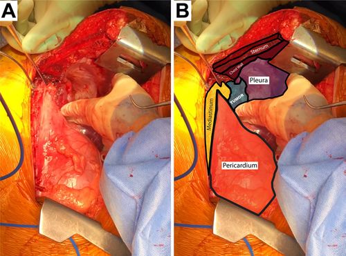
Burke et al.

FIG. 3. Bilateral thoracotomy and en bloc tumor resection. A: The bilat-
eral thoracotomy. B: Several structures are labeled, including the pleura,
the right lung, the pericardium, the tumor, and the chest wall. C: The
tumor after en bloc resection.                                               FIG. 4. Postoperative radiographs. Postoperative anterior-posterior
                                                                             (A) and lateral (B) radiographs of the instrumentation, as well as radio-
                                                                             graphs of the resected tumor (C and D).

was achieved. Once the tumor was completely exposed,
the anterior longitudinal ligament and residual annulus at
T1–2 and T4–5 was resected to release the T2, T3, and                        5-month follow-up, the incision was well healed, motor
T4 vertebral bodies. The tumor was then dissected off the                    strength was at least 4+/5 in the arms and legs (assessed
pleura on the posterior chest wall. After complete dissec-                   over telehealth), and there was residual dysesthesia in the
tion laterally, the T2, T3, and T4 vertebral bodies were                     patient’s left arm radiating to her left fifth digit.
excised in an en bloc fashion (Fig. 3C and Video 1).
    VIDEO 1. Intraoperative video of the bilateral clamshell thoracotomy     Discussion
    for en bloc resection of 3-level thoracic chordoma. Copyright UCSF           Thoracic chordomas are very uncommon and represent
    Department of Neurological Surgery. Published with permission.           an estimated 1% of all chordomas.4 Although these tumors
    Click here to view.                                                      tend to arise at a much earlier age than other chordomas,
    To reconstruct the anterior column, an expandable cage                   our 20-year-old patient was still well below the mean age
filled with rib autograft was then placed under intraopera-                  of presentation (37.5 years).2 Early diagnosis of thoracic
tive fluoroscopy. The bilateral hemiclamshell thoracotomy                    chordomas is generally difficult because of lack of spe-
was closed by thoracic surgery in the standard fashion.                      cific symptoms.6,7 The time from onset of symptoms to
Postoperative radiographs show that the instrumentation                      diagnosis is almost always more than 8 months.8,9 MRI
was in a good position (Fig. 4A and B). Radiographs of the                   is the radiological gold standard for diagnosis and surgi-
resected tumor show the vertebral bodies resected with                       cal planning. These tumors are isointense to hypointense
the tumor, verifying en bloc resection (Fig. 4C and D).                      on T1-weighted MRI, with variable enhancement. On T2-
Pathological analysis showed a neoplasm invading bone                        weighted MRI, chordomas are hyperintense but may have
and soft tissues composed of bland epithelioid cells with                    some heterogeneity in signal intensity because of calcifi-
eosinophilic cytoplasm and occasional cells with cyto-                       cation and bony sequestration.
plasmic vacuolations, arranged in cords and trabeculae in                        En bloc excision of chordomas is currently the most
a background of myxoid matrix. Neoplastic cells stained                      effective way to prevent local recurrence.10,11 Although
positive for epithelial membrane antigen, S-100, pancy-                      en bloc resection was completed in this case, it is often
tokeratin, and brachyury, consistent with a diagnosis of                     difficult to achieve en bloc excision without significant
chordoma. There was a marginal margin against the dura,                      morbidity. Moreover, chordomas are considered radiore-
and the patient was referred for proton beam radiation.                      sistant, rendering most radiation therapies ineffective.12
Two months after surgery, the patient experienced a 3–4-                     Chemotherapy is for the most part ineffective. En bloc re-
cm wound breakdown of the incision inferior to the right                     section during the initial surgery gives the best chance to
breast, which was revised by thoracic surgery. The patient                   achieve long-term local control. According to comprehen-
was placed on oral antibiotics and recovered well. At the                    sive population-based studies, the median overall survival

                                                                                                 Neurosurg Focus Volume 49 • September 2020              3

                                                                                                        Unauthenticated | Downloaded 10/13/20 11:23 AM UTC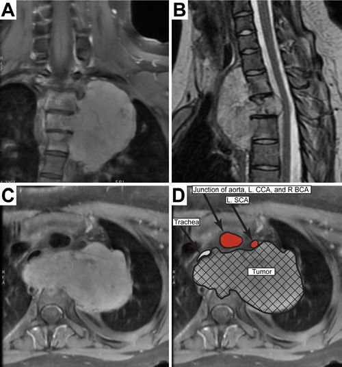
Burke et al.
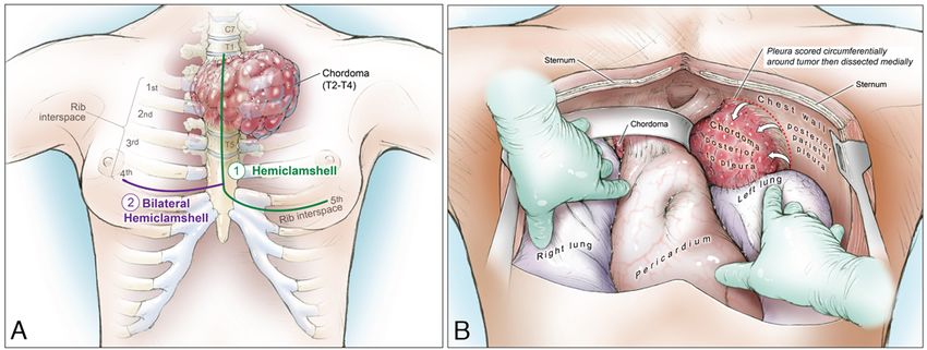
FIG. 5. Illustration of the bilateral hemiclamshell thoracotomy. A: Sequence of bony cuts employed in this case. B: Overall expo-
sure afforded by the bilateral hemiclamshell thoracotomy. Copyright Kenneth Probst. Published with permission.
for chordoma patients is just over 7 years, and the overall dal osteotomy, and a left-sided thoracotomy (with interval
5- and 10-year survival rates are 68%, and 40% respec- patient repositioning) to complete the rostral osteotomy
tively.12,13 and excise the tumor.9 Lau et al. reported the use of unilat-
The clamshell thoracotomy was first described by eral hemiclamshell thoracotomies to excise large spine tu-
Kortz in 1958, who used a transverse thoracosternotomy.14 mors invading the mediastinum and chest cavity, but they
The procedure was further developed and popularized for did not report the use of bilateral hemiclamshell thora-
bilateral lung transplantations by the Washington Uni- cotomies.29 The traditional formal clamshell thoracotomy
versity group.15,16 Although the midline sternotomy had does not involve an upper sternotomy.30 However, because
been previously described for this purpose,17,18 the bilat- in our patient the sternum was split for the hemiclamshell
eral clamshell thoracotomy (referred to as a “cross-bow” thoracotomy, the present case is technically a bilateral
thoracotomy at the time) was favored because it provided hemiclamshell thoracotomy (midline sternal split), not a
superior access to the left and right pleural spaces, as traditional clamshell thoracotomy (no midline sternoto-
well as the posterior mediastinum.15,18 Since these initial my). Regardless of the nomenclature, the clamshell thora-
descriptions, the procedure has been modified to include cotomy allowed simultaneous access to both chest cavities
the unilateral “hemiclamshell” thoracotomy,19,20 as well and the mediastinum. Figure 5 illustrates the sequence of
as the sternum-sparing variant in which a transverse ster- bony cuts (Fig. 5A), as well as the final thoracic exposure
notomy is avoided.21 Although the indications and relative afforded by the bilateral hemiclamshell thoracotomy (Fig.
advantages of the median sternotomy versus the bilateral 5B). Although attempts were made to access the entire tu-
clamshell approach for cardiopulmonary transplantation mor using a hemiclamshell thoracotomy, the contralateral
are still debated,22,23 the principal advantage of the bilat- chest cavity invasion precluded en bloc excision through
eral clamshell thoracotomy is superior exposure of both this approach only. Intraoperative details of the steps of
pleural spaces with simultaneous bilateral access to the the approach and tumor resection are provided in Video 1.
posterior mediastinum.15,23,24 The clamshell thoracotomy Although a morbid approach, the bilateral clamshell tho-
is therefore indicated when wide exposure of these struc- racotomy can be a useful approach in cases which require
tures is necessary, and it has been used for a variety of simultaneous access to both chest cavities for en bloc exci-
purposes, such as repair of cardiac defects25 and treatment sion of tumors of the thoracic spine.
of infection,26 pulmonary metastatic lesions,19 traumatic
conditions requiring emergent thoracic access,27 and ap- Acknowledgments
proaches to large intrathoracic tumors.28 In our case, the We would like to thank Dr. Melike Pekmezci for help inter-
presence of the tumor in bilateral pleural spaces as well as preting the pathology specimens and Kenneth Probst for illustra-
the need to dissect the tumor away from the posterior me- tions and artwork.
diastinum warranted the bilateral clamshell thoracotomy.
In general, the clamshell thoracotomy is rarely used in References
spinal surgery; however, there are instances in which the
morbidity of this approach is outweighed by the benefit. 1. George B, Bresson D, Herman P, Froelich S. Chordomas:a
review. Neurosurg Clin N Am. 2015;26(3):437–452.
For instance, Sciubba et al. reported a case of an en bloc 2. Hamilton K, Rebsamen S, Salamat S, Ahmed R. Pediatric
resection of a T1–5 ventral chordoma using a 3-staged ap- extraosseous sacral chordoma:case report and literature
proach consisting of a posterior approach to instrument review of embryonic derivation and clinical implications. J
the spine, a right-sided thoracotomy to complete the cau- Neurosurg Pediatr. 2019;23(5):628–633.
4 Neurosurg Focus Volume 49 • September 2020
Unauthenticated | Downloaded 10/13/20 11:23 AM UTCBurke et al.
3. Inci S, Palaoğlu S, Onol B, Erbengi A. Low cervical chor- 23. Shudo Y, Rinewalt D, Lingala B, et al. Impact of surgical
doma:case report. Spinal Cord. 1996;34(6):358–360. approach in double lung transplantation:median sternotomy
4. Fontes R, O’Toole JE. Chordoma of the thoracic spine in an vs clamshell thoracotomy. Transplant Proc. 2020;52(1):
89-year-old. Eur Spine J. 2012;21(4)(suppl 4):S428–S432. 321–325.
5. Boriani S, Bandiera S, Biagini R, et al. Chordoma of the mo- 24. Cooper JD. The evolution of techniques and indications for
bile spine:fifty years of experience. Spine (Phila Pa 1976). lung transplantation. Ann Surg. 1990;212(3):249–256.
2006;31(4):493–503. 25. Luciani GB, Starnes VA. The clamshell approach for the
6. O’Neill P, Bell BA, Miller JD, et al. Fifty years of experience surgical treatment of complex cardiopulmonary pathology in
with chordomas in southeast Scotland. Neurosurgery. 1985; infants and children. Eur J Cardiothorac Surg. 1997;11(2):
16(2):166–170. 298–306.
7. Rena O, Davoli F, Allegra G, et al. Giant chordoma of the 26. Ris H-B, Banic A, Furrer M, et al. Descending necrotizing
upper thoracic spine with mediastinal involvement:a surgical mediastinitis:surgical treatment via clamshell approach. Ann
challenge. Asian Spine J. 2014;8(3):353–356. Thorac Surg. 1996;62(6):1650–1654.
8. Reddy EK, Mansfield CM, Hartman GV. Chordoma. Int J 27. Simms ER, Flaris AN, Franchino X, et al. Bilateral anterior
Radiat Oncol Biol Phys. 1981;7(12):1709–1711. thoracotomy (clamshell incision) is the ideal emergency tho-
9. Sciubba DM, Gokaslan ZL, Black JH III, et al. 5-Level spon- racotomy incision:an anatomic study. World J Surg. 2013;
dylectomy for en bloc resection of thoracic chordoma:case 37(6):1277–1285.
report. Neurosurgery. 2011;69(2)(Suppl Operative):E248– 28. Odell DD, Macke RA, O’Shea MA. Clamshell thoracotomy:
E256. a unique approach to a massive intrathoracic schwannoma.
10. Pan Y, Lu L, Chen J, et al. Analysis of prognostic factors for Ann Thorac Surg. 2011;91(1):298–301.
survival in patients with primary spinal chordoma using the 29. Lau D, Yarlagadda J, Jahan T, et al. Desmoplastic fibroma
SEER Registry from 1973 to 2014. J Orthop Surg Res. 2018; of the spine causing severe mediastinal compression and
13(1):76. brachial plexus encasement:report of 2 cases. J Neurosurg
11. Stacchiotti S, Gronchi A, Fossati P, et al. Best practices for Spine. 2013;19(4):515–520.
the management of local-regional recurrent chordoma:a po- 30. Panchabhai TS, Chaddha U, McCurry KR, et al. Historical
sition paper by the Chordoma Global Consensus Group. Ann perspectives of lung transplantation:connecting the dots. J
Oncol. 2017;28(6):1230–1242. Thorac Dis. 2018;10(7):4516–4531.
12. Topsakal C, Bulut S, Erol FS, et al. Chordoma of the thoracic
spine—case report. Neurol Med Chir (Tokyo). 2002;42(4):
175–180. Disclosures
13. Radaelli S, Stacchiotti S, Ruggieri P, et al. Sacral chordoma: The current work was supported by the UCSF Department of
long-term outcome of a large series of patients surgically Neurosurgery. Dr. Chan reports receiving research support for
treated at two reference centers. Spine (Phila Pa 1976). 2016; a nonrelated study from Orthofix, Inc. Dr. Chou reports being a
41(12):1049–1057. consultant and receiving royalties from Globus. Dr. Mummaneni
14. Kortz AB. Experimental bilateral transsternal thoracotomy: reports being a consultant for DePuy Synthes, Globus, and
factors improving survival. J Thorac Surg. 1958;35(3):305– Stryker; having direct stock ownership in Spinicity/ISD; receiv-
308. ing non–study-related clinical and research grant support from
15. Pasque MK, Cooper JD, Kaiser LR, et al. Improved tech- NREF, AO Spine, and ISSG; and receiving royalties from DePuy
nique for bilateral lung transplantation:rationale and initial Spine, Thieme Publishers, and Springer Publishers.
clinical experience. Ann Thorac Surg. 1990;49(5):785–791.
16. Trulock EP, Cooper JD, Kaiser LR, et al. The Washington Author Contributions
University-Barnes Hospital experience with lung transplanta-
tion. JAMA. 1991;266(14):1943–1946. Conception and design: Burke. Acquisition of data: Burke, Chan,
17. Cooper JD, Patterson GA, Grossman R, Maurer J. Double- Mayer, Garcia, Mann, Berven, Chou, Mummaneni. Analysis and
lung transplant for advanced chronic obstructive lung dis- interpretation of data: all authors. Drafting the article: all authors.
ease. Am Rev Respir Dis. 1989;139(2):303–307. Critically revising the article: all authors. Reviewed submitted
18. Patterson GA, Cooper JD, Goldman B, et al. Technique of version of manuscript: all authors. Approved the final version of
successful clinical double-lung transplantation. Ann Thorac the manuscript on behalf of all authors: Burke. Statistical analy-
Surg. 1988;45(6):626–633. sis: Burke, Mann, Berven, Chou. Administrative/technical/mate-
19. Bains MS, Ginsberg RJ, Jones WG II, et al. The clamshell rial support: Burke. Study supervision: Burke.
incision:an improved approach to bilateral pulmonary and
mediastinal tumor. Ann Thorac Surg. 1994;58(1):30–33. Supplemental Information
20. Korst RJ, Burt ME. Cervicothoracic tumors:results of resec- Videos
tion by the “hemi-clamshell” approach. J Thorac Cardiovasc Video 1. https://vimeo.com/439339108.
Surg. 1998;115(2):286–295.
21. Meyers BF, Sundaresan RS, Guthrie T, et al. Bilateral se- Correspondence
quential lung transplantation without sternal division elimi-
John F. Burke: University of California, San Francisco, CA.
nates posttransplantation sternal complications. J Thorac
john.burke@ucsf.edu.
Cardiovasc Surg. 1999;117(2):358–364.
22. Macchiarini P, Ladurie FL, Cerrina J, et al. Clamshell or
sternotomy for double lung or heart-lung transplantation?
Eur J Cardiothorac Surg. 1999;15(3):333–339.
Neurosurg Focus Volume 49 • September 2020 5
Unauthenticated | Downloaded 10/13/20 11:23 AM UTCYou can also read