Evaluation of bone availability for grafts in different donor sites, through computed tomography - SciELO
←
→
Page content transcription
If your browser does not render page correctly, please read the page content below
Original Article
                                                                                                                http://dx.doi.org/10.1590/1678-7757-2019-0435

                                                         Evaluation of bone availability for
                                                         grafts in different donor sites, through
                                                         computed tomography

                                                         Abstract

     Géssyca Moreira Melo de Freitas                         Objective: To quantify the bone volume that can be safely withdrawn from

                                GUIMARÃES1               3 donor sites: (1) the mandibular symphysis, (2) the oblique mandibular
                                                         line and (3) the skullcap. Methodology: For the symphysis, 200 tomographic
                 Gabriel Fiorelli BERNINI1
                                                         exams were evaluated by the extension of the anterior loop of mental foramen,
           Dayane Kemp GRANDIZOLI1
                                                         by the nerve, by the distance of the foramens, by the distance between the
   Paulo Sergio Perri de CARVALHO1                       vestibular cortical and the lingual plates and by the distance between the
       Eduardo Sanches GONÇALES1                         apexes, or lower anterior teeth, and the mandibular base, using the “distance”
              Osny FERREIRA JUNIOR1                      tool of the I-CAT Vision, in the panoramic and parasagittal reformations. For
                                                         the oblique line, 70 TCFC exams were analyzed retrospectively in panoramic
                                                         and parasagittal reformations, evaluating the thickness of the vestibular
                                                         cortical and the distance between the cortical and the mandibular canal.
                                                         For the cranial bone, a hexagonal donor site located in parietal area was
                                                         considered. Results: The average dimensions of the bone blocks that can
                                                         be safely removed from the region of the mandibular symphysis are: 32.27
                                                         mm in length, 4.87 mm in height and 4 mm in thickness, providing a volume
                                                         of 628.61 mm3 available for grafting. In the oblique line, the available bone
                                                         volume for grafting was 859.61 mm3. In the region of the cranial vault,
                                                         multiplying the average bone thickness by the area of the hexagon, an
                                                         average volume of 2,499 mm3 was obtained. Conclusions: Comparing the
                                                         donor sites, the bone availability in the cranial vault is 3 times greater than
                                                         in the mandibular posterior region, and at least 2 times greater than in the
                                                         mandibular symphysis.

                                                             Keywords: Tomography. Surgery. Skull. Mandible.

                     Submitted: August 14, 2019
                   Modification: October 16, 2019
                     Accepted: October 22, 2019

                             Corresponding address:      1
                                                           Universidade de São Paulo, Faculdade de Odontologia de Bauru, Departamento de Cirurgia,
       Géssyca Moreira Melo de Freitas Guimarães         Estomatologia, Patologia e Radiologia, Bauru, São Paulo, Brasil.
 Departmento de Cirurgia, Estomatologia, Patologia
e Radiologia - Faculdade de Odontologia de Bauru -
                         Universidade de São Paulo.
              Al. Dr. Octávio Pinheiro Brisola, 9-75 -
                      17012-901- Bauru - SP - Brasil.
                              Phone: (14) 3235-8000
           e-mail: gessycaguimaraes@hotmail.com

                                                                 J Appl Oral Sci.   1/9   2020;28:e20190435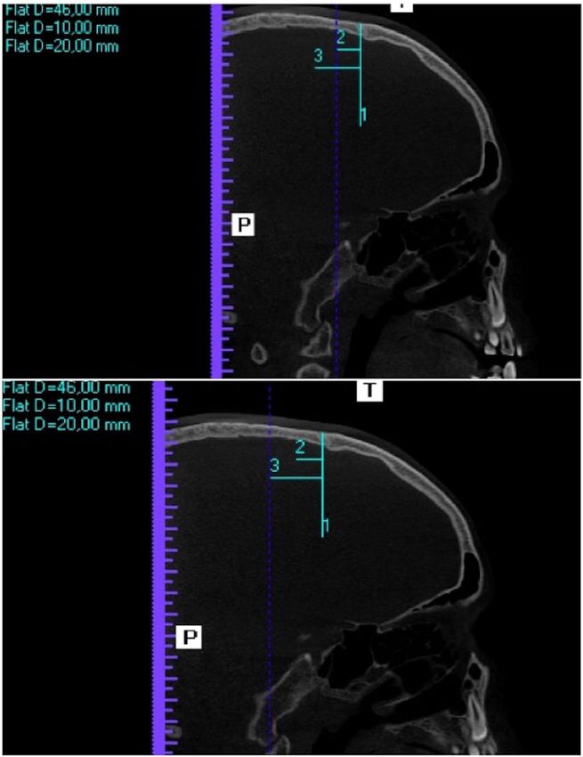
Evaluation of bone availability for grafts in different donor sites, through computed tomography

Introduction                                                                    graft size and shape,6 impairing the bone volume.7
                                                                                      Most studies on this subject 1,8-12 report an

   The rehabilitation of edentulous patients has                                advantage of the skullcap toward the other sites

occupied a prominent place in Dentistry. Implantology                           because it is a corticalized bone that undergoes less

offers excellent options for patients without enough                            resorption, leading to more predictable results for

bone to use a conventional prosthesis. However, for                             the installation of implants, both in the maxilla and

those with severe alveolar bone resorption, there                               mandible, with lower postoperative morbidity. The

is not enough bone for an implant installation. In                              disadvantages are related to the need for general

these cases, bone grafts are required.1 For larger                              anesthesia, to the potential complications and to the

reconstructions, donor sites in extraoral bones are                             patient acceptance of cranial surgery more than to its

the most viable options due to the greater amount of                            surgical difficulty.13

bone available.                                                                       As in any type of surgery, careful planning is

   Autogenous bone grafts are often used to correct                             essential; therefore, three-dimensional analysis

defects related to the bone volume of the recipient                             using computed tomography is very useful.1 Cone-

site, mainly because they are still considered the                              beam computed tomography (CBCT) is a diagnostic

gold standard when compared with biomaterials. In                               imaging method, especially indicated to examine

individuals who have lost permanent teeth due to                                the dentomaxillofacial complex, 4 which enables

trauma, caries or periodontal diseases and who lack                             the reformation of the maxillofacial bones without

the required bone volume, the symphysis can provide                             distortion and image-guided radiation dosing, with

an appropriate amount of bone for grafting, implant                             reduced costs.4 This examination technique improves

placement and prosthetic rehabilitation.2                                       the visualization of images and structures in a way that

   Bone grafts are influenced by factors such as                                was not possible with the conventional radiography.4

the surgical technique used, the bone quantity and                              Thus, this visualization capacity was used to quantify

quality of the donor site and the systemic conditions                           the bone availability, since studies that inform and

of the patients.1 The correct treatment planning, the                           discuss the bone volume that can be removed were

adequate revision of the medical history, the absence                           not found in the scientific literature.

of pathologies and deleterious habits, the proximity
of the alveolar process to the location of anatomical
structures – including maxillary sinus, nasal cavity,                           Methodology and Results
incisive canal (IC), mandibular canal (MC) and
mental foramen (MF) –, and a well-executed surgical                             Mandibular symphysis
technique will reduce complications during the surgical                               The sample size calculation was done according to
procedure and increase its success rate.          3,4
                                                                                some inclusion criteria. This study was approved by the
   The region of the mandibular body and ramus,                                 Research Ethics Committee of the University Center.
constituted by the cortical and trabecular bones, is                            A total of 200 CBCT exams of individuals of both
one of the most used intraoral donor sites for this                             genders, with at least 18 years of age, were obtained
purpose, primarily for its bone quality. This provides                          from the image archive of the surgery department of
osteogenesis, osteoconduction, osteoinduction and                               the University Center. An i-CAT Classic was performed
osteointegration, as well as low morbidity and few                              using the following parameters: flat panel detector,
postoperative sensorial complaints when compared                                0.3 voxel, 0.50 mm focal point, 120 V, 18.45 mAs,
with other donor sites. Besides having a high                                   20 s, (Imaging Science International, Hatfield,
concentration of bone morphogenetic proteins,5 this                             Pennsylvania, USA). All analysis and measurements
region has low volume loss and excellent incorporation                          were done in an appropriate room through a proper
in the short term. Another advantage is that the donor                          FlexScan S2000 monitor, 20” (Eizo Nanao Corporation,
and the recipient sites are in the same surgical field,                         Hakusan, Japan), by i-CAT Vision® Software.
reducing the surgical time and the necessary amount                                   In the parasagittal reformations, the following
of anesthetic and allowing the surgery to be performed                          elements were evaluated: (1) the interforaminal
at outpatient level. However, the access may reveal                             distance, (2) the distance between the apex of the
difficulties related to visibility and limitations on the                       anterior teeth and the beginning of the cortical base of

                                                 J Appl Oral Sci.         2/9   2020;28:e20190435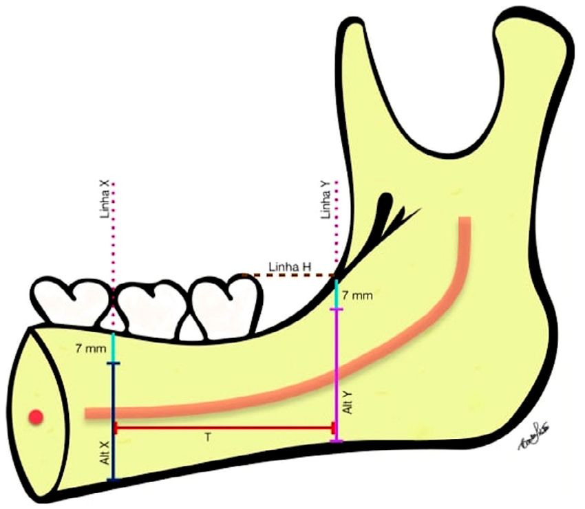
GUIMARÃES GM, BERNINI GF, GRANDIZOLI DK, CARVALHO PS, GONÇALES ES, FERREIRA JUNIOR O

the mandible and (3) the distance between the buccal                     age of 18 years old. They were obtained from the
surface of the cortical bone and the lingual surface of                  same database of i-CAT Classic equipment, flat panel
the lingual cortical.                                                    detector, 0.3 voxel, 0.50 mm focal point, 120 v, 18.45
   Panoramic reformation was used to aid in the                          mAs, 20 s, (Imaging Science International, Hatfield,
location of the parasagittal cuts, to visualize the                      Pennsylvania, USA). The sample size was calculated
mental foramen, canines and midline and to assess                        according to some inclusion criteria. This study was
the presence and extension of the anterior loop of the                   approved by the Research Ethics Committee of the
mental foramen (Figure 1).
                                                                         Table 1- Mean, minimum and maximum measures of all distance
                                                                         measurements in the parasagittal reformation, 0.30 mm thickness
Results
                                                                                             Averages        Minimum          Maximum
   Out of 200 patients whose exams were analyzed,
                                                                                                             average          average
105 were female and 95 were male, their ages varied
                                                                         IF                    42.27             33.3            55.2
from 18 to 78 years old and the average age was
                                                                         CB-D                  13.03             5.92            20.6
43.76 years old.
                                                                         ESPM-D                5.82              2.1             8.75
   The anterior loop of the mental nerve was
                                                                         ESPC-D                10.31             4.8             14.7
visualized in 47 images (23.5%), bilaterally in 36
                                                                         LM                    17.86             9.68           28.28
images (18.0%), unilaterally on the right side in 3
                                                                         ESPM-LM               5.93              2.4             9.3
images (1.5%) and unilaterally on the left side in 8                     ESPG-LM               10.5              5.6             16.5
images (4%). The mean distance measured between                          CB-E                  12.87             5.66            20.7
the anterior loop of the mental foramen and the base                     ESPM-E                5.54              2.4            10.25
of the mandible was 7.02 mm on the right side and                        ESPC-E                10.07             4.74           14.95
6.73 mm on the left side, the mean interforaminal                        AACB-D                7.02               3              13.8
distance was 42.27 mm and the mean height was 4.87                       AACB-E                6.73              3.3             14.7
mm, as can be seen in Table 1.
                                                                         IF - inter-foramen; BC-R – base-canine - right side; MT-R -
                                                                         medullary thickness - right side; CT-R - cortical thickness – right
External oblique line                                                    side; ML - midline-base; MT-ML medullary thickness - midline; CT-
                                                                         ML - cortical thickness - midline; BC-L – base-canine - left side;
Samples                                                                  MT-L - medullary thickness - left side; CT-L - cortical thickness
                                                                         – left side; ALBC-D - anterior loop of the mental foramen base-
   A retrospective study was conducted using CBCT
                                                                         canine - right side; ALBC-E - anterior loop of the mental foramen
exams of patients of both genders, with the minimum                      base-canine - left side

Figure 1- Axial, panoramic and parasagittal reformations and 3D reconstructions on i-CAT Vision® software screen

                                                J Appl Oral Sci.   3/9   2020;28:e20190435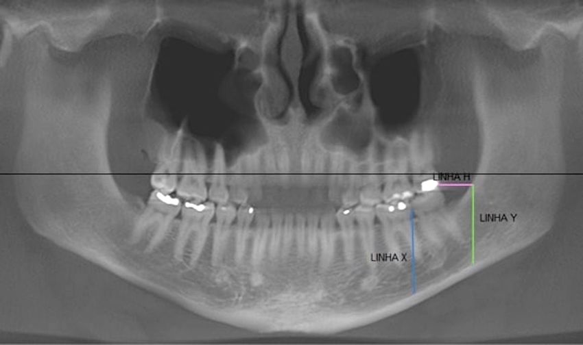
Evaluation of bone availability for grafts in different donor sites, through computed tomography

Figure 2- CBCT panoramic reformation showing the positions of Lines X, H and Y, which represent the limits of interest in the evaluation

                                                                                      University Center.

                                                                                      Measurements
                                                                                            All analyses and measurements were performed
                                                                                      in an appropriate room through a proper monitor
                                                                                      FlexScan S2000, 20” (Eizo Nanao Corporation,
                                                                                      Hakusan, Japan), by i-CAT Vision® Software. The area
                                                                                      submitted to the volumetric calculations was selected
                                                                                      based on the region of interest of the graft, being
                                                                                      established according to the following limits: Line X =
                                                                                      vertical line that tangents the distal of the crown of the
                                                                                      first lower molar; Line H = horizontal line that tangents
                                                                                      the highest cuspid of molars; Line Y = vertical line that
                                                                                      starts where line H crosses the anterior border of the
                                                                                      ascending mandibular ramus (Figure 2). The volume

Figure 3- Illustration of the height and length of the graft block                    was calculated through the expression V = H x L x T
                                                                                      where H = height, L = length and T = thickness.
                                                                                            Simulating a donor site for grafting, the height (H)
                                                                                      of the bone block was calculated on the panoramic
                                                                                      reformation through the distance from the alveolar
                                                                                      bone crest to the internal cortical bone of the mandible
                                                                                      base in Line X and, in Line Y, subtracting 7 mm to
                                                                                      the amount of bone required for the maintenance of
                                                                                      the molars and then calculating the average of these
                                                                                      measurements. After the measurements, the mean
                                                                                      between the heights X and Y was calculated. The
                                                                                      length (L) of the bone block was calculated through
                                                                                      the distance between Line X and Line Y (Figure 3). The
                                                                                      thickness of the hypothetical bone block was calculated
                                                                                      in the CBCT parasagittal reformations. The buccal
                                                                                      cortical bone thickness was measured both in Line X
                                                                                      and in Line Y, in three heights separated by 5 mm, that
                                                                                      is: (1) 7 mm, (2) 12 mm and (3) 17 mm below the
                                                                                      vestibular alveolar bone crest. After obtaining these
                                                                                      values, the average thickness on Line X and on Line
                                                                                      Y and the average thickness between X and Y were
Figure 4- Illustration of a CBCT parasagittal reformation showing
the positions of points 1, 2, 3 and of Line Z                                         calculated, resulting in the average thickness of the

                                                       J Appl Oral Sci.         4/9   2020;28:e20190435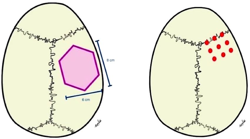
GUIMARÃES GM, BERNINI GF, GRANDIZOLI DK, CARVALHO PS, GONÇALES ES, FERREIRA JUNIOR O

buccal cortical bone. Through these measurements,                            Cranial bone
the bone volume available on the right side, on the                             Fifty CBCTs of individuals of both genders with the
left side and in total were calculated and expressed                         minimum age of 18 years old were obtained from
in cubic millimeters (mm3).                                                  the image files of the surgery department of the
    Furthermore, the distance from the center of the                         University Center. The sample size was calculated
upper cortical of the mandibular canal (Line Z) to the                       according to some inclusion criteria. This study was
buccal cortical bone was measured on Line X and on                           approved by the Research Ethics Committee of the
Line Y (Figure 4).                                                           University Center. All the exams were performed
                                                                             on an i-CAT Classic (Imaging Science International,
Results                                                                      Hatfield, Pennsylvania, USA), which has a flat panel
    The samples used in this study were images                               detector, with the following acquisition protocol: voxel
obtained from 70 patients, with ages between 18 and                          0.3 mm, focal point 0.50 mm, 120 V, 18.45 mAs, 20
68 years old (an age average of 29.61 years old), of                         s. All analyses and measurements were performed in
which 46 were women and 24 were men. The average                             a suitable room on a FlexScan S2000, 20" monitor
values of the measurements were: between the linear                          (Eizo Nanao Corporation, Hakusan, Japan) using the
distances X and Y: 18.98 mm; height on X and on Y =                          Software i-CAT Vision®.
17.33 mm and, considering the thickness of the cortical                         In order to calculate the bone volume that can be
bone, the average of the region (X and Y) was 2.6 mm.                        obtained, an area of hexagonal shape, 8 cm long and
The minimum, maximum and average values and the                              6 cm wide, similar to that described by De Ceulauer
standard deviation are shown in Table 2. Regarding                           and Abelos14 (2012) (Figure 5), was considered as
the volume determination through linear values, the                          corresponding to the donor site .
average bone volume available in the posterior region                           Initially, the area of this hexagon was calculated.
of the mandible was 859.26 mm3                                               Next, the cortical, medullary and total bone thickness
                                                                             (cortical + medullary) were measured at 9 points
                                                                             (Figure 5), obtaining the mean bone thickness. By
Table 2- Cortical, medullary and total (cortical + medullary) bone           multiplying the area of the hexagon by the bone
thickness and volumes, considering the mean of the 9 points of
the site studied. The table shows the result of the correlation test         thickness, the volume of bone that can be removed
between the bone thickness and the age of the individuals                    for grafts from that region was obtained.
                                                                                For the thickness measurements, on the MPR
                             Measurement            Standard
                              averages              deviation                screen of the software, in the window corresponding
    Distance of linear         18.98 mm             18.9±0.12                to the sagittal reformations, the blue line, which
    lengths X and Y
                                                                             determines the coronal reformations, was positioned
   Heights at X and Y          17.33 mm            17.33±3.00
                                                                             exactly on the coronal suture (Figure 6). Therefore, a
 Cortical bone thickness         2.6 mm             2.60±0.01
                                                                             coronal reformation was obtained at the level of the
Bone volume average             859.26 mm3                                   coronal suture, in which a vertical line corresponding

Figure 5- Illustration of the donor site of hexagonal shape described in the study by De Ceulaer, et al.31 (2012). Illustration of the 9 points
where the measurements were made

                                                   J Appl Oral Sci.    5/9   2020;28:e20190435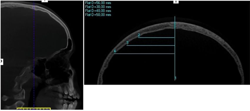
Evaluation of bone availability for grafts in different donor sites, through computed tomography
to the median sagittal suture was drawn using the determines the coronal reformations, was moved
distance tool (vertical line) with 30, 40 and 50 mm first 10 mm, then 20 mm posteriorly and the
from the right side, respectively, obtaining a distance cortical, medullary and total (cortical + medullary)
guide to the median sagittal suture, to perform the bone thickness were measured again (Figure 6). In
thickness measurements (Figure 7). summary, cortical, medullary and total (cortical +
In each of these positions, cortical, medullary medullary) bone thickness were measured at 3 points
and total (cortical + medullary) bone thickness at the level of the coronal suture, at 3 points 10 mm
measurements were performed at the level of the posteriorly and at 3 points 20 mm posteriorly, as
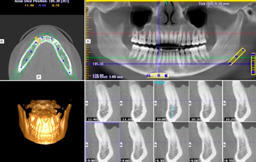
coronal suture. Afterwards, the blue line, which shown in Figure 2.
Figure 6- Blue line on the coronal suture. Guide for the measurement of bone thickness, 30, 40 and 50 mm to the right of the sagittal
suture
Figure 7- Blue line displaced 10 mm posterior to the coronal suture. Blue line displaced 20 mm posterior to the coronal suture
J Appl Oral Sci. 6/9 2020;28:e20190435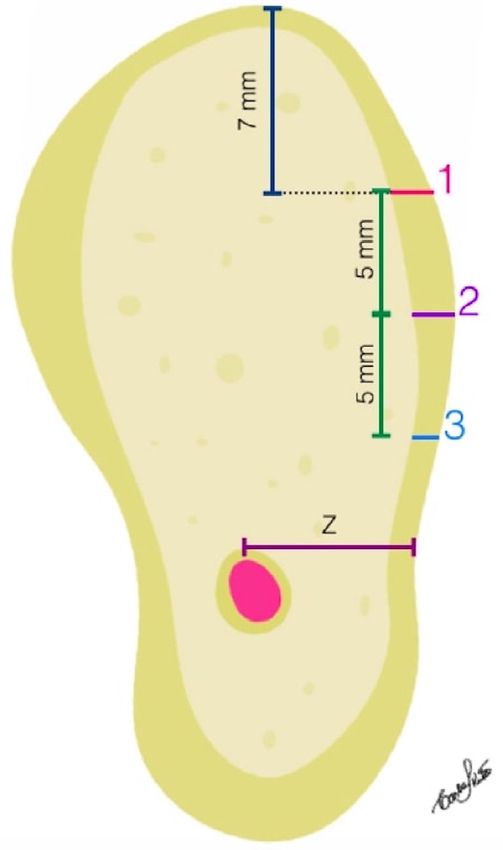
GUIMARÃES GM, BERNINI GF, GRANDIZOLI DK, CARVALHO PS, GONÇALES ES, FERREIRA JUNIOR O

Table 3- Comparison of the cortical, medullary and total (cortical          reported that none of the patients complained about
+ medullary) bone thickness in the 9 points of the area studied,
                                                                            morphology alteration of the chin after the removal of
by gender. The table shows the averages of measurements of the
cortical, medullary and total (cortical + medullary) bone thickness         grafts from the mandibular lower anterior region when
according to the gender of individuals
                                                                            these recommendations were respected.2
                         Mean E      Volume         Standard                   This study used as safety margins (1) an 8.00 mm
                                                    Deviation
                                                                            distance from the apexes of the roots of the anterior
   Cortical Bone        2.78 mm      1.167.60         158.76
                                                                            teeth, (2) the total preservation of the cortical at
  Medullary Bone        3.00 mm       1.260          443.004
                                                                            the base of the mandible, (3) a distance of 5.00 mm
     C-M Bone           5.95 mm       2.499          694.992
                                                                            anteriorly to the mental foramen and (4) a depth
*Statistically significant                                                  limited to 4.00 mm from the cortical vestibular. The

Results                                                                     average amount of available bone in the mandibular

    The ages of the 50 patients (25 women and 25                            symphysis region obtained was 628.61 mm.4

men) whose exams were used in this study ranged                                With the use of the CBCT, considering these safety

from 18 to 71 years old, with an average age of 35.6                        margins and a correct planning, our study reveals

years.                                                                      that an adequate patient selection and a reduction

    Table 3 shows the averages of cortical, medullary                       in postoperative complications are predictable.16-19,21

and total (cortical + medullary) bone thickness                                The symphysis may provide adequate bone grafts

measurements used to calculate the bone volume.                             to increase a site previously occupied by two to six
                                                                            teeth. It will never offer enough bone to raise an arch.
                                                                            If the increase in the complete dental arch is required
                                                                            or if the extent of the alveolar bone loss is significant,
Discussion                                                                  another source of bone should be considered.13

Mandibular symphysis
                                                                            External oblique line
    The imaging test of the symphysis is necessary to
                                                                               The use of autogenous bone from the mandibular
verify if there is enough bone to be used as graft.13
                                                                            body and ramus has been proved to be effective in
With the frequent use of CBCT, which offers more
                                                                            reconstructive surgeries of the maxillary bones.22
precision and detail, a great variation in the anatomy
                                                                            However, no studies report safe bone volume obtained
and dimensions of this region is identified. This proves
                                                                            in this region.5,22 Furthermore, the posterior region
it to be an important instrument for surgical planning,
                                                                            of the mandible, unlike the mandibular symphysis,
minimizing intercurrences and complications.1
                                                                            does not present defined limits for bone removal, so
    The removal of bone from the mandibular
                                                                            no protocol delimits the exact donor site and there is
symphysis for grafting is a surgical procedure and the
                                                                            no standard for the available volume.
region is completely repaired after 24 months, with
                                                                               In this study, we used the molar teeth as reference
the formation of a new cortical and the stabilization of
                                                                            for the anterior limit,23-25 specifically the distal of first
the bone remodeling. It is possible, then, to perform a
                                                                            molar,6,22 which is considered a safe limit to prevent
new intervention in the same region if necessary.13,15
                                                                            interference with the mental nerve ramus. As for the
    A safety margin of at least 5.00 mm to the apex of
                                                                            upper limit,24, Capelli6 (2003) indicates a distance from
the lower anterior teeth is indicated to avoid sensitivity
                                                                            4 to 6 mm medially to the oblique line; and Haggerty,
loss in these teeth.2 Experiments with animals have
                                                                            et al.25 (2015) says that the superior margin of the
shown that the safety margin should be at least 8.00
                                                                            graft coincides with the external oblique line. However,
mm.16 The main advantage of the 8 mm safety margin
                                                                            in this study, a 7 mm safety margin to the alveolar
to the apex of the roots is the 75% reduction of injury
                                                                            bone crest was recommended so that the removed
possibility in the incisive nerve.17,18
                                                                            bone would not be close to the cervical of the teeth. For
     One recommends to maintain the total integrity of
                                                                            the posterior limit, the reference was the exact place
the base of the mandible, preserving the preoperative
                                                                            where the occlusal plane touches the anterior edge
contour of the chin region and the facial profile,
                                                                            of the ascending mandibular ramus. If the removal of
leaving the inferior margin of the symphysis intact
                                                                            the patch was too high, the osteotomy could injure
and maintaining the midline protrusion, avoiding
                                                                            the buccal artery or expose adipose tissue. Fujita and
deformations and irregularities.16,19,20 A 2004 study

                                                   J Appl Oral Sci.   7/9   2020;28:e20190435
Evaluation of bone availability for grafts in different donor sites, through computed tomography

Shintani22 (2015) consider the mandibular lingula as                                 using a goniometer. The average thickness observed
the posterior limit. In the studies by Capelli6 (2003),                              was 4.8 mm, 4.5 mm, 6.1 mm, 4.2 mm, respectively,
incisions were made at the base of the coronoid                                      at the 4 evaluated points.28 Bernardino Junior, et
process, as well as in the reports by Haggerty et                                    al.29 (2011) measured the thickness of the skullcap
al.25 (2015), in which the extension in the posterior                                at the most protruding point of the parietal tuber.
direction can also include this region.                                              They measured 60 macerated human skulls at the
   For the lower limit, the reference considered                                     Federal University of Uberlândia, obtaining an average
is the junction between the anterior and posterior                                   thickness of 5.16 mm.
osteotomies, with an average height of 1 cm or the             6
                                                                                           The most comprehensive study on the subject
junction of the osteotomies that extend from 10 to 12                                measured 40 points on 281 dry skulls from the
mm below the external oblique line or 4 mm above the                                 Cleveland Museum of Natural History. The mean
mandibular canal.   25
                         In this study, the internal cortical                        thickness found was 6.3 mm, with values ranging from
of the mandibular base is considered the lower limit. In                             5.3 mm to 7.5 mm. The site of greatest thickness was
Line X, or anterior limit, the average height was 16.31                              the posterior parietal region.28
mm and in Line Y, or posterior limit, it was 18.36 mm.                                     Comparing the results of this study with those
The resulting average graft height was 17.33 mm.                                     found in the literature, a significant difference in
   The average distance between the anterior and                                     the methodology should be considered, since in all
posterior limits (Line X and Line Y) was 18.98 mm, as                                the previously mentioned studies performed direct
listed in Table 2. The average cortical thickness was                                measurements in dry skulls. This means that these
2.6 mm, ranging from 1.05 to 4.65 mm. Based on the                                   thickness measurements considered the external
linear values, the resulting average of bone volume                                  cortical, the medullary bone and the inner cortical
available in the posterior region of the mandible was                                bone. In the methodology of this study, on the other
859.26 mm3 (Table 2).                                                                hand, only the external cortical and the medullary
   Some authors          22
                              performed a very similar                               layer were measured, since they are the ones that
methodology, using the same references of this study                                 are effectively used in the grafts. As it can be seen in
(the distal of the first molar, then the distal of the second                        Table 3, the mean thickness of the cortical + medullary
molar, 10 mm distally to the second molar and 15 mm                                  bone of the 9 evaluated points was 5.95 mm.
distally to the second molar). The resulting values of                                     The bone volume of the other donor sites were
length, height and thickness were respectively: 26                                   628.61 mm3 in the mandibular symphysis and 859.33
mm, 10 mm and 2 mm. When comparing them to the                                       mm3 in the external oblique line region. The available
values of our study, the difference comes from the fact                              bone volume in the skullcap region, calculated in this
that they evaluate site located a little further in the                              study was 2,499 mm3 (Table 3). Comparing it with the
posterior direction; therefore, these authors present                                volumes available in the intraoral donor sites of the
higher length values and lower thickness values.                                     symphysis and of the posterior region of the mandible,
                                                                                     it is reported that the skullcap can offer bone volume
Cranial bone                                                                         almost 3 times greater than the latter and at least 2
   The selection of the graft donor site is based on (1)                             times more than the former. In addition, as it allows
the amount of bone needed in the recipient bed, (2)                                  the withdrawal of several blocks, the skullcap can be
the number and location of the implants and (3) the                                  used for reconstructions that need more extension.
acceptance of the risk of complications by the patient.                  25

   Pensler and McCarthy 26 (1985) studied the
thickness of the skullcap in the region of the parietal
                                                                                     Conclusion
and occipital bones and found it varied from 6.80
mm to 7.72 mm. In another study carried out in the
                                                                                           All sites discussed in this article are excellent
Anatomy laboratory of the School of Dentistry of the
                                                                                     options for the removal of autogenous bone grafts
Universidade Estadual Paulista, 49 dry skulls of adult
                                                                                     for the reconstruction of defects and for the bone
individuals were evaluated.27 In that study, all skulls
                                                                                     resorption of the jaws. The choice of the site will
had the cranial vault sectioned at the height of the
                                                                                     depend on the type of defect.
temporal bone and measured at 4 different points
                                                                                                Compared with intraoral donor sites, the bone

                                                      J Appl Oral Sci.         8/9   2020;28:e20190435
GUIMARÃES GM, BERNINI GF, GRANDIZOLI DK, CARVALHO PS, GONÇALES ES, FERREIRA JUNIOR O
availability of the skullcap is 3 times greater than that 13- Davies JE, Matta R, Mendes VC, Perri de Carvalho PS. Development,
characterization and clinical use of a biodegradable composite scaffold
of the posterior region of the mandible and at least 2
for bone engineering in oromaxillofacial surgery. Organogenesis.
times greater than that of the mandibular symphysis. 2010;6(3):161-6.
14- De Ceulaer J, Swennen G, Abeloos J, De Clercq C. Presentation of a
Acknowledgements cone-beam CT scanning protocol for preprosthetic cranial bone grafting
of the atrophic maxilla. Int J Oral Maxillofac Surg. 2012;41(7):863-6.
This study was financed in part by Coordenação de doi: 10.1016/j.ijom.2012.03.012.
Aperfeiçoamento de Pessoal de Nível Superior - Brasil 15- Esposito M, Grusonvin MG, Rees J, Karasoulos D, Felice P, Alissa
(CAPES) - Finance Code 001. R, et al. Effectiveness of sinus lift procedures for dental implant
rehabilitation: a Cochrane systematic review. Eur J Oral Implantol.
2010;3(1):7-26.
16- Hounsfield GN. Computerized transverse axial scanning
(tomography): Part I. Description of system. 1973. Br J Radiol.
References 1995;68(815):H166-72.
17- Hemmy DC, Tessier PL. CT of dry skulls with craniofacial
1- Carvalho PS, Carvalho MC, Ponzoni D. Reconstruction of alveolar deformities: accuracy of three dimensional reconstruction. Radiology.
bone defect with autogenous bone particles and osseointegrated 1985;157(1):113-6. doi: 10.1148/radiology.157.1.3929326.
implants: histologic analysis and 10 years monitoring. Ann Maxillofac 18- Hirsch JM, Ericsson I. Maxillary sinus augmentation using
Surg. 2015;51(1):135-9. doi: 10.4103/2231-0746.161145. mandibular bone grafts and simultaneous installation of implants.
2- Al-Ani O, Nambiar P, Ha KO, Ngeow WC. Safe zone for bone A surgical technique. Clin Oral Implant Res. 1991;2(2):91-6. doi:
harvesting from the interforaminal region of the mandible. Clin 10.1034/j.1600-0501.1991.020207.x.
Oral Implants Res. 2013(Supp A100):115-21. doi: 10.1111/j.1600- 19- Hausamen JE, Neukam FW. Transplantation of bones. Eur Arch
0501.2011.02393.x. Otorhinolaryngol Suppl. 1992;1:163-77.
3- Anderson LC, Kosinsk TF, Mentag PJ. A review of the intraosseous 20- Hofschneider U, Tepper G, Gahleitner A, Ulm C. Assessment
course of the nerves of the mandible. J Oral Implantol. 1991;17(4):394- of the blood supply to the mental region for reduction of bleeding
403. complications during implant surgery in the interforaminal region. Int
4- Angelopoulous C, Thomas SL, Hechler S, Parissis N, Hlavacek M. J Oral Maxillofac Implants. 1999;14(3):379-83.
Comparison between digital panoramic radiography and cone-beam 21- Hunt DR, Jovanovic SA. Autogenous bone harvesting: a chin
computed tomography for the identification of the mandibular canal as graft technique for particulate and monocortical bone blocks. Int J
part of presurgical dental implant assessment. J Oral Maxillofac Surg. Periodontics Restorative Dent. 1999;19(2):165-73.
2008;66(10):2130-5. doi: 10.1016/j.joms.2008.06.021. 22- Fujita A, Shintani S. Computed tomographic analysis of the
5- Verdugo F, Simonian K, Smith McDonald R, Nowzari H. Quantitation mandibular body and ramus in Japanese patients: relevance to bone
of mandibular ramus volume as a source of bone grafting. Clin Implant harvesting from the mandibular ramus. Implant Dent. 2015;24(4):402-
Dent Relat Res. 2009;11(Suppl 1):e32-7. doi: 10.1111/j.1708- 6. doi: 10.1097/ID.0000000000000271.
8208.2009.00172.x. 23- Misch CM. Ridge augmentation using mandibular ramus bone grafts
6- Capelli M. Autogenous bone graft from the mandibular ramus: a for the placement of dental implants: presentation of a technique. Pract
technique for bone augmentation. Int J Periodontics Restorative Dent. Periodont Aesthetic Dent. 1996;8(2):127-35; quiz 138.
2003;23(3):277-85. 24- Clavero J, Lundgren S. Ramus or chin grafts for maxillary sinus
7- Soehardi A, Meijer GJ, Strooband VF, de Koning M, Stoelinga PJ. The inlay and local onlay augmentation: comparison of donor site morbidity
potential of the horizontal ramus of the mandible as a donor site for and complications. Clin Implant Dent Relat Res. 2003;5(3):154-60.
block and particular grafts in pre-implant surgery. Int J Oral Maxillofac 25- Haggerty CJ, Vogel CT, Fisher GR. Simple bone augmentation
Surg. 2009;38(11):1173-8. doi: 10.1016/j.ijom.2009.07.006. for alveolar ridge defects. Oral Maxillofac Surg Clin North Am.
8- Friedlaender GE. Bone grafts. The basic science rationale for clinical 2015;27(2):203-26. doi: 10.1016/j.coms.2015.01.011
applications. J Bone Joint Surg Am. 1987;69(5):786-90. 26- Pensler J McCarthy JG. The calvarial donor site: an anatomic
9- Genutis S. Calvarial bone grafts in oral and maxillofacial surgery. study in cadavers. Plast Reconstr Surg. 1985;75(5):648-51. doi:
Grand Rounds: University of Illinois; 2003. 10.1097/00006534-198505000-00005.
10- Almeida-Junior P, Esper HR, Garcia-Junior IR, Sottovia AD, Magro 27- Monnazzi, Marcelo Silva, et al. Avaliação da espessura do osso
Filho O. Enxerto ósseo de calota craniana em reconstruções para parietal como região doadora de enxertos ósseos. Rev Bras Cir
implantodontia. Innovations J. 2004;8:13-6. Traumatol Buco Maxilo Fac. 2010;10(1):33-8.
11- Block MS, Jackson WC. Techniques for grafting the extraction site 28- Gonzalez AM, Papay FE, Zins JE. Calvarial thickness and its relation
in preparation for dental implant placement. Atlas Oral Maxillofac Surg to cranial bone harvest. Plast Reconstr Surg. 2006;117(6):1964-71.
Clin North Am. 2006;14(1):1-25. doi: 10.1016/j.cxom.2005.11.006. doi: 10.1097/01.prs.0000209933.78532.a7
12- Barone, A Covani U. Maxillary alveolar ridge reconstruction with 29- Bernardino Júnior R, Queiroz MM, Máximo RO, Teixeira E, Lizardo
nonvascularized autogenous block bone: clinical results. J Oral Maxillofac FB, Vilarinho GS, et al. Mensuração da espessura do osso da calota em
Surg. 2007;65(10):2039-46. doi: 10.1016/j.joms.2007.05.017. parietais de crânios macerados. Biosci J. 2011;27:995-1003.
J Appl Oral Sci. 9/9 2020;28:e20190435You can also read