Anatomy of the Skull of Saanen Goat. An anesthesiology and Stereology Approach
←
→
Page content transcription
If your browser does not render page correctly, please read the page content below
Int. J. Morphol.,
39(2):423-429, 2021.

                        Anatomy of the Skull of Saanen Goat.
                       An anesthesiology and Stereology Approach

Anatomía del Cráneo de la Cabra Saanen. Un Enfoque desde la Anestesiología y la Estereología

                          Xiangyang Wang1; Anjun Liu1; Jing Zhao2; Fathy M. Elshaer3,4 & Diaa Massoud3,5

WANG, X.; LIU, A.; ZHAO, J.; ELSHAER, F. M. & MASSOUD, D. Anatomy of the skull of Saanen goat. An anesthesiology and stereology
approach. Int. J. Morphol., 39(2):423-429, 2021.

         SUMMARY: The Saanen goat is known as the greatest milk producer among small ruminat breeds. However, its morphometric
features still remain unclear. Therefore, the present work aimed to investigate the functional anatomy of the upper and lower jaws as well as the
volumetric properties of the male and female Saanen goat for clinical applications. The heads of 20 adult animals (10 males and 10 females)
were included. Totally, 22 morphometric parameters were measured on three dimensional computed tomographic images using RadiAnt
DICOM Viewer software and some parameters were measured on hot macerated sample. The mean volume of paranasal sinuses as well as
conchal sinuses were estimated using stereological method. Based on the results, the differences between males and females were not significant
(p>0.05) in all desired parameters.The frontal and lacrimal sinuses were the largest and smallest paranasal sinus in both sexes. Also, the dorsal
and middle conchal sinuses were the largest and smallest ones, respectively. The common nasal meatus was the smallest and ventral meatus
was the largest meatus in the nasal cavity.In conclusion, these findings provide a basic data that would be useful in blocking terminal branches
of the cranial nerves in this breed for surgical purpose or teeth injuries treatment.

        KEY WORDS: Applied anatomy; CT scan; Morphometry; Ruminant; Stereology.

INTRODUCTION

        The superficial landmarks of anatomical features in                           Computed tomography (CT) is a reliable, and
the skull region can be attributed to the genetic and                         noninvasive procedure for evaluating different pathological
environmental factors which would be helpful in interpreting                  lesions or diseases in the head region as compared to the
extensive variety in the phenotyping between and within                       traditional radiography (Frazho et al., 2008). Although, this
breeds or species. Accurate knowledge of the anatomical                       modality has been used widely for describing the normal
structure of the skull can be an effective aid in ontogenic                   structure of head and other body regions in animals world
studies as well as the determination of sexual polymorphisms.                 (Morrow et al., 2000; Frazho et al.), however, the combination
(Olopade & Onwuka, 2008).                                                     of imaging techniques and stereological procedures has
                                                                              received little attention in veterinary practice.
        Previously, skull morphometry and its applied clinical
anatomy have been performed on different wild species (Onar                          The Saanen goat which originates Saanen Valley of
et al., 2005; Sarma, 2006), domestic animals (Onar, 1999;                     Switzerland, is known as the greatest milk producer among
Baranowski, 2010) and farm animals (Jakubowski et al., 2008;                  small ruminat breeds (Kurtul & Atalgin. 2008). Despite the
Parés I Casanova et al., 2010). In addition, different breeds                 great values of this breed, there is a little data about the
of goat including West African Dwarf goat (Olopade &                          morphometric and anatomical features of the skull in the
Onwuka, 2005), Nigerian Red Sokoto goat, (Olopade &                           Saanen goat. Therefore, this work was designed to present
Onwuka, 2008), Iranian native goat (Monfared et al., 2013),                   comprehensive morphometric data of the skull in this breed
Black Bengal goat (Uddin et al., 2009), and Markhoz goat                      with special reference to its regional anesthesia as well as to
(Goodarzi & Hoseini, 2014) have been previously subjected                     present the normal values of the paranasal and conchal sinuses
to skull morphometric analysis.                                               volume. The obtained results will compare to the other relevant

1
  Department of Rehabilitation Medicine, the 960th Hospital of the Joint Service of the Chinese People's Liberation Army, Jinan,Shandong, 250031, China.
2
  Department of Anesthesiology, the 960th Hospital of the Joint Service of the Chinese People's Liberation Army, Jinan, Shandong, 250031, China.
3
  Department of Biology, College of Science, Jouf University, P.O. Box 2014, Sakaka, Al-Jouf, Saudi Arabia.
4
  Department of Zoology, Faculty of Science, Al-Azhar University, Nasr City, Cairo, Egypt.
5
  Department of Zoology, Faculty of Science, Fayoum University, Fayoum, Egypt.

                                                                                                                                                 423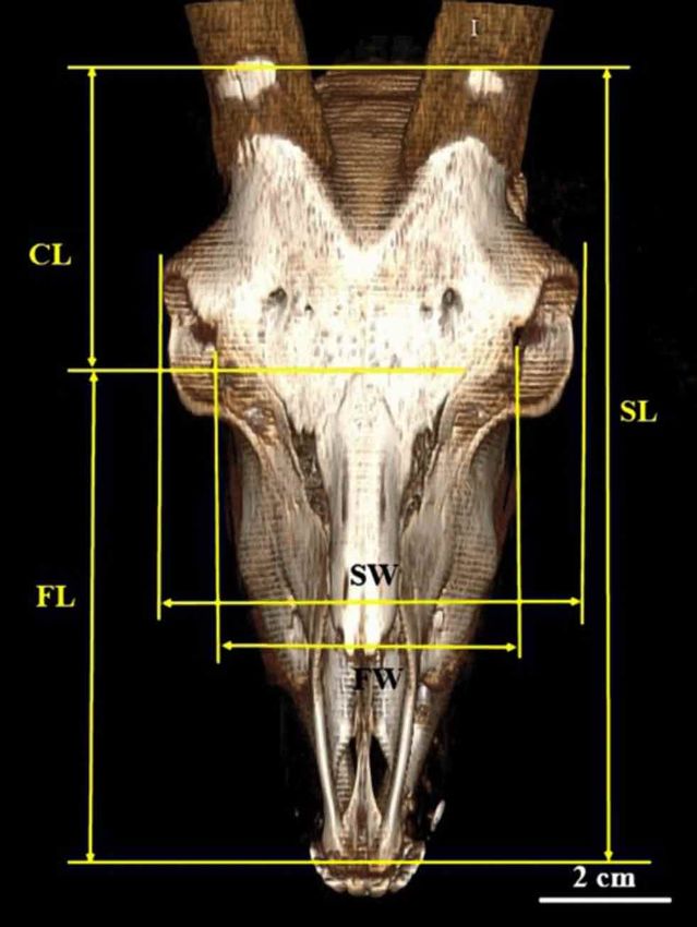
WANG, X.; LIU, A.; ZHAO, J.; ELSHAER, F. M. & MASSOUD, D. Anatomy of the skull of Saanen goat. An anesthesiology and stereology approach. Int. J. Morphol., 39(2):423-429, 2021.
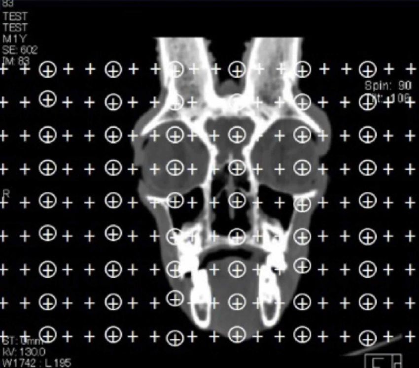
results. This information would be useful for veterinarians who used for estimating the absolute volume of the paranasal
are involved in small ruminant clinical practices. sinuses. For this purpose, Cavalieri principle was performed
on the obtained CT scan images. On average, 121 images
were selected from each animal. A point probe constituted
MATERIAL AND METHOD of 220points (+) was superimposed on each CT scan image
and the fractional volume of the cavities was estimated using
the following formula:
The heads of 20 adult Saanen goats (10 males and 10
females) were used in this work. The heads were separated Vv(structure/reference) = ∑Pstructure / ∑Preference
from the goats that died because of diseases unrelated to the
head or neck and transferred to the dissection room, Zoology In this formula, ∑Pstructure isthe sum of the points hit
Department, Faculty of Science, Fayoum University, the desired cavities and ∑Preference is thesum of the points hit
Fayoum, Egypt. The present research was performed in the total section area (Fig. 1). The fractional volume was
accordance with the recommendations of the National multiplied by the total volume of the skull. Therefore, the
Institutes of Health’s Guide for the Care and Use of following formula was applied to determine the reference
Laboratory Animals. The research protocol was approved volume:
by the Scientific Ethics Committee of Fayoum University.
Ten heads were used for computed tomographic (CT) Vtotal = ∑P × [SU] × d/SL]2 × t
imaging and another ten samples were processed by hot
maceration technique to measure some parameters that could In this formula. ∑P is the sum of the points hit the
not be investigated with CT images. whole sections, SU is the scale unit, d is the distance between
two points, SL is the scale length, and t is the section interval.
Computed tomographic imaging. The head samples were
used for CT scan imaging with a helical scanner The coefficient of error (CE) of the Cavalieri estimate
(SiemensSomatom®- 2 detectors, Germany/ Kvp: 105 V- of volume was predicted using the following formula
mAs: 130 and slice thickness: 1.5 mm). All CT scan images (Gocmen-Mas et al., 2009):
were investigated using an image analysis workstation (Clear
Canvas by Synaptive Medical, Toronto, Canada) and
Afterward, 3D reconstruction was done using the using
RadiAnt DICOM Viewer software.
Volume estimation. In the present work a combination of
un-biased and designed-based stereology and CT scan were
Morphometric measurements. The 3D images underwent
morphometric measurements in RadiAnt DICOM
Viewer software and totally twenty- two parameters in
upper and lower skull were described as below (Figs. 2 to 4):
1. Skull length (SL); was measured as a distance between the rostral
point of the incisive bone to the external occipital protuberance
of the occipital bone.
2.Skull height (SH); was measured as a distance from the summit
of the frontal bone to the tip of the paracondylar process.
3. Skull width (SW); was measured as a distancethe distance
between the lateral margins of the eyes.
4. Cranial length (CL); was the distance between the nuchal crest
and the caudal rims of the eyes.
5. Cranial width (CW); was the distance between the most lateral
points of the cranial cavity at the level of the external acoustic
meatus.
6. Facial length (FL); was the distance between the nasofrontal
Fig. 1.Transverse CT scan image of the Saanen goat skull suture and the most rostral point of the incisive bone.
superimposed with a compound point grid for estimating the volume 7. Facial width (FW); was the distance between the rostral rims of
density of desired cavities using Cavalieri principle. the eyes.
424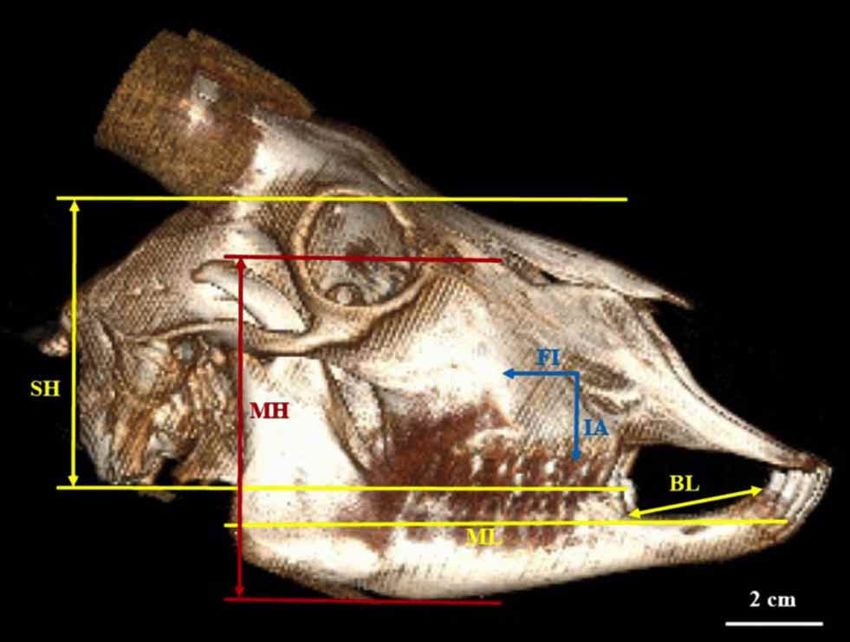
WANG, X.; LIU, A.; ZHAO, J.; ELSHAER, F. M. & MASSOUD, D. Anatomy of the skull of Saanen goat. An anesthesiology and stereology approach. Int. J. Morphol., 39(2):423-429, 2021.

8. Mandibular height (MH); was a perpendicular line which
   connected the tip of the coronoid process to the ventral margin
   of the mandible.
9. Mandibular length (ML) was measured as a distance from the
   caudal margin of the ramus to the incisive teeth.
10. Bar length (BL); was the distance between the most lateral
   incisive tooth and first premolar teeth.
11. FI the distance between the facial tuberosity and infraorbital
   foramen.
12. MFIDwas measured as a distancefrom the mental foramen to
   the most lateral incisive tooth.
13. MFPD was measured as a distancefrom the mental foramen to
   the cranial margin of the first premolar tooth.
14. MFVD was measured as a distance from the mental foramen
   to the ventral margin of the mandibular body.
15. MFC was measured as a distance from the mental foramen to
   the caudal margin of the mandibular ramus.
                                                                                            Fig. 3. 3D reconstruction of the Saanen goat skulls shown on
16. M1; was the distance between the ventral margin of the
                                                                                            lateralview. SH: skull height, ML: mandibular length, MH:
   mandibular foramen and ventral margin of the mandible.
                                                                                            mandibular height, BL: bar length, FI: facial tuberosity to
17. M2; was the distance between the mandibular foramen and
                                                                                            infraorbital foramen, IA: Infraorbital foramen to alvelolar tooth.
   caudal margin of the ramus.
18. M3; was the shortest distance between the mandibular fora-
   men and caudal angle of the mandible.
19. Skull index; Skull width / skull length × 100
20. Facial index; Facial width / facial length × 100
21. Cranial index; Cranial width / cranial length × 100

                                                                                            Fig. 4. (a) Lateral and (b) medial views of the mandible of the
                                                                                            Saanen goat.MFID: mental foramen to the most lateral incisive
                                                                                            tooth, MFPD: mental foramen to the first premolar tooth, MFVD:
                                                                                            mental foramen to the ventral margin of the mandibular body, MFC:
                                                                                            mental foramen to the caudal margin of the mandibular ramus.
Fig. 2. 3D reconstruction of the Saanen goat skull is shown on                              M1: mandibular foramen to the ventral margin of the mandible,
dorsal view. SL: Skull length, FL: facial length, CL: cranial length,                       M2: mandibular foramen to the caudal margin of the mandible,
SW: skull width, FW: facial width.                                                          M3: mandibular foramen to the most caudal angle of the mandible.

                                                                                                                                                                              425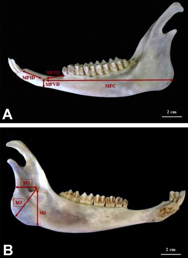
WANG, X.; LIU, A.; ZHAO, J.; ELSHAER, F. M. & MASSOUD, D. Anatomy of the skull of Saanen goat. An anesthesiology and stereology approach. Int. J. Morphol., 39(2):423-429, 2021.

Statistical analysis. The obtained data are presented as mean                               The distance between the mental foramen and caudal margin
± standard deviation (SD). The difference between males and                                 of the mandibular ramus was measured to be 15.35±0.34
females animals was analyzed using Student t-test in the SPSS                               cm. The mandibular foramen was placed 2.14±0.11 cm away
software version 20.0 (SPSS, Inc., Chicago, IL) and differences                             from the caudal margin of the mandible and 4.19±0.1 cm
were considered significant at P value less than 0.05.                                      away from the ventral margin of the mandible, on the medial
                                                                                            surface of the ramus. The skull index, facial index and cranial
                                                                                            index were estimated to be 52.15±2.11, 65.26±4.1 and
RESULTS                                                                                     101.62±15.44, respectively.

                                                                                            Table II. Cranialand facialmorphometric parameters (cm) of Saanen
Stereological fidings. The total volume of the head cavities,                               goat (n=20) are expressed as Mea±SD.
and paranasal sinuses are presented in Table I. The frontal                                   Parameters                   Male                   Female                 P-value
sinus was the largest paranasal sinusin Saanen goat with                                      SL                        22.17±3.55              24.21±1.17                0.19
volume 186.16±0.77 cm3 in males and 174.6±2.26 cm3 in                                         SH                        11.46±1.56              10.58±0.51                0.82
femals. The dorsal nasal concha contained a sinus with                                        SW                        11.86±0.25              11.22±0.33                0.99
volume 25.15±1.69 cm3 in male goats and 26.8±1.77 cm3 in                                      CL                        8.18±1.75               8.55±0.24                 0.75
female ones, which was larger than middle and ventral                                         CW                        8.51±1.25               7.26±0.75                 0.63
conchal sinuses. Estimation of the volume of nasal meatuses                                   FL                        14.54±3.12              14.77±0.88                0.27
                                                                                              FW                        9.52±0.56               9.12±0.57                 0.65
showed that ventral nasal meatus with volume 5.3±0.29 cm3
                                                                                              FI                        1.61±0.22               1.44±0.25                 0.18
in males and 4.82±0.5 cm3 in females was the largest meatus
                                                                                              IA                        3.11±0.23               3.18±0.14                 0.22
in the nasal cavity of the Saanen goat.
                                                                                            Skull length (SL), Skull height (SH), Skull width (SW), Cranial length
                                                                                            (CL), Cranial width (CW), Facial length (FL), Facial width (FW).Facial
Table I. Total volume (cm3) of paranasal and conchal sinuses in the                         tuberosity to infraorbital canal (FI), Infraorbital canal to root of the alveolar
Saanen goat (n=20) are expressed as Mea±SD.                                                 tooth (IA).
                      Male                       Female                 P-value
 Ms                186.61±0.65                 175.94±0.28               0.22               Table III. Mandibular morphometric parameters (cm) of Saanen
 Fs                221.2±9.27                  227.73±6.75               0.17               goat (n=20) are expressed as Mea±SD.
 Ls                35.51±4.77                  35.22±3.85                0.35                P aramete rs         Male         Female        P-value
 Dcs               26.55±2.84                  27.15±2.14                0.19                MH                9.44±1.33      9.12±0.33        0.25
 Mcs               13.57±1.22                  13.35±1.15                0.15                ML               17.56±2.81     17.79±0.25        0.66
 Vcs               22.84±1.12                  21.19±1.44                0.25                BL                4.52±0.81      4.63±0.21        0.13
 Dm                 3.26±0.95                  318.2±0.74                0.18                MFID              2.51±0.33      2.55±0.71        0.51
 Mm                 2.35±0.27                   2.11±0.38                0.08                MFPD              2.18±0.08      2.22±0.17        0.22
 Vm                 5.04±0.18                   4.88±0.25                0.12                MFVD              0.91±0.05      0.94±0.81        0.18
 Cm                 1.58±0.16                   1.33±0.19                0.15                MFC              15.11±0.28     15.53±0.29        0.54
                                                                                             M1                4.18±0.75      3.95±0.66        0.78
                                                                                             M2                2.11±0.63      2.07±0.45        0.26
Morphometric fidings. The obtained morphometric data                                         M3                4.01±0.21      4.15±0.56        0.15
are presented in the Tables II-V. Based on statistical analysis,
                                                                                            Mandibular height (MH), Mandibular length (ML), Bar length (BL),
the difference of osteometric parameters between male and                                   distance from the mental foramen to the most lateral incisive tooth (MFID),
female Saanen goat was not significant (p>0.05). The skull                                  distance from the mental foramen to the cranial margin of the first premolar
length in Saanen goat was as much as 22.67±0.93 cm, which                                   tooth (MFPD), distance from the mental foramen to the ventral margin of
                                                                                            the mandibular body (MFVD), distance from the caudal margin of the
14.3±0.44 cm of this length was related to the facial length
                                                                                            mandibular ramus to the mental foramen (MFC), distance from the ventral
and 8.37±0.87 cm of this was composed by cranial length.                                    margin of the mandible to the ventral margine of the mandibular foramen
The skull height and width were measured as long as                                         (M1), diastance from thecaudal margin of the ramus to the mandibular
11.47±0.68 and 11.81±0.28 cm, respectively. The height and                                  foramen (M2), distance between the mandibular foramen and caudal angle
                                                                                            of the mandible (M3).
length of the lower jaw were 9.41±0.59 and 17.3±0.99 cm,
respectively, whereas, the length of Bar was recorded to be
4.5±0.31 cm. The distance between the facial tuberosity and                                 Table IV. Cranial and facial indices of Saanen goat (n=20) are
                                                                                            expressed as Mea±SD.
infraorbital canal was 1.57±0.18 cm and the distance between
                                                                                              Indices no                    Male                   Female                P-value
the infraorbital canal and alveolar teeth was 3.28±0.17 cm.                                   S kull index               53.45±1.55               46.34±1.07              0.49
The mental foramen was located on the bar region with a                                       F acial index              65.47±1.22               61.74±0.35              0.74
2.45±0.38 cm distance from the most lateral incisive tooth                                    Cranial index              104.94±6.88              84.81±3.18              0.02
and 2.15±0.09 cm distance from the first premolar tooth.                                      Nasal index                36.17±1.07               35.56±1.13              0.22

426
WANG, X.; LIU, A.; ZHAO, J.; ELSHAER, F. M. & MASSOUD, D. Anatomy of the skull of Saanen goat. An anesthesiology and stereology approach. Int. J. Morphol., 39(2):423-429, 2021.
Table V. Comparison of mandibular morphometric parameters in Saanen goat and other goat breeds. Data are expressed as Mea±SD.
Male Saanen goat Female Saanen goat Gwembe valley Markhoz goat Black Bengal
goat(Kataba et (Goodarzi and goat (Uddin et
al., 2014 Hosseini, 2013) al., 2009)
MH 9.44±1.33 9.12±0.33 6.64±0.44 8.94±0.43 8.83±0.57
ML 17.56±2.81 17.79±0.25 11.24±0.52 13.37±0.67 14.21±0.98
BL 4.52±0.81 4.63±0.21
MFID 2.51±0.33 2.55±0.71 1.58±0.19 1.58±0.11 2.11±0.17
MFPD 2.18±0.08 2.22±0.17 - - -
MFVD 0.91±0.05 0.94±0.81 - - -
MFC 15.11±0.28 15.53±0.29 9.26±0.49 11.42±0.42 11.69±0.40
M1 4.18±0.75 3.95±0.66 2.35±0.26 3.43±0.25 3.64±0.23
M2 2.11±0.63 2.07±0.45 1.10±0.07 1.19±0.17 1.47±0.25
M3 4.01±0.21 4.15±0.56 - - -
DISCUSSION
In the present study, the morphometric parameters first or upper premolar. This observation was in line with
of the upper and lower jaws were selected using the most the black Bengal goat (Uddin et al.). From a clinical view,
relevant superficial and palpable landmarks including detecting and blocking this nerve leads to anesthetizing the
infraorbital foramen, mental foramen, facial tuberosity, upper lip, nostril and skin of the face at the level of the
diastema and mandibular foramen. infraorbital foramen. Therefore, this data can be applied
directly by clinicians who are involved in ruminant medici-
Comparing the measured morphometric parameters ne (Hall et al., 2000).
between the male and female Saneen goats showed no
significant differences. This result was in agreement with The distance from the mental foramen to the lateral
other previous reports in various goat breeds (García- alveolar root in the Saanen goat (2.45 ± 0.38 cm) was not
González & Barandalla, 2002; Samuel et al., 2013). comparable to those observed in the West African Dwarf
goat (Olopade & Onwuka, 2005), Markhoz goat (Goodarzi
The mean total skull length of the Saanen goat was & Hoseini), Maradai goat of Nigeria (Olopade & Onwuka,
found to be 22.67±0.93 cm. Previous studies reported 16.99 2007), and Iranian native goat (Monfared et al.). This
± 1.59 cm for West African Dwarf goat (Olopade & Onwuka, landmark is also clinically important for detecting the site
2005) and 18.67 ± 0.66 cm for Markhoz goat (Goodarzi & of the mental nerve. Successful blocking of this nerve results
Hoseini). This shows that the total skull length of the Saanen in analgesia of the lower incisive and premolar teeth and
goat is longer than that those goat species. lower lip (Hall et al.).
The skull index of the Saanen goat was estimated In addition to the mental nerve, the mandibular nerve
52.15 ± 2.11 %. This value was more than those stated for is also of clinical importance. Therefore, its distance from
Tuj sheep (Özcan et al., 2010) and Markhoz goat (Goodarzi mandibular angle, caudal margin of the mandibular ramus
& Hoseini). This data indicate that the wider skull in the and ventral margin of the mandibular body would be helpful
Saanen goat than Markhoz goat and Tuj sheep. to determine the exact site of the nerve. In this regard, other
morphometric parameters such as mandibular length
In the present work, the distance between the (17.3±0.99 in male and 17.93±0.54 in female) and mandibular
infraorbital foramen and facial tuberosity as well as the height (9.41±0.59 in male and 9.23±0.71 in female) are
distance between the root of the alveolar tooth and effective for localizing the mandibular nerve (Hall et al.).
infraorbital foramen was longer than those provided in
Markhoz goat (Goodarzi & Hoseini), West African Dwarf The differences observed in the morphometric
goat (Olopade & Onwuka, 2005) and Iranian native goat measurements of the head of the Saanen goat and other goat
(Monfared et al.). In ruminants, the facial tuberosity is the species reported in literature could be due to the adaptations
most palpable prominence which can be used as a superfi- of skull structures to the environmental factors of various
cial landmark to explore the infraorbital nerve. In the Sa- geographic locations where the goats arise (Albarella et al.,
neen goat, infraorbital foramen was located dorsal to the 2009).
427WANG, X.; LIU, A.; ZHAO, J.; ELSHAER, F. M. & MASSOUD, D. Anatomy of the skull of Saanen goat. An anesthesiology and stereology approach. Int. J. Morphol., 39(2):423-429, 2021.
The paranasal sinuses are air-filled spaces lined with purpose or teeth injuries treatment as well as for diagnosis
a thin layer of respiratory mucosa which make them suscep- pathological conditions which alter the volume of the
tible to chronic infections and inflammation. In spite of the paranasal sinuses.
sensitive structure (Kawarai et al., 1999). Any pathological
conditions such as hypoplasia, atelectasia, and sinusitis can
be interpreted by knowing the normal volumetric properties ACKNOWLEDGEMENTS
of the sinuses. Moreover, the paranasal sinus anatomy should
be considered for endoscopic sinus surgeries (Bargbrouth et
al., 2002). The financial supports of the 960th Hospital of the
Joint Service of the Chinese People's Liberation Army, Jinan,
In another part of the present work, we applied the Shandong, China, and Fayoum University, Fayoum, Egypt,
sterological methods in combination with CT scan imaging are highly appreciated.
to provide a basic data for volumetric properties of the
paranasal and conchal sinuses of Saanen goat in normal
condition. The combination of imaging techniques such as WANG, X.; LIU, A.; ZHAO, J.; ELSHAER, F. M. &
CT scan and MRI (Magnetic Resonance Imaging) with MASSOUD, D. Anatomía del cráneo de la cabra Saanen. Un en-
Cavalieri principles were used frequently in human medici- foque desde la anestesiología y la estereología. Int. J. Morphol.,
ne for volume estimations in normal and pathological 39(2):423-429, 2021.
conditions (Kawarai et al.; Emirzeoglu et al., 2005, 2007).
RESUMEN: La cabra Saanen es conocida como la mayor
In veterinary medicine, squamous cell carcinoma of the productora de leche entre las razas de pequeños ruminos, sin em-
paranasal sinuses, alveolitis, sinusitis, sinonasal neoplasia, bargo, sus características morfométricas aún permanecen sin reve-
and cerebral abscess are some of the pathological lesions lar. Por lo tanto, el presente trabajo tuvo como objetivo investigar
that were previously subjected to diagnosis in goats using la anatomía de los huesos del cráneo y cara, así como sus propie-
CT scan imaging (Gerros et al., 1998). In a study, DeVilbiss dades volumétricas, en la cabra Saanen, tanto del macho como de
et al. (2013) used of CT imaging to investigate the symptoms la hembra con la finalidad de contribuir con las aplicaciones clíni-
of leukoencephalomyelitis due to caprine arthritis cas. Se incluyeron las cabezas de 20 animales adultos (10 machos
encephalitis virus in a 5-year-old Australian Cashmere goat. y 10 hembras). Se midieron 22 parámetros morfométricos en imá-
genes de tomografía computarizada tridimensionales utilizando el
The only study on dimension of the cranial cavity in goat
software RadiAnt DICOM Viewer; algunos parámetros se midie-
was done by Rodrigues et al. (2010). These authors did not ron en muestra macerada en calor. El volumen medio de los senos
use CT scan imaging or stereological methods for their paranasales y concales se estimó mediante método estereológico.
evaluation. Bahar et al. (2014) investigated the two and three En base a los resultados, las diferencias entre machos y hembras
dimensional anatomy of paranasal sinuses in Arabian foals no fueron significativas (p> 0.05) en todos los parámetros desea-
on CT scan, but did not use Cavalieri principles. Another dos. Los senos frontal y lagrimal eran de mayor y menor volumen
study was performed to determine the volume of the fron- en ambos sexos, respectivamente. Además, los senos conchal dor-
tal sinus in giraffe and some artiodactyls species using three sal y medio eran los más grandes y los más pequeños, respectiva-
dimensional reconstruction without using stereological mente. El meato nasal común fue el más pequeño y el meato ven-
tral el más grande en la cavidad nasal. En conclusión, estos hallaz-
methods (Badlangana et al., 2011). The present results
gos brindan un dato básico que sería útil en el bloqueo de las ra-
showed that the frontal sinus was the largest one in male or mas terminales de los nervios craneales en esta raza, con fines qui-
female Saanen goats. The size of the frontal sinus in rúrgicos o tratamiento de lesiones dentales.
ruminants is closely related to the size of the frontal bone,
but are less dependent to the overall cranial size or horn PALABRAS CLAVE: Anatomía aplicada; Tomografía
size. This indicates a purely structural role for the frontal computarizada; Morfometría; Rumiante; Estereología.
sinuses in horned ruminants (Farke, 2010). The ventral na-
sal meatus with volume 5.3±0.29 cm3 was the largest meatus
in the nasal cavity. This finding indicates that vental meatus REFERENCES
in the Saanen goat is the best suitable passage for inserting
nasal or endoscopic tubes.
Albarella, A.; Dobney, K. & Rowley-Conwy, P. Size and shape of the
Eurasian wild boar (Sus scrofa), with a view to the reconstruction of its
In conclusion, as far as we know, there is a few Holocene history. Environ. Archaeol., 14(2):103-36, 2009.
information about these morphometric and volumetric Badlangana, N. L.; Adams, J. W. & Manger, P. R. A comparative assessment
parameters in the Saanen goat skull. Therefore, these findings of the size of the frontal air sinus in the giraffe (Giraffa camelopardalis).
Anat. Rec. (Hoboken), 294(6):931-40, 2011.
provide a basic data that would be useful for blocking ter-
Bahar, S.; Bolat, D.; Dayan, M. O. & Paksoy, Y. Two- and three-dimensio-
minal branches of the cranial nerves in this breed for surgical
428WANG, X.; LIU, A.; ZHAO, J.; ELSHAER, F. M. & MASSOUD, D. Anatomy of the skull of Saanen goat. An anesthesiology and stereology approach. Int. J. Morphol., 39(2):423-429, 2021.
nal anatomy of paranasal sinuses in Arabian foals. J. Vet. Med. Sci., (Maradi) goats (Capra hircus): implications for regional anaesthesia
76(1):37-44, 2014. of the head. Int. J. Morphol., 25(2):407-10, 2007.
Baranowski, P. Morphometric analysis of early medieval dog skulls from Onar, V. A morphometric study on the skull of the German shepherd dog
Pomerania allowing for forehead position index and dorsal notch of (Alsatian). Anat. Histol. Embryol., 28(4):253-6, 1999.
the foramen magnum. E. J. P. A. U., 13(4):16-28, 2010. Onar, V.; Belli, O. & Owen, P. R. Morphometric examination of red fox
Bargbrouth, G.; Prior, J. O.; Lepori, D.; Duvoisin, B.; Schnyder, P. & (Vulpes vulpes) from the Van-Yoncatepe Necropolis in Eastern Anatolia.
Gudinchet, F. Paranasal sinuses in children: size evaluation of maxillary, Int. J. Morphol., 23(3):253-60, 2005.
sphenoid, and frontal sinuses by magnetic resonance imaging and Özcan, S.; Aksoy, G.; Kürtül, I.; Aslan, K. & Özüdogru, Z. A comparative
proposal of volume index percentile curves. Eur. Radiol., 12(6):1451- Morphometric Study on the skull of the Tuj and Morkaraman sheep. J.
8, 2002. Kafkas Univ. Vet. Fak. Derg., 16(1):111-4, 2010.
DeVilbiss, B.; Neelis, D.; Ochoa, J.; Ziegler, J.; Barrington, G. & Allen, A. Parés I Casanova, P. M.; Kamal, S. & Jordana, J. On biometrical aspects of
Computed tomography findings in a 5-year-old Australian Cashmere the cephalic anatomy of Xisqueta sheep (Catalunya, Spain). Int. J.
goat (Capra hircus) suffering leukoencephalomyelitis due to caprine Morphol., 28(2):347-51, 2010.
arthritis encephalitis virus. Can. Vet. J., 54(10):960-4, 2013. Rodrigues, R. T. S.; Matos, W. C. G.; Walker, F. M.; Costa, F. S.; Wanderley,
Emirzeoglu, M.; Sahin, B.; Bilgic, S.; Celebi, M. & Uzun, A. Volumetric C. W. S.; Pereira Neto, J. & Faria, M. D. Dimensions of the cranium
evaluation of the paranasal sinuses in normal subjects using computer and of the cranial cavity and intracranial volume in goats (Capra hircus
tomography images: a stereological study. Auris Nasus Larynx, LINNAEUS, 1758). J. Morphol. Sci., 27(1):6-10, 2010.
34(2):191-5, 2007. Samuel, O. M.; Olopade, J. O.; Korzerzer, M. R. & Onwuka, S. K.
Emirzeoglu, M.; Sahin, B.; Selcuk, M. B. & Kaplan, S. The effects of section Craniometric evaluation ofsome cranial indices of clinical significance
thickness on the estimation of liver volume by the Cavalieri principle ingoats (Capra hircus) from the Middle-Belt Region of Nigeria – case
using computed tomography images. Eur. J. Radiol., 56(3):391-7, 2005. for population surveillance and ecomigration. Eur. J. Wildl. Res.,
Farke, A. A. Evolution and functional morphology of the frontal sinuses in 2(4):89-97, 2013.
Bovidae (Mammalia: Artiodactyla), and implications for the evolution Sarma, K. Morphological and craniometrical studies on the skull of Kagani
of cranial pneumaticity. Zool. J. Linn. Soc. Lond., 159(4):988-1014, goat (Capra hircus) of Jammu Region. Int. J. Morphol., 24(3):449-55,
2010. 2006.
Frazho, J. K.; Tano, C. A. & Ferrell, E. A. Diagnosis and treatment of Uddin, M. M.; Ahmed, S. S. U.; Islam, K. N. & Islam, M. M. Clinical
dynamic closed-mouth jaw locking in a dog. J. Am. Vet. Med. Assoc., anatomy of the head region of the black Bengal goat in Bangladesh.
233(5):748-51, 2008. Int. J. Morphol., 27(4):1269-73, 2009.
García-González, R. & Barandalla, I. Sexual dimorphism of pyrenean
chamois (Rupicapra p. pyrenaica) based on skull morphometry. Piri-
neos, 157:25-37,2002
Gerros, T. C.; Mattoon, J. S. & Snyder, S. P. Use of computed tomography
in the diagnosis of a cerebral abscess in a goat. Vet. Radiol. Ultrasound,
39(4):322-4, 1998.
Gocmen-Mas, N.; Pelin, C.; Canan, S.; Yasici, A. C.; Zagyapan, R.; Senan, Corresponding author:
S.; Karabekir, H. S. & Sahin, B. Stereological evaluation of volumetric Jing Zhao
asymmetry in healthy human cerebellum. Surg. Radiol. Anat., Department of Anesthesiology
31(3):177-81, 2009.
the 960th Hospital of the Joint Service of the Chinese People's
Goodarzi, N. & Hoseini, T. S. Morphologic and osteometric analysis of the
skull of Markhoz goat (Iranian angora). Vet. Med. Int., 2014:972682,
Liberation Army
2014. Jinan, Shandong, 250031
Hall, L. W.; Clarke, K. W. & Trim, C. M. Wright’s Veterinary Anaesthesia CHINA
and Analgesia. 10th ed. London, ELBS and Bailliere Tindall, 2000.
Jakubowski, H.; Komosa, M. & Frackowiak, H. Allometric analysis of
cranial parameters of American mink, including bones of masticatory Email: zjkfk960@sina.com
apparatus. E. J. P. A. U., 11(3):2-10, 2008.
Kawarai, Y.; Fukushima, K.; Ogawa, T.; Nishizaki, K.; Gunduz, M.;
Fujimoto, M. & Masuda, Y. Volume quantification of healthy paranasal
cavity by three-dimensional CT imaging. Acta Otolaryngol. Suppl.,
Received: 07-09-2020
540:45-9, 1999. Accepted:02-10-2020
Kurtul, I. & Atalgin, S. H. Scanning electron microscopic study on the
structure of the lingual papillae of the Saanen goat. Small Rumin. Res.,
80(1-3):52-6, 2008.
Monfared, A L..; Naji, H. & Sheibani, M. T. Applied anatomy of the head
region of the iranian native goats (Capra hircus). Glob. Vet., 10(1):60-
4, 2013.
Morrow, K. L.; Park, R. D.; Spurgeon, T. L.; Stashak, T. S. & Arceneaux,
B. Computed tomographic imaging of the equine head. Vet. Radiol.
Ultrasound, 41(6):491-7, 2000.
Olopade, J. O. & Onwuka, S. K. A craniometric analysis of the skull of the
Red Sokoto (Maradi) goat (Capra hircus). Eur. J. Anat., 12(1):57-62,
2008.
Olopade, J. O. & Onwuka, S. K. Some aspects of the clinical anatomy of
the mandibular and maxillofacial regions of the West African dwarf
goat in Nigeria. Int. J. Morphol., 23(1): 33-6, 2005.
Olopade, O. & Onwuka, S. K. Osteometric studies of the skull of red sokoto
429You can also read