Stability as a Key Factor for Bone Repair in Nonunion of Mandibular Osteotomy - SciELO
←
→
Page content transcription
If your browser does not render page correctly, please read the page content below
Int. J. Morphol.,
38(2):309-315, 2020.

                       Stability as a Key Factor for Bone Repair
                          in Nonunion of Mandibular Osteotomy

    Estabilidad como Factor Clave en la Reparación de la Nonunión de la Osteotomía Mandibular

          Priscila Alves dos Santos1; Alexandre Elias Trivelato2; Sergio Olate3; Luciana Asprino1 & Márcio de Moraes1

DOS SANTOS, P. A.; TRIVELATO, A. E.; OLATE, S.; ASPRINO, L. & DE MORAES, M. Stability as a key factor for bone repair
in nonunion of mandibular osteotomy. Int. J. Morphol., 38(2):309-315, 2020.

         SUMMARY: Stability is necessary to ensuring proper bone repair after osteotomies and fractures. The aim of this research was
to analyze how the repair of pseudoarthrosis sites was affected by different conditions in related to soft tissue. An experimental study was
designed with 18 New Zealand rabbits. Six study groups were formed. An osteotomy was performed on the mandibular body of each
animal and muscle was installed at the osteotomy site to model pseudoarthrosis. Fixation by surgery was then carried out, using plates
and screws. The animals were submitted to euthanasia after 21, 42 and 63 days to make a descriptive comparison of the histological
results. No animal was lost during the experiment. In all the samples, bone formation was observed with different degrees of progress.
Defects treated with or without removal of the tissue involved in pseudoarthrosis presented comparable bone repair, showing that
stability of the bone segments allows the repair of adjacent tissue. In some samples cartilaginous tissue was associated with greater bone
formation. Stabilization of the fracture is the key in bone repair; repair occurs whether or not the pseudoarthrosis tissue is removed.

        KEY WORDS: Nonunion; Bone repair; Mandibular fracture; Mandibular osteotomy.

INTRODUCTION

       Mandible fracture is close to 25 % of maxillofacial                           Thus rigid or stable internal fixation using plates and
fractures (Gassner et al., 2003). The etiology and clinical                 screws has been a big pillar in the repair of maxillofacial
presentation is related to certain variables, such as the force             fractures and osteotomies. Including the many advantages
of the trauma, the point to which it was applied, local                     that it presents, its ability to stabilize segments with precise
conditions such as tooth presence, the patient's systemic                   anatomical positioning help the bone repair in different
conditions and the use of some drugs (Madsen et al., 2009;                  clinical scenarios.
Tucker et al., 2013).
                                                                                    A complication in bone repair of osteotomies or frac-
        Andreassen et al. (1998) analyzed bone repair in                    tures is the nonunion. Although the incidence is low, when
subjects treated with rigid internal fixation (RIF) or under                it occurs it is difficult to treat and frequently requires further
maxillomandibular block (MMB), finding that faster bone                     surgery. Lamphier et al. (2003) presented 4.8 % of cases
repair was observed in subjects with RIF, although after two                with nonunion, while Olate et al. (2013) presented only a
months, both presented the same bone repair quality. Dodson                 single case in their sequence of 66 patients treated for frac-
et al. (1990) found no differences in complications when                    tures in the mandibular body. Seemann et al. (2010) also
they compared patients with RIF and BMM. A logical                          presented 4.8 % cases with pseudoarthrosis; they determined
conclusion from these two studies is that stability of the bone             that age, gender (including hormonal elements), presence
segments allows bone repair irrespective of where the                       of multiple fractures or the use of MMB had no correlation
stabilization is applied; bone repair is also observed in                   with the presence of pseudoarthrosis. This suggests that other
subjects treated with an external splint on the mandible                    local variables, or systemic variables specific to the patient,
(Tucker et al.).                                                            may be related with the occurrence of nonunion, one of which

1
  Department of Oral Diagnosis, Piracicaba Dental School, State University of Campinas, Brazil.
2
  Department of Oral and Maxillofacial Surgery and Periodontology, Riberao Preto Dental School, Sao Paulo University, Brazil.
3
  Centre of Excellence in Morphological and Surgical Studies & Division of Oral, Facial and Maxillofacial Surgery, Universidad de La Frontera, Chile.

                                                                                                                                               309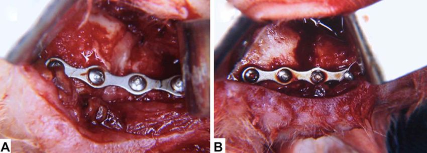
DOS SANTOS, P. A.; TRIVELATO, A. E.; OLATE, S.; ASPRINO, L. & DE MORAES, M. Stability as a key factor for bone repair in nonunion of mandibular osteotomy.
                                                              Int. J. Morphol., 38(2):309-315, 2020.

is tobacco (Chen et al., 2018). In an economic analysis, Lee                           Group 4. Osteotomy, immediate reduction and RIF;
et al. (2019) indicated that nonunion was more frequent in                             euthanasia at 42 days.
the mandibular body of elderly subjects, and that these ca-
ses involved higher treatment costs and use of hospital                                Group 5. Osteotomy and induction of pseudoarthrosis; at
resources than other types of mandibular fractures.                                    21 days a new surgical approach was created and the site
                                                                                       fixed with plates and screws and without any kind of
       Failure of segment stability (Madsen et al.) and the                            curettage; euthanasia at 63 days.
presence of large spaces between the stabilized fragments
(Olate et al. 2019) are associated with absence of bone repair                         Group 6. Osteotomy and induction of pseudoarthrosis; at
and with the presence of inflammatory infiltrate or                                    21 days a new surgical approach was created, with extensive
connective tissue, and such cases develop into nonunion.                               curettage of the pseudoarthrosis zone, followed by fixation
The aim of this research was to analyze bone repair in a                               with plates and screws; euthanasia at 63 days.
pseudoarthrosis model generated in rabbit mandibles,
determining the importance of stabilization of the fragments                           Histological analysis. After the experimental times of 21,
in bone repair.                                                                        42 and 63 days after the first surgery, the animals were
                                                                                       submitted to euthanasia. The surgical pieces were obtained
                                                                                       by total resection and fixed in a solution of formalin 4 %.
MATERIAL AND METHOD                                                                    They were decalcified and routine histological processing
                                                                                       was carried out; 6mm slices were cut, with hematoxylin-
                                                                                       eosin staining. Descriptive analysis was performed and
       Eighteen New Zealand rabbits aged between 3 and 6                               comparison between groups was realized.
months were selected and separated into 6 groups of 3 animals
each. The animals were kept with water and food ad libitum,
and their daily functioning was cared for.                                             RESULTS

        Fracture and pseudoarthrosis model. Mandibular frac-
ture was realized by surgery with submandibular approach                                      The experimental animals were kept without
following the protocols of Thomsen & Ericson (1987) and                                complications during the study period; no animal was lost.
Alister at al. (2017). The approach allowed a vertical osteotomy                       The following analyses were carried out for each group:
3 mm wide in the mandibular body, using a low-velocity drill.
                                                                                       Group 1. Connective tissue and abundant chondrocytes were
       The method for inducing pseudoarthrosis consisted in                            observed, with absence of granulation tissue and accompanied
interposing muscle tissue into the osteotomy space (platysma                           by osteogenic activity with immature bone tissue; tissue with
muscle) for 21 days after the osteotomy, following observations                        organized structure and incipient bone repair (Fig. 4).
described by Haug & Schwimmer (1994) (Fig. 1).
                                                                                       Group 2. Cartilaginous tissue was observed at the periphery
       For RIF was used a 1.5 straight plate and 4 screws                              of the defect, with osteogenic activity showing the presence
(Fig. 2). The subjects were assigned to 6 groups with                                  of islands of immature bone tissue and discrete formation of
methodologies for the osteotomy, pseudoarthrosis model and                             bone callus; in all areas the presence of blood vessels and
euthanasia as presented below (Fig. 3).                                                centres of ossification was observed (Fig. 5).

Group 1. Osteotomy, immediate reduction and RIF;                                       Group 3. Close to the osteosynthesis plate, remnants of
euthanasia at 21 days.                                                                 cartilaginous tissue were observed in the absence of
                                                                                       granulation tissue; advanced osteogenic activity was
Group 2. Osteotomy and induction of pseudoarthrosis; 21                                observed with presence of mainly mature bone tissue in the
days a new surgical approach was created and the site fixed                            analysis zone. The presence of well structured bone callus
with plates and screws and without any kind of curettage;                              was observed in the majority of the sample (Fig. 5).
euthanasia at 42 days.
                                                                                       Group 4. The presence of cartilage was observed, with
Group 3. Osteotomy and induction of pseudoarthrosis; at                                absence of connective tissue and absence of granulation
21 days a new surgical approach was created, with extensive                            tissue. Well organized bone tissue was observed with
curettage of the pseudoarthrosis zone, followed by fixation                            presence of bone callus, blood vessels and centres of
with plates and screws; euthanasia at 42 days.                                         ossification in the majority of the samples (Fig. 4).
310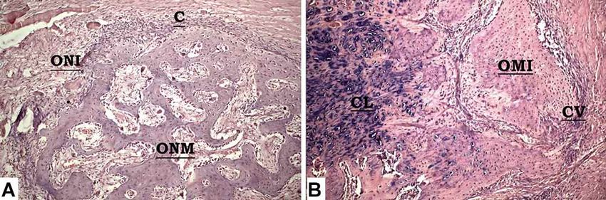
DOS SANTOS, P. A.; TRIVELATO, A. E.; OLATE, S.; ASPRINO, L. & DE MORAES, M. Stability as a key factor for bone repair in nonunion of mandibular osteotomy.
Int. J. Morphol., 38(2):309-315, 2020.
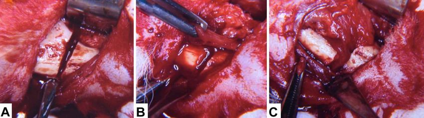
Fig. 1. Surgical model used in this investigation. A) Approach and osteotomy of the mandible; B) rotated flap of peripheral muscle ready
to be installed on the osteotomy site; C) muscle installed to model pseudoarthrosis.
Fig. 2. Rigid internal fixation installed in the animals. A) group in which all the pseudoarthrosis tissue between the two bone segments
was removed; B) group in which the tissue between the bone segments was left in place. In both groups, stabilization with plates and
screws ensured the immobility of the segments.
Fig. 3. Diagram of the experi-
mental model used in this
investigation. Analysis was
carried out in both groups in
accordance with this protocol.
311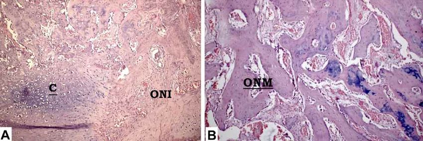
DOS SANTOS, P. A.; TRIVELATO, A. E.; OLATE, S.; ASPRINO, L. & DE MORAES, M. Stability as a key factor for bone repair in nonunion of mandibular osteotomy.
                                                              Int. J. Morphol., 38(2):309-315, 2020.

           Fig. 4. Histological images in H-E (250mm) obtained from group 1 (A) and group 4 (B), treated with RIF immediately
           after the osteotomy and euthanasia after 21 days (A) and 42 days (B). (C): presence of homogeneous cartilaginous
           tissue; ONI: presence of immature new bone tissue; ONM: presence of mature new bone tissue.
Group 5. The presence of cartilage was observed at the                                 Group 6. No presence of cartilaginous tissue was observed;
periphery of the osteotomy, with presence of fibrous tissue; in                        there was evidence of the presence of initial bone tissue,
one sample, presence of adipose tissue was observed; mature                            with the cortical in the active remodeling phase and empty
bone tissue was not observed in any of the samples, and tissue                         spaces typical of incipient repair stages. Blood vessels and
was maintained with some areas of connective tissue and areas                          centres of ossification were observed, indicating bone
of ossification, with presence of blood vessels indicative of                          formation (Fig. 6).
structure in the ossification process (Fig. 6).

           Fig. 5. Histological images in H-E (250 mm) obtained from group 2 (A) and group 5 (B), treated with RIF 21 days
           after provoking the pseudoarthrosis model and without removal of any type of tissue at the time of the second
           surgery to install the fixation. Euthanasia after 42 days (A) and 63 days (B). (C): presence of homogeneous
           cartilaginous tissue; ONI: presence of immature new bone tissue; ONM: presence of mature new bone tissue.

           Fig. 6. Histological images in H-E (250mm) obtained from group 3 (A) and group 6 (B), treated with RIF 21 days after
           provoking the pseudoarthrosis model and with removal of all the tissue between the bone fragments at the time of the second
           surgery to install the fixation. Euthanasia after 42 days (A) and 63 days (B). (C): presence of cartilaginous tissue in the lateral
           regions of the defect; ONI: presence of immature new bone tissue; ONM: presence of mature new bone tissue.

312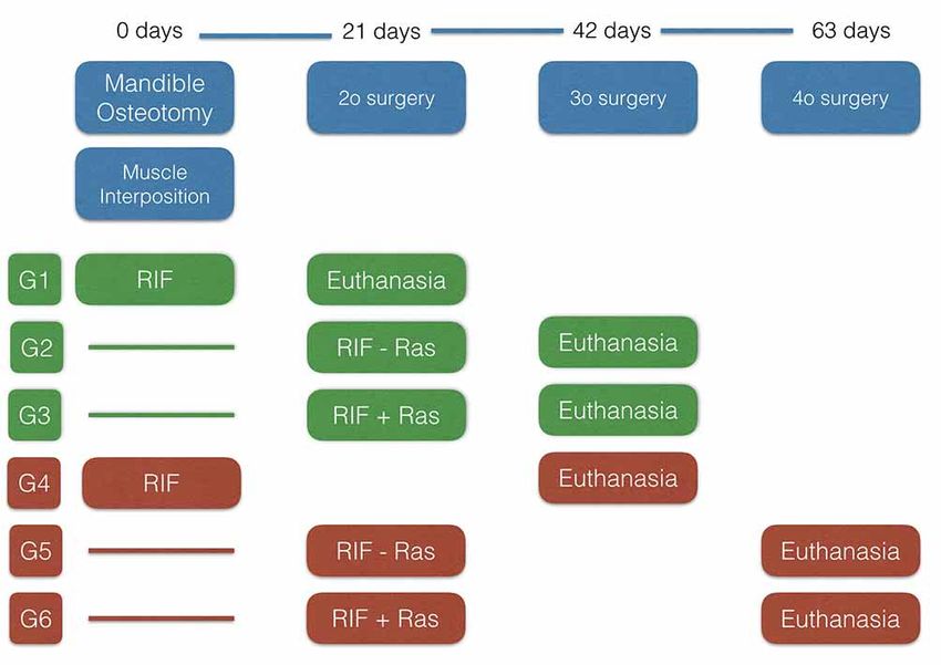
DOS SANTOS, P. A.; TRIVELATO, A. E.; OLATE, S.; ASPRINO, L. & DE MORAES, M. Stability as a key factor for bone repair in nonunion of mandibular osteotomy.
                                                              Int. J. Morphol., 38(2):309-315, 2020.

DISCUSSION                                                                             principally of fibrous tissue with abundant fibroblasts and
                                                                                       no osteoblast integration. Bone repair in defects of the fa-
                                                                                       cial skeleton is influenced by the distance between the bone
        This work presents the results of bone repair in cases                         segments, their stability (immobilization) and environmental
of pseudoarthrosis model in rabbits. The investigation was                             conditions (infection, quality of the periosteum, etc.) (Spanjer
carried out smoothly and no animal was lost during the study.                          et al., 2017).

        When a fracture is treated in conditions of optimum                                    Thus the repair zone between the bone segments is
stability using anatomical reduction and interfragmentary                              highly active, since the cells present in nonunion areas are
compression, bone repair occurs by primary or direct                                   viable and versatile development. In an in vitro study, Boyan
ossification, without formation of a periosteal callus (Del-                           et al. (1992) reported that cells present in this structure are
gado-Martínez & Alcántara-Martos, 2006). However, when                                 capable of responding to bone morphogenetic protein in the
the fracture or osteotomy is not completely stabilized and                             same way as mesenchymal cells. Iwakura et al. (2009)
space or mobility exists between the fragments, secondary                              showed that the hypertrophic tissue present in the nonunion
or indirect ossification occurs, which is the commonest form                           serves as a reservoir of cells with the potential to develop
of repair (Gerstenfeld et al., 2006). If the mobility exceeds                          into chondrocytes or osteoblasts. The conclusions of these
the tolerance to deformation of the tissue present in the callus,                      two works are related with our conclusions and suggest that
alterations in the consolidation will occur such as                                    the nonunion can develop into a bone repair site without the
pseudoarthrosis or hypertrophic nonunion (Green et al.,                                need for this tissue to be removed.
2005).
                                                                                               Wu & Chen (1997) compared open and closed
        The speed of consolidation is influenced by the type                           techniques for treating cases of nonunion. They reported that
of bone, the type of fracture, the method of treatment, and                            the closed technique produced faster bone repair, suggesting
the patient's age and general state (Panteli et al., 2015;                             that a new operation of the nonunion site might damage
Ricketts et al., 2016). Local factors such as separation of                            vascular support, making repair slower. Muller et al. (2007)
the bone ends may also have an impact; this may be related                             also reported that bone repair can occur when a new
with bone loss or resorption at the fracture site, interposition                       osteosynthesis system is installed without any need to treat
of soft tissues between the bone ends, excessive traction or                           (remove) the hypertrophic tissue at the nonunion site.
the use of internal fixation (Wraighte & Scammell, 2007;
Jahagirdar & Scammell, 2009).                                                                  The versatility of the tissue involved in bone repair
                                                                                       at osteotomy sites allows distraction osteogenesis to be
        In the facial skeleton, the repair of fractures and                            developed over a long provisional period. Likewise
osteotomies generally occurs by endochondral ossification,                             contraction osteogenesis was confirmed by the study of
based on the formation of bone callus from the periosteum                              Alkan et al. (2011), who reduced the mandible size in an
and the endosteum. The process includes an inflammatory                                animal model, achieving reduction and nonunion at the end
initial phase, cartilage formation, mineralisation and bone                            of the mechanical procedure; the histological images showed
remodeling (Marsell & Einhorn, 2011). Insufficient or                                  that 3 months after movement was stopped, normal bone
interrupted immobilization and excessive separation of the                             formation was obtained.
bone fragments could produce pseudoarthrosis (Macki et
al., 2016) and maintain a condition in which the bone                                          All these conditions indicate that the nonunion tissue
segments function as a joint and not a solid mineralized                               between bone fragments presents a great potential; for this
structure.                                                                             reason it is currently the focus of new treatment methods to
                                                                                       improve the predictability of bone union in cases of complex
        The results of the recent study published by Olate et                          pathologies or sequelae, and radiotherapy (Donneys et al.,
al. (2019) show the importance of the position and condition                           2015). The application of low intensity pulsed ultrasound
of the bone segments when the repair process of a fracture                             has also been investigated as a strategy in the fracture zone,
or osteotomy is in the initial stage. The distance between                             increasing the differentiation of cells derived from the bone
the bone ends established in the different groups defined the                          callus (Imai et al. 2014).
type of repair and the evolution of the repair process. When
the separation between the mandible segments did not exceed                                    These results show that the stability and proximity
5 mm, a clear endochondral repair was observed, while the                              of the bone fragments play great importance in proper bone
group with a separation of 8mm between the bone fragments                              repair. Factors like a long waiting time before stabilization
presented consolidation of the fracture; the cicatrize consisted                       of the fracture, use of tobacco and tooth extractions have
                                                                                                                                                                     313
DOS SANTOS, P. A.; TRIVELATO, A. E.; OLATE, S.; ASPRINO, L. & DE MORAES, M. Stability as a key factor for bone repair in nonunion of mandibular osteotomy.
Int. J. Morphol., 38(2):309-315, 2020.
been cited as risk factors for complications in fracture REFERENCES
healing, including nonunion (Hsieh et al., 2019). For these
reasons, the quality and quantity of the fixation material are
critical through their ability to support the immobilization Alister, J. P.; Veuthey, C.; Uribe, F.; Vásquez, B.; del Sol, M. & Olate, S.
Experimental model for the study of mandibular reconstruction. Options
of the segments and provide sufficient mechanical stability
in rabbits Oryctolagus cuniculus. Int. J. Morphol., 35(3):1185-90, 2017.
for the patient and the type of fracture beingtreated Alkan, A.; Kilic, E.; Ocak, H.; Ozturk, M. & Gunhan, O. Is it possible to
(Rodriguez Chessa et al., 2014; de Medeiros et al., 2016; shorten the jaws using contraction osteogenesis? J. Oral Maxillofac.
Meram et al., 2018). Surg., 69(6):e195-200, 2011.
Boyan, B. D.; Schwartz, Z.; Swain, L. D.; Khare, A. G.; Heckman, J. D.;
Ramirez, V.; Peters, P. & Carnes Jr., D. L. Initial effects of partially
purified bone morphogenetic protein on the expression of
CONCLUSION glycosaminoglycan, collagen, and alkaline phosphatase in nonunion
cell cultures. Clin. Orthop. Relat. Res., (278):286-304, 1992.
Chen, C. C.; Zenaga, J.; Patel, R. & Branham, G. Complications and
reoperations in mandibular angle fractures. JAMA Facial Plast. Surg.,
It is concluded that this model to induce 20(3):238-43, 2018.
pseudoarthrosis was appropriate; the use of RIF allowed bone de Medeiros, R. C.; Sigua, E. A.; Navarro, P.; Olate, S. & Albergaria Barbosa,
repair to occur even in groups in which pseudoarthrosis was J. R. In vitro mechanical analysis of different techniques of internal
fixation of combined mandibular angle and body fractures. J. Oral
provoked before the installation of RIF, and also in groups
Maxillofac. Surg., 74(4):778-85, 2016.
in which curettage of the zone was not carried out and the Delgado-Martínez, A. & Alcántara-Martos, T. Agentes sistémicos que
tissue between the fragments was left unaltered. Studies with modifican la consolidación de las fracturas. Rev. Esp. Cir. Ortop.
a larger number of animals are necessary to review different Traumatol., 50(Supl. 1):5-12, 2006.
Dodson, T. B.; Perrott, D. H.; Kaban, L. B. & Gordon, N. C. Fixation of
protocols in the animal model and improve treatment of
mandibular fractures: a comparative analysis of rigid internal fixation
complex patients. and standard fixation techniques. J. Oral Maxillofac. Surg., 48(4):362-
6, 1990.
Donneys, A.; Nelson, N. S.; Page, E. E.; Deshpande, S. S.; Felice, P. A.;
Tchanque-Fossuo, C. N.; Spiegel, J. P. & Buchman, S. R. Targeting
DOS SANTOS, P. A.; TRIVELATO, A. E.; OLATE, S.;
angiogenesis as a therapeutic means to reinforce osteocyte survival
ASPRINO, L.; DE MORAES, M. Estabilidad como fac- and prevent nonunions in the aftermath of radiotherapy. Head Neck,
tor clave en la reparación de la nounión de osteotomía 37(9):1261-7, 2015.
mandibular. Int. J. Morphol., 38(2):309-315, 2020 Gassner, R.; Tuli, T.; Hächl, O.; Rudisch, A. & Ulmer, H. Cranio-
maxillofacial trauma: A 10 year review of 9,543 cases with 21,067
injuries. J. Craniomaxillofac. Surg., 31(1):51-61, 2003.
RESUMEN: La estabilidad de las osteotomías y de Gerstenfeld, L. C.; Alkhiary, Y. M.; Krall, E. A.; Nicholls, F. H.; Stapleton,
las fracturas son fundamentales para asegurar la adecuada S. N.; Fitch, J. L.; Bauer, M.; Kayal, R.; Graves, D. T.; Jepsen, K. J.; et
reparación ósea; el objetivo de esta investigación fue analizar la al. Three-dimensional reconstruction of fracture callus morphogenesis.
reparación presente en sitios de pseudoartrosis realizando la lim- J. Histochem. Cytochem., 54(11):1215-28, 2006.
Green, E.; Lubahn, J. D. & Evans, J. Risk factors, treatment, and outcomes
pieza de la zona previo a la fijación o manteniendo el tejido de la
associated with nonunion of the midshaft humerus fracture. J. Surg.
nounión en el mismo lugar durante la osteosíntesis. Se diseñó un Orthop. Adv., 14(2):64-72, 2005.
estudio experimental incluyendo 18 conejos de raza Neozelandesa. Haug, R. H. & Schwimmer, A. Fibrous union of the mandible: a review of
Se formaron 6 grupos de estudios a quienes se relizó una osteotomía 27 patients. J. Oral Maxillofac. Surg., 52(8):832-9, 1994.
en el cuerpo mandibular y posterior instalación de músculo en el Hsieh, T. Y.; Funamura, J. L.; Dedhia, R.; Durbin-Johnson, B.; Dunbar, C.
lugar de la osteotomía para fabricar un modelo de pseudoartrosis. & Tollefson, T. T. Risk factors associated with complications after
En cirugía posterior se fijó con placa y tornillos. Se realizaron eu- treatment of mandible fractures. JAMA Facial Plast. Surg., 21(3):213-
tanasias a los 42 y 63 días para comparar los resultados de forma 20, 2019.
Imai, Y.; Hasegawa, T.; Takeda, D.; Akashi, M.; Lee, S. Y.; Niikura, T.;
descriptiva mediante estudio histológico. No fue perdido ningún
Shibuya, Y.; Kurosaka, M. & Komori, T. The osteogenic activity of
animal durante el experimento. En todas las muestras evaluadas se human mandibular fracture haematoma-derived cells is stimulated by
observó formación ósea en diferentes niveles de avance; defectos low-intensity pulsed ultrasound in vitro. Int. J. Oral Maxillofac. Surg.,
tratados con o sin el retiro del tejido involucrado en la pseudoartrosis 43(3):367-72, 2014.
presentaron una condición de reparación ósea comparables, deter- Iwakura, T.; Miwa, M.; Sakai, Y.; Niikura, T.; Lee, S. Y.; Oe, K.; Hasegawa,
minando que la estabilidad de los segmentos óseos permite la re- T.; Kuroda, R.; Fujioka, H.; Doita, M.; et al. Human hypertrophic
paración del tejido adyacente. El tejido cartilaginoso se presentó nonunion tissue contains mesenchymal progenitor cells with
en algunas muestras asociadas a sectores con mayor presencia de multilineage capacity in vitro. J. Orthop. Res., 27(2):208-15, 2009.
Jahagirdar, R. & Scammell, B. E. Principles of fracture healing and disorders
formación ósea. La estabilización de la fractura es clave en la re-
of bone union. Surgery (Oxford), 27(2):63-9, 2009.
paración ósea; la reparación se produce manteniendo o retirando Lamphier, J.; Ziccardi, V.; Ruvo, A. & Janel, M. Complications of
el tejido presente en la pseudoartrosis. mandibular fractures in an urban teaching center. J. Oral Maxillofac.
Surg., 61(7):745-9, 2003.
PALABRAS CLAVE: No unión; Reparación ósea; Lee, K. C.; Chuang, S. K. & Koch, A. The healthcare cost of mandibular
Fractura mandibular; Osteotomía mandibular. nonunions. J. Craniofac. Surg., 30(8):2539-41, 2019.
314DOS SANTOS, P. A.; TRIVELATO, A. E.; OLATE, S.; ASPRINO, L. & DE MORAES, M. Stability as a key factor for bone repair in nonunion of mandibular osteotomy.
Int. J. Morphol., 38(2):309-315, 2020.
Macki, M.; Syeda, S.; Kerezoudis, P.; Bydon, A.; Witham, T. F.; Sciubba, Corresponding author:
D. M.; Wolinsky, J. P.; Bydon, M. & Gokaslan, Z. rhBMP-2 protects Dr. Sergio Olate
against reoperation for pseudoarthrosis and/or instrumentation failure: Facultad de Odontología
a matched case-control study of 448 patients. J. Clin. Neurosci., 32:99-
Universidad de La Frontera
103, 2016.
Madsen, M. J.; Haug, R. H.; Christensen, B. S. & Aldridge, E. Management
Claro Solar 115, Oficina 414
of atrophic mandible fractures. Oral Maxillofac. Surg. Clin. North Am., Temuco
21(2):175-83, 2009. CHILE
Marsell, R. & Einhorn, T. A. The biology of fracture healing. Injury,
42(6):551-5, 2011.
Meram, A. T.; Olate, S. & Palmieri Jr., C. F. Is the three-dimensional strut Email: sergio.olate@ufrontera.cl
plate an adequate fixation technique for mandibular symphysis fractu-
res? J. Oral Maxillofac. Surg., 76(1):140-5, 2018.
Olate, S.; de Assis, A. F.; Pozzer, L.; Cavalieri-Pereira, L.; Asprino, L. &
de Moraes, M. Pattern and treatment of mandible body fracture. Int. J.
Received: 10-09-2019
Burns Trauma, 3(3):164-8, 2013. Accepted: 10-12-2019
Olate, S.; Vásquez, B.; Sandoval, C.; Vasconcellos, A.; Alister, J. P. & del
Sol, M. Histological analysis of bone repair in mandibular body
osteotomy using internal fixation system in three different gaps without
bone graft in an animal model. BioMed Res. Int., 2019:8043510, 2019.
Panteli, M.; Pountos, I.; Jones, E. & Giannoudis, P. V. Biological and
molecular profile of fracture non-union tissue: current insights. J. Cell.
Mol. Med., 19(4):685-713, 2015.
Ricketts, S.; Gill, H. S.; Fialkov, J. A.; Matic, D. B. & Antonyshym, O. M.
Facial fractures. Plast. Reconstr. Surg., 137(2):424e-444e, 2016.
Rodriguez-Chessa, J.; Olate, S.; Netto, H. D.; Noia, C.; de Moraes, M. &
Mazzonetto, R. In vitro resistance of titanium and resorbable (poly L-
co-DL lactic acid) oteosynthesis in mandibular body fracture. Int. J.
Oral Maxillofac. Surg., 43(3):362-6, 2014.
Seemann, R.; Schicho, K.; Wutzl, A.; Koinig, G.; Poeschl, W. P.; Krennmair,
G.; Ewers, R. & Klug, C. Complication rates in the operative treatment
of mandibular angle fractures: a 10-year retrospective. J. Oral
Maxillofac. Surg., 68(3):647-50, 2010.
Spanjer, E. C. K.; Bittermann, G. K. P.; van Hooijdonk, I. E. M.; Rosenberg,
A. J. W. P. & Gawlitta, D. Taking the endochondral route to
craniomaxillofacial bone regeneration: a logical approach? J.
Craniomaxillofac. Surg., 45(7):1099-106, 2017.
Thomsen, P. & Ericson, L. E. Titanium implants in rabbit knee joints. Acta
Orthop. Scand., 58(3):265-9, 1987.
Tucker, D. I.; Zachar, M. R.; Chan, R. K. & Hale, R. G. Characterization
and management of mandibular fractures: lessons learned from Iraq
and Afghanistan. Atlas Oral Maxillofac. Surg. Clin. North Am.,
21(1):61-8, 2013.
Wraighte, P. J. & Scammell, B. E. Principles of fracture healing. The
foundation years. Acute Care, 3(6):243-51, 2007.
Wu, C. C. & Chen, W. J. Treatment of femoral shaft aseptic nonunions:
comparison between closed and open bone-grafting techniques. J. Trau-
ma, 43(1):112-6, 1997.
315You can also read