Glenohumeral joint capsular tissue tension loading correlates moderately with shear wave elastography: a cadaveric investigation
←
→
Page content transcription
If your browser does not render page correctly, please read the page content below
Glenohumeral joint capsular tissue
tension loading correlates moderately
with shear wave elastography: a cadaveric
investigation
                                                                                                    ORIGINAL ARTICLE
Charles W. Nichols1, Jean-Michel Brismée2, Troy L. Hooper2,
                                                                                                    https://doi.org/10.14366/usg.19032
Antony Bertrand-Grenier3,4,5, Kerry K. Gilbert2, Marc-Olivier St-Pierre6, Jeegisha Kapila7,         pISSN: 2288-5919 • eISSN: 2288-5943
Stéphane Sobczak6,8                                                                                 Ultrasonography 2020;39:114-120

*Author affiliations appear at the end of this article.

                                                                                                    Received: June 5, 2019
Purpose: The purpose of this study was to investigate changes in the mechanical properties of       Revised: July 31, 2019
capsular tissue using shear wave elastography (SWE) and a durometer under various tensile           Accepted: August 1, 2019
loads, and to explore the reliability and correlation of SWE and durometer measurements to          Correspondence to:
                                                                                                    Charles W. Nichols, PT, DPT, MEd, ScD,
evaluate whether SWE technology could be used to assess tissue changes during capsule tensile       Department of Physical Therapy, School
loading.                                                                                            of Health Professions, University of
                                                                                                    North Texas Health Science Center,
Methods: The inferior glenohumeral joint capsule was harvested from 10 fresh human cadaveric        3500 Camp Bowie, MET 533, Ft.
specimens. Tensile loading was applied to the capsular tissue using 1-, 3-, 5-, and 8-kg weights.   Worth, TX, USA
Blinded investigators measured tissue stiffness and hardness during loading using SWE and           Tel. +1-817-735-2681
                                                                                                    Fax. +1-817-735-2518
a durometer, respectively. Intraobserver reliability was established for SWE and durometer          E-mail: charles.nichols@unthsc.edu
measurements using intraclass correlation coefficients (ICCs). The Pearson product-moment
correlation was used to assess the associations between SWE and durometer measurements.
Results: The ICC3,5 for durometer measurements was 0.90 (95% confidence interval [CI], 0.79 to
                                                                                                    This is an Open Access article distributed under the
0.96; P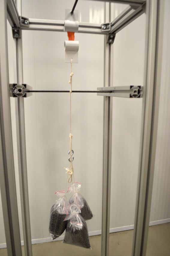
Shear wave elastography of glenohumeral capsule
Introduction one female, with a mean age of 77.8±5.4 years, height of 1.65±0.09
m, weight of 59.6±15.67 kg, and body mass index of 21.6±5.31
The glenohumeral joint (GHJ) is a frequent site of pain and kg/m2.
pathology with a reported prevalence from 7% to 23% in adults The inferior capsular attachment to the humerus and scapula was
[1,2]. GHJ osteoarthritis can affect up to 33% of people over 60 excised along with the GHJ capsule to allow for testing. The samples
years of age [3,4] and adhesive capsulitis affects 2%-5% of the were then trimmed to a 12-mm width for testing. The specimens
general population and 10%-15% of individuals with diabetes [5,6]. were then placed in containers and frozen in storage until needed
In adhesive capsulitis, the capsule becomes contracted and thicker, at -20°C. Tissue specimens were thawed for 1-2 hours and
particularly in the infraglenoid recess and rotator cuff interval [7- then tested at room temperature (18°C). Tissue specimens were
9], which limits joint mobility [10]. Inferior capsule thickening of connected to a standing frame using a custom clamp and a rope
more than 3.5 mm recorded using ultrasound is 66.7% sensitive with the attached load (Fig. 1) along with a random selection
and 92.5% specific for the diagnosis of shoulder adhesive capsulitis of 1-, 3-, 5-, and 8-kg loads for measurements using SWE and
[11]. Capsular tissue elasticity affects range of motion (ROM), or the the durometer. Investigators recording the durometer and SWE
allowed amount of joint plane-specific motion (e.g., GHJ flexion), measurements were blinded to the loads applied to the tissue
more than thickness, indicating a need for capsular tissue stiffness samples.
measurements [12]. Capsular tightness restricts joint ROM and
increases joint contact pressures, leading to intra-articular pathology SWE Measurements
[13] and limiting the ability to complete basic activities, such as The SWE procedures were performed at a regional health and
dressing and hygiene [3]. Clinicians commonly manage patients with social services center (Centre Intégré Universitaire de Santé et
ROM limitations, functional impairments, and pain using stretching de Services Sociaux de la Mauricie-et-du-Centre-du-Québec). An
and joint mobilization techniques [14-16]. Several authors [17-19] Aixplorer (SuperSonic Imagine, Aix-en-Provence, France) diagnostic
reported that GHJ mobilization using loads of 20-80 N increased device was used, with an XL-15-4 linear transducer that allowed the
capsule extensibility or elongation and improved patient symptoms investigator to quantify tissue elasticity without probe compression.
[20,21]. The proposed mechanical and neurophysiological changes
responsible for increased joint ROM following joint mobilizations
are not well understood and require further investigation [22]. Shear
wave elastography (SWE) measurements are a reproducible method
of evaluating muscle and tendon [23-26], with good repeatability,
as demonstrated by intraclass coefficients (ICCs) of 0.81-0.91. This
allows SWE to provide information on tissue diagnosis, injury, and/
or healing states [27,28]. However, no study has validated SWE
readings in capsular tissue against a reference standard. Therefore,
the purpose of this study was to investigate: (1) the reliability of
SWE measurements of GHJ capsular tissue under various loads;
and (2) the degree of correlation between measurements of GHJ
capsular tissue tension properties under various loads made using a
durometer versus SWE.
Materials and Methods
This study was approved by the Ethics Sub-committee of the
Department of Anatomy at the Université du Québec à Trois-Rivières,
Trois-Rivières, Québec and exempted by the University of North
Texas Regional Institutional Review Board. A convenience sample of
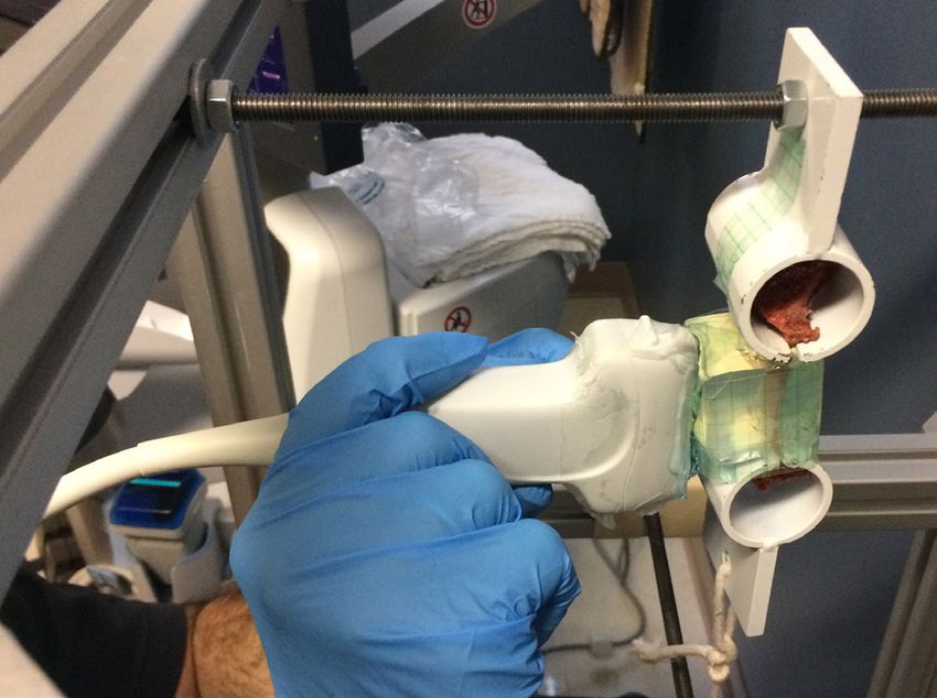
Fig. 1. Testing frame set-up. A standing frame with custom clamps
10 frozen GHJ specimens (5 right and 5 left) was acquired from five secured superiorly and inferiorly with a suspended 3-kg weight and
fresh cadavers through the Department of Anatomy at the Université stabilizing bar. The arrow indicates the location of the glenohumeral
du Québec à Trois-Rivières. The specimens included four males and joint capsular tissue prior to application of the gelatin pad.
e-ultrasonography.org Ultrasonography 39(2), April 2020 115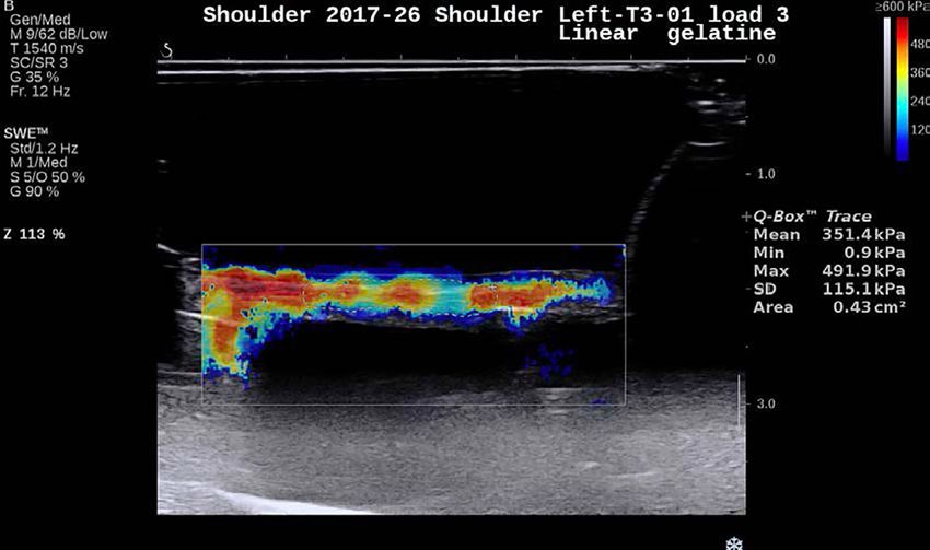
Charles Wayne Nichols, et al.

The investigator performing the SWE measurements was a medical             the Q-box tracer and used to acquire a measurement of elasticity
physicist with over 8 years of experience using SWE and 10 years of        (kPa) within the Q-box data area. Data were obtained for five
image processing.                                                          measurements from each tissue specimen using loads of 1, 3, 5, and
   Ultrasound gel was placed on the sample tissue followed by              8 kg for the shoulder in three trials to assess reliability (Fig. 3). The
application of a conforming gelatin pad, and the specimen was              reliability and correlation analyses were calculated using the mean
secured with Opsite Flexifix (Smith & Nephew, Andover, MA, USA)            measurements obtained for each load of each trial.
(Fig. 2). The ultrasound gel was applied to the Opsite covering and
then visualized with B-mode ultrasound. Once an acceptable image           Durometer
of the capsular tissue was achieved, the SWE was activated and             The Shore A durometer (HT-6510A Shore A, Tongbao, Shenzhen,
the image scanned for best resolution. Once the region of interest         China), a digital, hand-held, spring-loaded device (Fig. 4),
(ROI) was identified in the Q-box, which provides the field of view,       was calibrated, and the pressor was applied parallel to mid-
the probe was removed. The ROI was then marked and saved using             portion of tissue specimens and held in contact for 1-2 seconds
                                                                           until a measurement was obtained. This was repeated for five
                                                                           measurements using randomly selected 1-, 3-, 5-, and 8-kg loads
                                                                           in two trials separated by at least 5-minute intervals by a blinded
                                                                           investigator to assess reliability. Data were recorded for five
                                                                           measurements from the tissue specimens at each load in three trials
                                                                           to assess reliability (Fig. 4).
                                                                              Descriptive statistics including mean, median, standard deviation,
                                                                           ranges, and minimum/maximum values were calculated for the
                                                                           durometer and SWE measurements. The reliability of the durometer
                                                                           and SWE measurements was established using the intraclass
                                                                           correlation coefficient (ICC3,5). Data normality was assessed using
                                                                           the Shapiro-Wilk test, which indicated that the SWE and durometer
                                                                           measurements were normally distributed for the 1-, 3-, and 5-kg
                                                                           loads (SWE, P=0.572-0.960; durometer, P=0.441-0.730), and the
                                                                           kurtosis and skewness coefficients were lower than ±1. Therefore,
Fig. 2. Measurement set-up for tissue. The shear wave elastography         the associations between SWE and durometer measurements were
measurements of capsular tissue were made using a linear                   assessed with Pearson correlation coefficients. Statistical significance
transducer over tissue surrounded by gelatin and secured by Opsite         was set at P
Shear wave elastography of glenohumeral capsule

                                                                       Table 1. Mean durometer and SWE measurements under loads
                                                                       of 1, 3, and 5 kg
                                                                        Load (kg)        Durometer (HA)                    SWE (kPa)
                                                                            1                 9.4±1.8                      277.1±30.0
                                                                            3               15.6±2.5                       354.2±33.0
                                                                            5               18.7±2.5                       410.2±37.2
                                                                       Values are presented as mean±SD.
                                                                       SWE, shear wave elastography; HA, hardness unit of Shore A durometer; SD,
                                                                       standard deviation.

                                                                                                   Discussion
                                                                       This is the first study to assess the reliability of SWE and correlations
                                                                       between the durometer and SWE for GHJ capsular tissue properties
                                                                       under clinically applicable tensile loads using cadaveric tissue to
                                                                       eliminate neuromuscular influences. Tensile loads were selected
                                                                       based on pilot testing of the durometer for capsular tissue with good
                                                                       reliability (ICC3,5, 0.87; 95% CI, 0.68 to 0.95) and a high correlation
                                                                       between the tensile load applied and durometer measurements for
                                                                       loads of 1, 2, and 5 kg, as well as other prior studies of GHJ tissue
                                                                       properties [17,18,29]. The SWE measurements for reliability and
Fig. 4. Shore A durometer. The arrow indicates the device indenter.    correlation in this study were performed by one observer in 1 day.
                                                                       The mean of five successive measurements was used to determine
                                                                       reliability. This design provided the best conditions to determine
were performed using IBM SPSS statistics version 23 software (IBM      SWE reliability values. In this SWE study, the machine's Q-box
Corp., Armonk, NY, USA).                                               provided measurement selection through identification of the shear
                                                                       ROI. However, when the 5- and 8-kg loads were applied, saturation
                            Results                                    in some specimens due to the 800-kPa maximum SWE limit affected
                                                                       the mean tissue shear measurements, thereby creating a ceiling
The SWE and durometer measurement values are shown in Table 1.         effect, which could have affected measurement validity at higher
   The intra-day reliability of the durometer measurements was         loads. Our findings showed that GHJ capsular tissue measurements
0.90 (95% confidence interval [CI], 0.79 to 0.96; P
Charles Wayne Nichols, et al.

   A durometer measures a material’s hardness or resistance to           to clear other tissue from around the capsule, any remaining non-
deformation by applying an indentation load on the specimen,             capsular tissue could have contributed to some load resistance.
giving a measure of tissue hardness based on an arbitrary Shore          Studies evaluating whether such properties are maintained over
unit (HA) [30,31]. Durometers have been used in various medical          time and using other joint capsular tissue would be valuable. Further
applications, such as in dermatology [32,33] and for measurements        research is needed to determine the effects of tensile loading on
of organ [34-36], breast [37], and muscle [38] tissue. The intra-        capsular tissue and to provide insight into the effects of stretching
rater and inter-rater reliability measures of durometer measurements     and joint mobilization loads on joint ROM changes in vivo .
of epidermal tissue were good to excellent [32,33]. However,                In conclusion, SWE is a simple and reliable method of measuring
reliability has not been established in capsular tissue. Likewise,       the elastic properties of the GHJ capsule in cadaveric tissue.
GHJ capsule hardness measurements with a durometer had high              Additional research is required for the evaluation of capsular tissue
intra-rater reliability (ICC, 0.90) and could be used as a control       tension properties during and following various loads, without the
method to validate diagnostic methods such as SWE [34]. These            interference of the neuromuscular system, using SWE.
findings establish the durometer as a reliable and simple tool for
measuring capsular tissue hardness in vitro . Selected durometer         ORCID: Charles W. Nichols: https://orcid.org/0000-0001-6379-2445; Jean-Michel
measurements at higher loads were difficult to obtain due to tissue      Brismée: https://orcid.org/0000-0002-1037-7704; Troy L. Hooper: https://orcid.org/0000-
                                                                         0003-3436-2993; Antony Bertrand-Grenier: https://orcid.org/0000-0001-5965-9254;
sample thinness, which affected the consistency of the measurement
                                                                         Kerry K. Gilbert: https://orcid.org/0000-0003-4899-4617; Marc-Olivier St-Pierre:
location in some specimens. This may explain why the reliability         https://orcid.org/0000-0003-1638-2915; Jeegisha Kapila: https://orcid.org/0000-
coefficients were not as high as those reported by Kissin et al. [33];   0003-1430-1149; Stéphane Sobczak: https://orcid.org/0000-0002-9223-7982
however, our results were at the higher part of the range reported
by Merkel et al. [32] for epithelial tissue measurements.                Author Contributions
   A modest correlation was observed between SWE and durometer           Conceptualization: Nichols CW, Brismée JM, Hooper TL, Kapila J,
measurements of GHJ capsular tissue stiffness and hardness for           Gilbert KK. Data acquisition: Nichols CW, Brismée JM, Bertrand-
the 1-kg load and the 1- and 3-kg loads combined, and a small            Grenier A, St-Pierre MO, Sobczak S. Data analysis or interpretation:
correlation was found with the 3-kg tensile loads. This indicates that   Nichols CW, Brismée JM, Sobczak S. Drafting of the manuscript:
as the load increased, GHJ capsule stiffness as measured by SWE          Nichols CW, Brismée JM, Sobczak S. Critical revision of the
and hardness as measured by the durometer increased at lower             manuscript: Brismée JM, Hooper TL, Kapila J, Bertrand-Grenier
loads. Similar correlation values between B-mode ultrasonography         A, Gilbert KK, Sobczak S. Approval of the final version of the
and durometer measurements were reported in muscle tissue [38],          manuscript: all authors.
indicating that both modalities measure different parameters that
are closely related to the modulus of elasticity. Achilles tendon        *Author affiliations
tensile loads and SWE have been moderately correlated under              1
                                                                           Department of Physical Therapy, School of Health Professions,
progressive loads [39]. In the current study, the 5-kg tensile load      University of North Texas Health Science Center, Ft. Worth, TX;
resulted in a moderate negative correlation between the durometer        2
                                                                           Department of Rehabilitation Sciences, Center for Rehabilitation
and SWE measurements; these results should be viewed with                Research, Texas Tech University Health Sciences Center, Lubbock, TX,
caution due to tissue thinness, inconsistencies in the location of the   USA; 3Centre Intégré Universitaire de Santé et de Services Sociaux
durometer and SWE measurement locations, and SWE saturation              de la Mauricie-et-du-Centre-du-Québec (CIUSSS MCQ), Trois-
levels at the 5-kg tensile load.                                         Rivières, Québec; 4Centre Hospitalier Affilié Universitaire Régional,
   There are some advantages of using SWE to evaluate joint              Centre Intégré Universitaire de Santé et de Services Sociaux de la
capsule properties. First, it is a reliable measurement modality and     Mauricie-et-du-Centre-du-Québec (CIUSSS MCQ), Trois-Rivières,
can conveniently and quickly assess the elastic properties of a joint    Québec; 5Département de Chimie, Biochimie et Physique, Université
capsule. In the present study, the time required for scanning and        du Québec à Trois-Rivières, Trois-Rivières, Québec; 6Département
evaluating the capsule was only a few minutes. These advantages          d’Anatomie, Université du Québec à Trois-Rivières, Trois-Rivières,
make SWE a promising modality to diagnose capsular pathology             Québec, Canada; 7Texas Tech University Health Sciences Center,
and to evaluate treatment progression and the efficacy of different      Lubbock, TX, USA; 8Unité de Recherche en Anatomie Clinique et
interventions.                                                           Fonctionnelle (URACEF), Trois-Rivires, Québec, Canada
   There were some limitations to our study. Soft tissues around the
capsule were removed by hand and although great care was taken

118                                                   Ultrasonography 39(2), April 2020                                           e-ultrasonography.org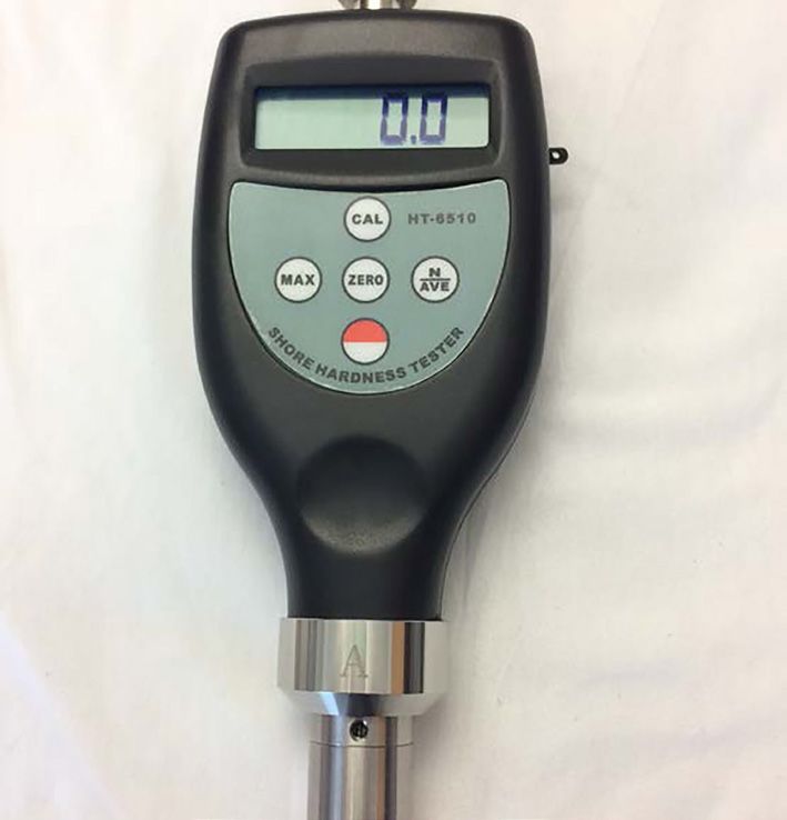
Shear wave elastography of glenohumeral capsule

Conflict of Interest                                                          12. Takenaga T, Sugimoto K, Goto H, Nozaki M, Fukuyoshi M, Tsuchiya
No potential conflict of interest relevant to this article was reported.          A, et al. Posterior shoulder capsules are thicker and stiffer in
                                                                                  the throwing shoulders of healthy college baseball players: a
Acknowledgments                                                                   quantitative assessment using shear-wave ultrasound elastography.
We would like to express our deepest gratitude to the people and                  Am J Sports Med 2015;43:2935-2942.
their families who donate their bodies for educational and research           13. Richards DP, Burkhart SS. Arthroscopic debridement and capsular
                                                                                  release for glenohumeral osteoarthritis. Arthroscopy 2007;23:1019-
purposes. Without their contribution, studies like this one would
                                                                                  1022.
not be possible. We would like to acknowledge Dany Lemay and
                                                                              14. Kelley MJ, Shaffer MA, Kuhn JE, Michener LA, Seitz AL, Uhl TL, et
Jonathan St-Arnaud from the department of mechanical engineering
                                                                                  al. Shoulder pain and mobility deficits: adhesive capsulitis. J Orthop
for their help in the experimental set-up.
                                                                                  Sports Phys Ther 2013;43:A1-A31.
                                                                              15. Lin HT, Hsu AT, An KN, Chang Chien JR, Kuan TS, Chang GL.
                            References                                            Reliability of stiffness measured in glenohumeral joint and its
                                                                                  application to assess the effect of end-range mobilization in
 1. Wofford JL, Mansfield RJ, Watkins RS. Patient characteristics and             subjects with adhesive capsulitis. Man Ther 2008;13:307-316.
    clinical management of patients with shoulder pain in U.S. primary        16. Bang MD, Deyle GD. Comparison of supervised exercise with
    care settings: secondary data analysis of the National Ambulatory             and without manual physical therapy for patients with shoulder
    Medical Care Survey. BMC Musculoskelet Disord 2005;6:4.                       impingement syndrome. J Orthop Sports Phys Ther 2000;30:126-
 2. Luime JJ, Koes BW, Hendriksen IJ, Burdorf A, Verhagen AP, Miedema             137.
    HS, et al. Prevalence and incidence of shoulder pain in the general       17. Hsu AT, Ho L, Chang JH, Chang GL, Hedman T. Characterization
    population; a systematic review. Scand J Rheumatol 2004;33:73-                of tissue resistance during a dorsally directed translational
    81.                                                                           mobilization of the glenohumeral joint. Arch Phys Med Rehabil
 3. Menge TJ, Boykin RE, Byram IR, Bushnell BD. A comprehensive                   2002;83:360-366.
    approach to glenohumeral arthritis. South Med J 2014;107:567-             18. Hsu AT, Ho L, Ho S, Hedman T. Immediate response of glenohumeral
    573.                                                                          abduction range of motion to a caudally directed translational
 4. Chillemi C, Franceschini V. Shoulder osteoarthritis. Arthritis                mobilization: a fresh cadaver simulation. Arch Phys Med Rehabil
    2013;2013:370231.                                                             2000;81:1511-1516.
 5. Paul A, Rajkumar JS, Peter S, Lambert L. Effectiveness of sustained       19. Cereatti A, Calderone M, Buckland DM, Buettner A, Della Croce U,
    stretching of the inferior capsule in the management of a frozen              Rosso C. In vivo glenohumeral translation under anterior loading in
    shoulder. Clin Orthop Relat Res 2014;472:2262-2268.                           an open-MRI set-up. J Biomech 2014;47:3771-3775.
 6. Aydeniz A, Gursoy S, Guney E. Which musculoskeletal complications         20. Johnson AJ, Godges JJ, Zimmerman GJ, Ounanian LL. The effect
    are most frequently seen in type 2 diabetes mellitus? J Int Med Res           of anterior versus posterior glide joint mobilization on external
    2008;36:505-511.                                                              rotation range of motion in patients with shoulder adhesive
 7. Suh CH, Yun SJ, Jin W, Lee SH, Park SY, Park JS, et al. Systematic            capsulitis. J Orthop Sports Phys Ther 2007;37:88-99.
    review and meta-analysis of magnetic resonance imaging features           21. Tyler TF, Nicholas SJ, Lee SJ, Mullaney M, McHugh MP. Correction
    for diagnosis of adhesive capsulitis of the shoulder. Eur Radiol              of posterior shoulder tightness is associated with symptom
    2019;29:566-577.                                                              resolution in patients with internal impingement. Am J Sports Med
 8. Lewis J. Frozen shoulder contracture syndrome: aetiology, diagnosis           2010;38:114-119.
    and management. Man Ther 2015;20:2-9.                                     22. Talbott NR, Witt DW. In vivo measurements of humeral movement
 9. Sridharan R, Engle MP, Garg N, Wei W, Amini B. Focal uptake at the            during posterior glenohumeral mobilizations. J Man Manip Ther
    rotator interval or inferior capsule of shoulder on (18)F-FDG PET/CT          2016;24:269-276.
    is associated with adhesive capsulitis. Skeletal Radiol 2017;46:533-      23. Payne C, Watt P, Cercignani M, Webborn N. Reproducibility of shear
    538.                                                                          wave elastography measuresof the Achilles tendon. Skeletal Radiol
10. Lee SY, Lee KJ, Kim W, Chung SG. Relationships between capsular               2018;47:779-784.
    stiffness and clinical features in adhesive capsulitis of the shoulder.   24. Peltz CD, Haladik JA, Divine G, Siegal D, van Holsbeeck M, Bey MJ.
    PM R 2015;7:1226-1234.                                                        ShearWave elastography: repeatability for measurement of tendon
11. Cheng X, Zhang Z, Xuanyan G, Li T, Li J, Yin L, et al. Adhesive               stiffness. Skeletal Radiol 2013;42:1151-1156.
    capsulitis of the shoulder: evaluation with US-arthrography using a       25. Rosskopf AB, Ehrmann C, Buck FM, Gerber C, Fluck M, Pfirrmann
    sonographic contrast agent. Sci Rep 2017;7:5551.                              CW. Quantitative shear-wave US elastography of the supraspinatus

e-ultrasonography.org                                     Ultrasonography 39(2), April 2020                                                       119
Charles Wayne Nichols, et al.
muscle: reliability of the method and relation to tendon integrity Rheum 2008;59:699-705.
and muscle quality. Radiology 2016;278:465-474. 33. Kissin EY, Schiller AM, Gelbard RB, Anderson JJ, Falanga V, Simms
26. Tas S, Onur MR, Yilmaz S, Soylu AR, Korkusuz F. Shear wave RW, et al. Durometry for the assessment of skin disease in systemic
elastography is a reliable and repeatable method for measuring the sclerosis. Arthritis Rheum 2006;55:603-609.
elastic modulus of the rectus femoris muscle and patellar tendon. J 34. Belyaev O, Herden H, Meier JJ, Muller CA, Seelig MH, Herzog T, et
Ultrasound Med 2017;36:565-570. al. Assessment of pancreatic hardness: surgeon versus durometer. J
27. Youk JH, Son EJ, Park AY, Kim JA. Shear-wave elastography for Surg Res 2010;158:53-60.
breast masses: local shear wave speed (m/sec) versus Young 35. Hong TH, Choi JI, Park MY, Rha SE, Lee YJ, You YK, et al. Pancreatic
modulus (kPa). Ultrasonography 2014;33:34-39. hardness: correlation of surgeon's palpation, durometer
28. Ryu J, Jeong WK. Current status of musculoskeletal application of measurement and preoperative magnetic resonance imaging
shear wave elastography. Ultrasonography 2017;36:185-197. features. World J Gastroenterol 2017;23:2044-2051.
29. Muraki T, Yamamoto N, Berglund LJ, Sperling JW, Steinmann SP, 36. Yoon YC, Lee JS, Park SU, Kwon JH, Hong TH, Kim DG. Quantitative
Cofield RH, et al. The effect of cyclic loading simulating oscillatory assessment of liver fibrosis using shore durometer. Ann Surg Treat
joint mobilization on the posterior capsule of the glenohumeral Res 2017;93:300-304.
joint: a cadaveric study. J Orthop Sports Phys Ther 2011;41:311- 37. Mori H, Uemura N, Koga H, Okazaki M. Objective assessment of
318. reconstructed breast hardness using a durometer. Breast Cancer
30. Falanga V, Bucalo B. Use of a durometer to assess skin hardness. J 2018;25:81-85.
Am Acad of Dermatol 1993;29:47-51. 38. Kudo S, Nakamura S. Relationship between hardness and
31. Piaggesi A, Romanelli M, Schipani E, Campi F, Magliaro A, Baccetti deformation of the vastus lateralis muscle during knee flexion using
F, et al. Hardness of plantar skin in diabetic neuropathic feet. J ultrasound imaging. J Bodyw Mov Ther 2017;21:549-553.
Diabetes Complications 1999;13:129-134. 39. Sahr M, Sturnick DR, Nwawka OK. Quantitative ultrasound
32. Merkel PA, Silliman NP, Denton CP, Furst DE, Khanna D, Emery P, et assessment of the achilles tendon under varied loads. J Ultrasound
al. Validity, reliability, and feasibility of durometer measurements of Med 2018;37:2413-2418.
scleroderma skin disease in a multicenter treatment trial. Arthritis
120 Ultrasonography 39(2), April 2020 e-ultrasonography.orgYou can also read