Short Term Assessment of Surgical Treatment of Lisfranc Fracture Dislocation
←
→
Page content transcription
If your browser does not render page correctly, please read the page content below
The Egyptian Journal of Hospital Medicine (October 2021) Vol. 85 (1), Page 2945-2950

      Short Term Assessment of Surgical Treatment of Lisfranc Fracture Dislocation
                            Sameh Mohamed Holyl, Elsayed Abdelmoty Mohammed,
                           Alaeddin Mohamed Hadida*, Ahmed Hatem Farhan Imam
                      Department of Orthopedic, Faculty of Medicine, Zagazig University, Egypt
    *Corresponding Author: Alaeddin Mohamed Hadida, Mobile: (+20)1012109449, E-mail: alaeddinhadida@gmail.com

ABSTRACT
Background: Lisfranc fractures are rare injuries, with reported incidence of 0.2% of all fractures and 1/55,000 per year
incidence in the population. These are reported to occur 2–4 times more commonly in men, and the peak incidence is in
the third decade of life.
Objective: This study was aimed to assess the functional outcome of using open reduction and internal fixation in
management of ligamentous Lisfranc injuries.
Patients and Methods: This prospective operative study was conducted on 18 patients with Displaced Lisfranc injury
admitted to the Orthopedic Surgery Department, Faculty of Medicine, Zagazig University, during the period from
February 2021 to August 2021. The diagnosis was made by medical history taking, clinical examination and radiological
assessment.
Results: The mean operation time was 140.0±27.43 minutes with minimum 90 and maximum 200 minutes and mean
hospital stay was 4.38±1.33 with minimum 3 and maximum 8 days. The mean healing time 8.38±2.45 with minimum 6
and maximum 16 weeks. The most prevalent complication was infection (22.2%), Transient numbness (5.6%), Delay
healing (5.6%), Loss reduction (5.6%) and overall complicated cases were 5 cases 27.8%.
Conclusion: It could be concluded that anatomical reduction of Lisfranc injury can be achieved by open reduction and
internal fixation with the Kirschner wires (K-wires) and Cannulated Screws.
Keywords: Lisfranc, Midfoot, Open reduction, Internal fixation.

INTRODUCTION                                                      metatarsal bases. The notch sign, in which a small notch
     Lisfranc injury refers strictly to an injury in which        appears in the lateral aspect of the medial cuneiform,
one or more of the metatarsals are displaced with respect         might also be seen (7).
to the tarsus (1). Lisfranc injuries are infrequent, at                Conservative       treatment   includes    midfoot
approximately 0.2% of all fractures, although in 20% of           stabilization and movement restriction. For Lisfranc
cases they are not diagnosed or are diagnosed late (2).           injuries without displacement on weight bearing
     However, early, and accurate diagnosis of these              radiographs, the use of cast immobilization for 6 to 12
injuries are fundamental requirements for their                   weeks is common. Modern surgical treatments include
appropriate treatment and to prevent long-term                    closed reduction and immobilization, closed reduction
sequelae. Men are two to four times more likely to suffer         and percutaneous pinning, and open reduction with
a Lisfranc joint injury, possibly because they participate        percutaneous pinning or screw fixation (8).
more frequently in high-speed activities. These injuries               This study was performed to assess the functional
are common in the third decade of life. The majority              outcome of using open reduction and internal fixation in
(87.5%) are closed injuries and are becoming more                 management of ligamentous Lisfranc injuries.
frequent in athletes, in whom it is common to see subtle
Lisfranc injuries (3). These are injuries that have               PATIENTS AND METHODS
occurred in sports such as soccer, gymnastics and                     This prospective operative study included a total of
running (4).                                                      18 patients with displaced Lisfranc injury admitted to
     Lisfranc injuries, which disrupt the strong midfoot          the Department of Orthopedic, Faculty of Medicine,
ligaments supporting the arch, require immediate                  Zagazig University, during the period from February
surgical intervention to prevent complications such as            2021 to August 202. Patients were 4 males and 14
compartment syndrome and amputation. On clinical                  females, aged 39-70 years and the mean age was
examination, patients can present with edema, point               56.77±8.17 years.
tenderness, and decreased function (5). The dorsal
drawer test of the medial column will elicit a “clunk”            Ethical Consideration:
compared with the contralateral side, and the passive                  Written informed consent was obtained from
midfoot pronation abduction test will yield positive              all participants and the study was approved by the
results (6).                                                      Research Ethical Committee, Faculty of Medicine,
     On radiographic evaluation, Lisfranc injuries                Zagazig University (ZU-IRB). The work has been
commonly show an increased asymmetric joint space at              carried out in accordance with The Code of Ethics of
the naviculocuneiform joint and first and second

                      This article is an open access article distributed under the terms and conditions of the Creative
                      Commons Attribution (CC BY-SA) license (http://creativecommons.org/licenses/by/4.0/)
                                                             2945
   Received:17 /5 /2021
   Accepted:13 /7 /2021
https://ejhm.journals.ekb.eg/
the World Medical Association (Declaration of the sensory branches of the superficial peroneal nerve,
Helsinki) for studies involving humans. the deep peroneal nerve, and the interspace between the
bases of the first and second metatarsal and the dorsalis
Inclusion criteria: Age; 18-70 years old. Gender; both pedis artery Intraoperative fluoroscopy in multiple
male and female. Displaced Lisfranc injury when projections and application of stress in multiple
displacement is greater than 2 mm, between the first and directions at various joints in the midfoot help unmask
second metatarsals on weight bearing anteroposterior any instability not otherwise evident. After
foot radiograph. subperiosteal exposure of the involved joints, reduction
Exclusion criteria: Patient underwent a subsequent and stabilization is performed in a sequential manner.
surgery that confounded meaningful postoperative Reduction of joints sometimes requires
outcome analysis. Patients< 18 or >70 years. Infected debridement to remove any interposed fragments of
fractures. Medical co-morbidities such as diabetes, liver bone, cartilage, or soft tissues such as
disease, or chronic renal disease. capsuloligamentous structures or tendons. A large bone
clamp is applied with one limb of the clamp inserted
Pre-operative: over the medial aspect of the medial cuneiform through
All patients underwent full history taking, local and a small incision and blunt dissection down to the bone,
general clinical examination, Radiographic images and with the other limb of the clamp inserted through an
(weight bearing anteroposterior, oblique, and lateral x- existing wound or through a small incision and blunt
rays). Laboratory investigations included: Complete dissection over the lateral aspect of the base of the
blood count (CBC), PT, PTT, random blood sugar, second metatarsal. This clamp holds the reduction
Liver and Kidney function tests, HIV, HBsAg and HCV between the medial cuneiform and the base of the
Abs. Surgical procedure was recorded, including type of second metatarsal. Through the previous exposure, both
surgery, duration of operation and complications. the metatarsocuneiform and naviculocuneiform
Operative treatment was performed by open reduction articulations of the first ray must be stabilized if injured.
and internal fixation under regional block. The medial column is stabilized first and then the
middle column and lastly the lateral column.
Surgical technique: The first TMT joint is debrided and reduced, then
The operation was performed with the patient lying in a provisionally stabilized using a guidewire placed
supine position, the entire leg is prepared from the level dorsally 1.5 cm distal to the articulation and directed
of the knee down and draped to isolate it in a sterile plantarly and proximally.A guide wire for the
fashion. A single dose of an antibiotic was administered cannulated drill is inserted in a dorsolateral to plantar
intravenously for prophylaxis against infection, a sterile medial direction from the base of the second metatarsal
tourniquet was applied at the level of the ankle and into the medial cuneiform under fluoroscopic guidance,
inflated after exsanguination, a triangular support was a third pin was placed from medial to lateral between
used under the knee to keep the foot positioned the medial and middle cuneiforms if required.
plantigrade on the operating table, plantigrade If adequate reduction is seen, 4- or 4.5-mm cannulated
positioning of the foot enables the surgeon to achieve a screws are inserted over these pins starting with the
better orientation for intraoperative imaging and to "Lisfranc screw" , which is the screw between the
direct drill holes and insert implants, such as screws. medial cuneiform and the second metatarsal base.
This technique also helps the surgeon to work on the Screws placed into the metatarsal bases should be
foot, with minimal manual assistance required to hold countersunk to avoid fracture into the adjacent joints,
the foot. The pattern of injury dictates the exposure lag screws should not be excessively tightened to avoid
required and the placement and number of incisions. unnecessary compression of the joint surfaces. Screw
fixation has been shown to have lower rates of
Two or three longitudinal incisions over the midfoot. redisplacement and faster return to weight bearing
The first is over the medial border of the foot postoperatively compared with Kirschner wire (K-wire)
centered at the base of the first ray, the second is fixation, small fragment (3.5 mm) cortical screw
between the first and second metatarsal bases, and the fixation and 4- or 4.5-mm cannulated screws are
third over the fourth metatarsal base. recommended for the first, second, and third TMT
The skin bridges between these incisions are joints. The screws are removed at 4 months but
usually narrow, and the incisions must be kept short to occasionally can be left longer. Placing ‘‘Lisfranc’s
avoid vascular compromise. This can result in poor screw’’ from the second metatarsal into the medial
visualization of the joints and excessive retraction cuneiform provides stability to the tarsometatarsal joint
leading to neuromas and skin necrosis. Accurate complex equal to that achieved with the traditional
placement of the incision may be aided by the use of orientation of screw insertion in opposite direction.
fluoroscopy, the medial incision on the skin is placed Cannulated screws can also be used for fixation of
just lateral to the extensor hallucis longus tendon, the tarsometatarsal or intercuneiform joints of the medial
author avoids going through the tendon sheath. Further and middle columns. Guidewires for the cannulated
dissection should proceed with extreme care to protect screws are driven from the base of the first metatarsal to
2946https://ejhm.journals.ekb.eg/
the first cuneiform and similarly in the second ray, if such as the letter “H” are also available to span 2 or
necessary, and also between the cuneiforms. Fully more joints in the medial and middle columns (Fig. 1).
threaded screws avoid compression across the joints. They can be used to span joints more proximally,
such as the naviculocuneiform joint if instability
Staples/Locking Plates extends proximally. The stability of the lateral column
Extra-articular stabilization can be achieved by is assessed next. If necessary, these joints are reduced
implants that do not need to traverse for spanning and held with K wires.
adjacent bones, a compression staple or mini two-hole Dorsal plating can be considered for bridging
plate or staple with locking screws, such as the Claw fixation of comminuted fractures with bony fragments
Plate™, can then be used across the intercuneiform or in the TMT joints and total ligamentous injuries and as
tarsometatarsal joints of the medial or middle column. an alternative surgical treatment in certain cases, such
The Claw Plate™ is essentially a low-profile staple that as joint damage and loss of fixation. The concern for
has holes at either end instead of limbs, these holes take joint damage resulting from screw fixation across the
screws and lock their heads. The body of the staple has TMT joints is eliminated, plating provides prolonged
a diamond-shaped gap, which when stretched brings the fixation, wound problems are not more common with
screws together, the staples or screws should be inserted plating than with wire fixation. The surgeon can elect to
under fluoroscopic guidance, K wires are used to leave the plate in place, hand plating sets are often
stabilize the lateral 3 columns if necessary. Longer useful for this technique, and dorsal plate fixation has
locking plates available in different lengths or shapes, been shown to be as biomechanically sound as screw
fixation.
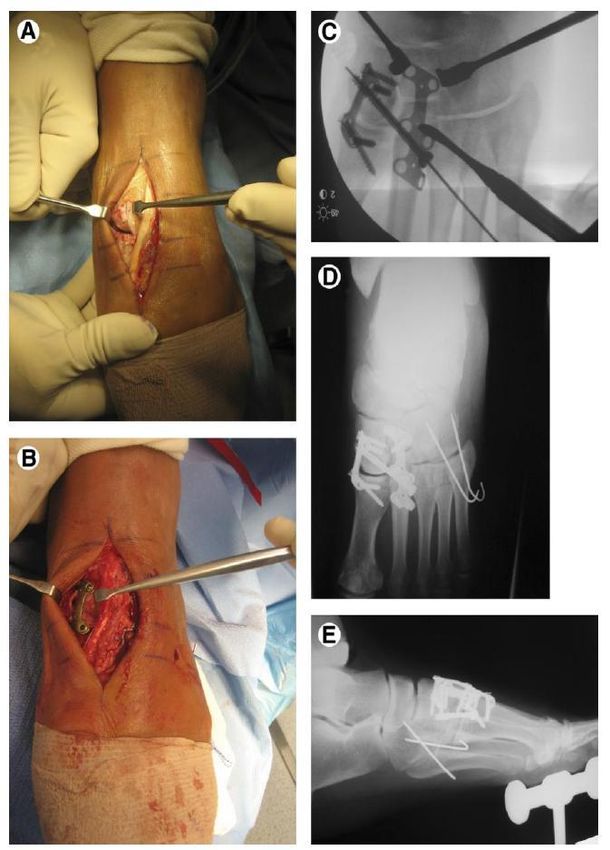
Fig. (1): Intraoperative images. Photographs showing (A) instability of the first ray at metatarso-cuneiform
joint(B) stabilization with a locking plate. (C) showing further stabilization with a cannulated screw and another
locking plate over the second ray. (D and E) after extra-articular stabilization of Lisfranc injury.
2947https://ejhm.journals.ekb.eg/
Post-operative follow up:
Following surgical treatment patients were splinted Table (2) shows that 33.3% had joint injury, and
for two weeks, nonweight-bearing in a bivalved cast regard Myerson classification the majority were class
and encouraged to remove the cast daily for ankle and IIA with 55.6% then grade I with 27.8% and IIB 11.1%
toe range of motion exercises. At six weeks post- and only one case III with 5.6%.
surgery, a standing x-ray was reviewed to check
maintained alignment. begin weight-bearing as Table (3): Operation time and hospital stay
tolerated. Twelve weeks postoperatively, a second x-ray distribution among studied group (N=18)
further was done to assess alignment and healing. Operation Hospital
patients were placed in a boot and weaned out as time/minutes stay DAY
tolerated. Formal physical therapy at this point was Mean± SD 140.0±27.43 4.38±1.33
started. On each follow-up, patients were subjected to Median 140.0 4.0
clinical and radiological assessment. Table (3) shows that the mean operation time was
140.0±27.43 minutes with minimum 90 and maximum
Statistical analysis 200 minutes and mean hospital stay was 4.38±1.33 with
Data collected throughout history, basic clinical minimum 3 and maximum 8 days.
examination, laboratory investigations and outcome
measures coded, entered and analyzed using Microsoft Table (4) : Healing time distribution among studied
Excel software. group (N=18)
Data were then imported into Statistical Package Healing time /weeks
for the Social Sciences (SPSS version 20.0) (Statistical
Mean± SD 8.38±2.45
Package for the Social Sciences) software for analysis.
According to the type of data qualitative represent as Median 8.0
number and percentage, quantitative continues group Table (4) shows that the mean healing time 8.38±2.45
represent by mean ± SD , the following tests were used with minimum 6 and maximum 16 weeks
to test differences for significance;. difference and
association of qualitative variable by Chi square test Table (5): Complication distribution among studied
(X2) . Differences between quantitative independent group (N=18)
groups by t test. P value was set athttps://ejhm.journals.ekb.eg/
73.55±6.55, after 6 months was 84.77±3.87 and after 12 compromised wound healing in one patient (3.7%) and
months was 92.61±3.95 with a high significant Transient numbness in one patient (3.7%).
difference Cochran et al. (16) reported that of the 18 patients
in the ORIF group, complications included 4 with
Table (7): satisfaction distribution among studied permanent deep peroneal nerve sensory changes and 2
group at different times (N=18) superficial infections that occurred after implant
N % removal, both were successfully treated with oral
Satisfaction Not 3 16.7 antibiotics.
Satisfied 15 83.3 The current study showed that the mean AOFAS
Total 18 100.0 score significantly increase with time of follow up
Table (7) shows that 15 patients (83.3%) were satisfied where it was 46.55±4.74 postoperative, after 3 months
and only 3 patients (16.7%) were not satisfied was 73.55±6.55, after 6 months was 84.77±3.87 and
after 12 months was 92.61±3.95 with a high significant
DISCUSSION difference. Which in agreement with the study of Fan
The current study included 18 patients with mean et al. (14), who reported that AOFAS score increased
age of 56.77±8.17 (rang 39-70), they were 14 female from 58.69 to 82.31 after follow with a highly
(77.8%) and 4 males (22.2%). The result was nearly significant differences. Also, Ren et al. (9) reported that
agreed with the study of Ren et al.(9) who reported that the median AOFAS score in the surgical treatment
he study population consisted of 38 (62.3%) male and group was 89.9± 3.7 (range 85-97) after 6 months
23 (37.7%) female patients, with a mean age of 39.4 follow up.
(range 19-64) years. While Li et al. (10) reported that Qu et al. (17), reported that the AOFAS midfoot
among 10 cases of Lisfranc injuries, there were 6 (60%) scoring system was applied for functional evaluation at
males and 4 (40%) females with mean age 32 years 6 and 12 months after surgery. The average scores at 6
ranging 25-45 years. and 12 months were 69.2 (55–86) and 88.2 (68–95) with
The current study showed that 33.3% had joint a high significant difference (P < 0.001).
injury, and regard Myerson classification the majority The current study showed that 15 patients (83.3%)
were class IIA with 55.6% then grade I with 27.8% and were satisfied and only 3 patients (16.7%) were not
IIB 11.1% and only one case III with 5.6%. satisfied. Which in agreement with the study of Ahmad
Wang et al. (11) found that of 15 patients with et al.(18), who studied the outcome of early open
Lisfranc injuries, according to Myerson classification reduction and internal fixation of 20 cases of Lisfranc
there was one patients (6.6%) with type A, 10 patients injuries using AOFAS-M score found that Good to fair
(66.6%) type B2, 3 patients (20%) type C1 and one results were seen in 90% cases (n=18).
patients (6.6%) with type C2. Pereira et al. (19) reported that among 19 patients
Kumaran et al. (12), reported that among 15 with Lisfranc injuries, 3 patients (15.8%) were
patients with Lisfranc injuries, fractures were classified Excellent, 6 patients (31.6%) were good, 3 patients
as Type A(n=2, 13.3%), type B (n=10, 66.7%), and (15.8%) were fair and 7 patients (36.8%) were poor
TYPE C(n=3, 20%)
The current study showed that the mean operation CONCLUSION
time was 140.0±27.43 minutes with minimum 90 and It could be concluded that anatomical reduction of
maximum 200 minutes and mean hospital stay was Lisfranc injury can be achieved by open reduction and
4.38±1.33 with minimum 3 and maximum 8 days. Liu internal fixation with the Kirschner wires (K-wires) and
et al. (13) found that the average time duration for the Cannulated Screws. Normal structure of Lisfranc joint
first-stage operation was 138.9 minutes while the mean is regained to a great extent; injured ligaments were also
hospital stay was 13.34 days repaired. Therefore, this method offers excellent
The current study showed that the mean healing curative effect and can avoid postoperative
time 8.38±2.45 with minimum 6 and maximum 16 complications and improve the patients' quality of life.
weeks. Fan et al. (14), reported that the mean fracture
healing time was 9.8 weeks (range: 8–13 weeks). RECOMMENDATIONS
The current study showed that the most prevalent Further study should be carried out with larger
complication was infection (22.2%), Transient groups of patients and with longer duration follow up
numbness (5.6%), Delay healing (5.6%), loss reduction are required to long term results and to validate the
(5.6%) and overall complicated cases were 5 cases results of this study.
27.8% which nearly similar to the study of Kohli et al.
(15)
who reported that of 27 patients with Lisfranc Financial support and sponsorship: Nil.
injuries complication were recorded in 6 patients
(22.2%), superficial wound infection in 2 patients Conflict of Interest: Nil.
(7.4%), loss of reduction (early postoperator) in one
patient (5.9%), delayed discharge in one patient (3.7%), REFERENCES
2949https://ejhm.journals.ekb.eg/
1. Bucholz R, Heckman J (2002): Rockwood and Green comminution of the second metatarsal base. Acta
Fractures in Adults. Philadelphia, PA: Lippincott Orthopædica Belgica, 83, 396-404.
Williams & Wilkins. Pp. 2182-245. 12. Kumaran J, Neelakrishnan R, Bharathiselvan V et al.
https://www.ncbi.nlm.nih.gov/nlmcatalog/101258565 (2018): A study of functional outcome of Lisfranc
2. Arntz C, Veith R, Hansen S (2002): Fractures and fracture dislocations managed by various operative
fracture-dislocations of the tarsometatarsal joints. J. methods in rural south Indian population. National
Bone Joint Surg., 70:173-181. Journal of Clinical Orthopaedics, 2(4): 85-89.
3. Palma L, Santucci A, Sabetta S et al. (1997): Anatomy 13. Liu X, An J, Chen Y et al. (2020): Staged surgical
of the Lisfranc joint complex. Foot Ankle Int., 18:356- treatment of open Lisfranc fracture dislocations using an
364. adjustable bilateral external fixator: A retrospective
4. Aitken A (1968): Dislocation of Tarsometatarsal Joint. review of 21 patients. Acta Orthopaedica et
JBJS., 20: 246-260. Traumatologica Turcica, 54(5): 488-493.
5. Aitken A, Preidler K, Wang Y et al. (2002): The 14. Fan M, Li X, Jiang X et al. (2019): The surgical
anatomy of the joint as a risk factor for Lisfranc's outcome of Lisfranc injuries accompanied by multiple
dislocation and fracture dislocation: J Bone Joint Surg., metatarsal fractures: A multicenter retrospective study.
84:981–985. Injury, 50(2): 571-578.
6. Quzonian T, Sheriffs M (1990): In vitro determination 15. Kohli S, Srikantharajah D, Bajaj S (2021): clinical and
of midfoot motion. Foot Ankle, 10:140–146. radiological outcomes after open reduction and internal
7. Gaines RJ, Wright G, Stewart J (2009): Injury to the fixation of Lisfranc injuries: a single-centre experience.
Tarsometatarsal Joint Complex During Fixation of In Orthopaedic Proceedings. The British Editorial
Lisfranc's Fracture-Dislocations: An Anatomic Study. J Society of Bone & Joint Surgery, 4: 103-106.
Trauma, 66(4):1125-8. 16. Cochran G, Renninger C, Tompane T et al. (2017):
8. Sarrafian S (1993): Anatomy of Foot and Ankle: Primary arthrodesis versus open reduction and internal
Philadelphia J.B. Lippincott, Pp. 779. fixation for low-energy Lisfranc injuries in a young
https://www.scribd.com/document/399391041/Sarrafian athletic population. Foot & Ankle International, 38(9):
-s-Anatomy-Foot-Ankle-3rd 957-963.
9. Ren W, Li H, Lu J et al. (2019): Undisplaced subtle 17. Qu W, Ni S, Wang Z et al. (2016): Severe open Lisfranc
ligamentous Lisfranc injuries, conservative or surgical injuries: one-stage operation through internal fixation
treatment with percutaneous position screws?. Chinese associated with vacuum sealing drainage. Journal of
Journal of Traumatology, 22(4): 196-201. Orthopaedic Surgery and Research, 11(1):1-7.
10. Li B, Zhao W, Liu L et al. (2015): Efficacy of open 18. Ahmad L, Reyaz A, Mubashir M et al. (2014):
reduction and internal fixation with a miniplate and Outcome after early open reduction and Kirschner wire
hollow screw in the treatment of Lisfranc injury. Chinese fixation of Lisfranc joint injuries. The Foot and Ankle
Journal of Traumatology, 18(1), 18-20. Online Journal, 7 (1): 1-5.
11. Wang L, Yang C, Huang J et al. (2017): Open 19. Pereira C, Espinosa E, Miranda I et al. (2008):
reduction and internal fixation versus primary partial Evaluation of the surgical treatment of Lisfranc joint
arthrodesis for Lisfranc injuries accompanied by fracture-dislocation. Acta Ortopédica Brasileira, 16: 93-
97.
2950You can also read