The Relative Localizing Value of Interictal and Immediate Postictal SPECT in Seizures of Temporal Lobe Origin
←
→
Page content transcription
If your browser does not render page correctly, please read the page content below
Downloaded from jnm.snmjournals.org by on February 17, 2020. For personal use only.

The Relative Localizing Value of Interictal and
Immediate Postictal SPECT in Seizures of
Temporal Lobe Origin
Kalarickal J. Oommen, MD1; Sadia Saba, MD1; Joseph A. Oommen, MD1; Paul C. Francel, MD, PhD2;
Charles D. Arnold, MD3; and Don A. Wilson, MD3
1Department of Neurology, University of Oklahoma Health Sciences Center, Oklahoma City, Oklahoma; 2Department of

Neurosurgery, University of Oklahoma Health Sciences Center, Oklahoma City, Oklahoma; and 3Department of Radiology,
University of Oklahoma Health Sciences Center, Oklahoma City, Oklahoma

Although interictal hypoperfusion and ictal hyperperfusion are
established localizing findings in partial epilepsy, their relative
                                                                            T    he site of onset of ictal events provides the best means
                                                                            of localization of the seizure focus (1,2). Since propagation
value is disputed. After a meta analysis of several published               of the ictal electrical discharge from the epileptogenic zone
articles on SPECT brain imaging in patients with epilepsy (with
                                                                            to the symptomatic zone may not be appreciable in the scalp
extractable data on at least 6 patients per article), institutions
                                                                            recording, intracranial elecrtrocorticography (ECoG) and
using SPECT for evaluation of epilepsy have been encouraged
to perform ictal scanning or interictal and postictal SPECT stud-           depth recordings are presumably the most accurate methods
ies. Methods: We compared the relative localizing values of                 of localization. Even such recordings have been shown to be
hypoperfusion in video-electroencephalographically (EEG) moni-              associated with lack of improvement in seizure control (3).
tored interictal SPECT (IISPECT) and hyperperfusion in immedi-              This led to the search for additional methods for the local-
ate postictal or periictal SPECT (PISPECT) in nonlesional pa-               ization of the surgical focus in the last quarter of the 20th
tients who underwent temporal lobectomies in our epilepsy                   Century (4). MRI can detect almost 100% of the structural
center from 1995 to 1998. We also evaluated the usefulness of               lesions that are associated with epilepsy and the structural
combined interpretation of IISPECT and PISPECT when avail-                  correlates of mesial temporal sclerosis (5). Other widely
able. Results: Our experience with continuous cerebral blood-
                                                                            used techniques include PET and SPECT, which can be
flow monitoring, published elsewhere, and SPECT results indi-
                                                                            positive even when MRI is negative. PET claims higher
cate that these recommendations are valid, but obtaining ictal
SPECT is often serendipitous. We found that (a) interictal hypo-            sensitivity and specificity but is more expensive and is not
perfusion was easier to demonstrate by SPECT but was less                   available except in selected epilepsy centers. SPECT, on the
often concordant with the EEG focus than hyperperfusion in                  other hand, is less expensive and is more widely available
PISPECT, but not significantly (P ⫽ 0.11) so; (b) the lower                 than PET.
incidence of hyperperfusion in PISPECT in our series was due to                However, SPECT is also the most controversial and,
the occurrence of hypoperfusion in PISPECT, which was seen in               according to some, possibly the least specific (6). The
34.5% of our patients; and (c) hypoperfusion in PISPECT did                 principle behind the technology is the perfusional abnor-
have localizing value when it occurred on the same side as the              malities that accompany the metabolic changes in epilepto-
hypoperfusion noted in IISPECT. Conclusion: On the basis of
                                                                            genic brain tissue in the interictal phase and the changes in
our findings, we recommend the use of 3 distinct perfusion
patterns that emerge from the combined interpretation of
                                                                            the blood flow in the periictal phase. In the 1970s, regional
IISPECT and PISPECT we proposed earlier (patterns 1–3), for                 cerebral blood flow measurements using the radioactive
localization purposes when possible, rather than ictal SPECT,               isotope of xenon (133Xe) documented the cerebral blood
IISPECT, or PISPECT by itself.                                              flow changes during seizures. Using this technique, inves-
Key Words: epilepsy; hypoperfusion; hyperperfusion; SPECT                   tigators found increased blood flow in the ictal phase (7) and
                                                                            decreased blood flow in the interictal phase at the site of the
J Nucl Med 2004; 45:2021–2025
                                                                            electroencephalographically (EEG) demonstrated focus (8).
                                                                            Several studies that followed, using PET (9) and SPECT
                                                                            (10), confirmed these early findings of interictal hypoper-
                                                                            fusion and ictal hyperperfusion in the epileptogenic region,
  Received Mar. 17, 2004; revision accepted Jul. 15, 2004.
                                                                            thus adding another dimension to the localization of the
  For correspondence or reprints contact: Kalarickal J. Oommen, MD, De-     epileptogenic focus in partial seizures. However, reports of
partment of Neurology, University of Oklahoma Health Sciences Center, 711
Stanton L. Young Blvd., Suite 215, Oklahoma City, Oklahoma, 73104.
                                                                            interictal increase in perfusion by some authors (11,12) as
  E-mail: Kalarickal-oommen@ouhsc.edu                                       well as ictal (13) and late ictal (14) hypoperfusion reported

                                                        WHEN     TO   DO SPECT     TO   LOCALIZE EPILEPSY? • Oommen et al.           2021
Downloaded from jnm.snmjournals.org by on February 17, 2020. For personal use only.

by others added to the controversy surrounding the useful-      epilepsy as a standard indication for brain SPECT (33) in
ness of SPECT for surgical localization in epilepsy.            their Procedure Guidelines. However, not enough studies
   As a result of subsequent technologic advances in CT and     have compared the value of IISPECT, ictal SPECT, and
more and more stable isotopes, SPECT, with its ability to       immediate postictal SPECT (PISPECT) (34), and their rel-
demonstrate localized changes in cerebral blood flow (CBF)      ative merit is thus yet to be established.
interictally, during ictus, and postictally became a valuable      In this study we compared the concordance of perfusional
tool for the evaluation of epilepsy (10,15). Its major disad-   changes in video-EEG monitored, semiquantitative IISPECT
vantage has been the controversy surrounding its sensitivity    and PISPECT studies in nonlesional patients with seizures of
and specificity. Although most reports favored interictal       temporal lobe onset who underwent temporal lobectomies at
hypoperfusion and ictal hyperperfusion, departures from         the Comprehensive Oklahoma Program for Epilepsy from
this principle were noted early on (11–14). Some claimed        1995 to 1998. We also attempted correlation of the previously
interictal studies to be as sensitive as PET (15,16), others    described perfusional patterns we proposed earlier (patterns
considered ictal studies to be more reliable (17,18). and yet   1–3), resulting from combined analysis and interpretation of
others doubted their usefulness (19,20). The reasons for the    IISPECT and PISPECT studies.
controversy are that the earlier perfusional studies—some
done with 133Xe, others with 123I-isopropyliodoamphet-          MATERIALS AND METHODS
amine, 99mTc-hexamethylpropyleneamine oxime (HMPAO),
                                                                   Forty-two patients underwent temporal lobectomies during the
yet others with 18F-FDG PET, and a small number of centers
                                                                study period. Twenty-one patients were male and 21 were female
using the more stable 99mTc-ethylcysteinate dimer (99mTc-       (mean age ⫾ SD, 32.14 ⫾ 10.95 y old). All patients underwent
ECD), using differing technologies, performed on varied co-     continuous video-EEG monitoring, MRI, and scalp EEG for lo-
horts— had little in common to justify comparisons. At-         calization of the seizure focus. Seizure foci were localized by
tempts at making sense of the enormous data accumulated         reviewing the edited video-EEG, of a minimum of 3 each, of each
worldwide, under such circumstances, only added to the          the patient’s typical seizures. In cases in which the scalp EEG was
controversy.                                                    nonlocalizing or was in conflict with the MRI findings, video-EEG
   Interictal hypometabolism and ictal hypermetabolism are      monitoring was repeated after implantation of subdural strip elec-
the most common PET findings in the epileptogenic cortex        trodes. IIPSPECT was performed after the patients had been sei-
in partial epilepsies (9,21,22). We also know that blood flow   zure free, under video-EEG surveillance for at least 24 h, to reduce
                                                                the effect of prior seizures (35,36) over the temporal lobe blood
is coupled to epileptiform activity and metabolism. Penfield
                                                                flow and, thereby, the SPECT results. A safety window of 24 h was
in 1933 (23), Penfield et al. in 1939 (24), and Plum et al.     set after the initial injection of the radioisotope before the second
(25) had already demonstrated that there is increased CBF       injection to allow for adequate decay of radioactivity produced by
in the region of the epileptogenic focus during and imme-       the first injection and to enhance radiation safety.
diately after partial seizures. PET studies in a few patients      The radioisotope used was 99mTc-ECD. PISPECT was accom-
during focal status epilepticus (26) and during or after a      plished by injection of 99mTc-ECD with either the clinical or
single complex partial seizure have confirmed the presence      electrical onset of seizures in the epilepsy-monitoring unit under
of periictal hyperperfusion and hypermetabolism.                combined closed-circuit video and EEG surveillance of the patient
   More recent evidence from long-term surface cortical         by specially trained EEG-monitoring technologists. The doses
CBF monitoring in temporal lobe epilepsy also has con-          were restocked twice a day at the bedside for easy availability and
firmed that the interictal epileptic foci are significantly     the patients had heparin-locks for immediate intravenous access
                                                                during the seizure. The injection was performed by specially
hypoperfused relative to the nonepileptic cortex (27,28).
                                                                trained nurses. SPECT image acquisition was done as soon as it
   The range of positive interictal SPECT (IISPECT) studies     was practically feasible after the ictal injection, in most cases
varies from 57% to 95% (12,29). From a meta analysis of 30      within 3 h of injection. Imaging data were acquired using a
published studies from a literature search with at least 6      Siemens triple-head scanner with a high-intensity fanbeam colli-
patients in each study, Devous et al. (30) concluded that       mator in a 128 ⫻ 128 matrix with a zoom factor of 1.23 (2.89
IISPECT, has a sensitivity of 0.75, which is similar to the     mm/pixel) with 120 stops or projections at 60,000 counts per stop,
results obtained with interictal PET, which shows interictal    for both the IISPECT and PISPECT scans. This required that some
focal abnormalities in 70% of patients with complex partial     patients had to stay longer in the scanner than others but ensured
seizures (9). But the relative validity of IISPECT and ictal    a measure of consistency in counts that was as standardized and as
SPECT has been the subject of a long controversy (6,31).        quantitative as possible.
Our own experience with SPECT has been that it is a                We were able to obtain images of excellent quality because the
                                                                brain distribution of 99mTc-ECD is stable over time (37) and the
valuable addition to MRI and video-EEG in reducing the
                                                                brain-to-background ratio is 17:1 for 5 h as opposed to 2:1 for
need for invasive monitoring, with its consequent morbidity     HMPAO (38). The images were interpreted by a nuclear medicine
and expense, similar to the experience of Rowe et al. (32)      physician who was unaware of the MRI and EEG results, and the
and, hence, in reducing the cost of presurgical evaluation of   studies were later reviewed and correlated with MRI and EEG, in
seizures of temporal lobe origin. SPECT has thus become a       conference with the electroencephalographer and the neuroradi-
routine procedure at epilepsy centers, and the Society of       ologist. Studies were reported as normal or as showing evidence of
Nuclear Medicine has included presurgical localization of       hypoperfusion or hyperperfusion in the various brain regions in the

2022     THE JOURNAL    OF   NUCLEAR MEDICINE • Vol. 45 • No. 12 • December 2004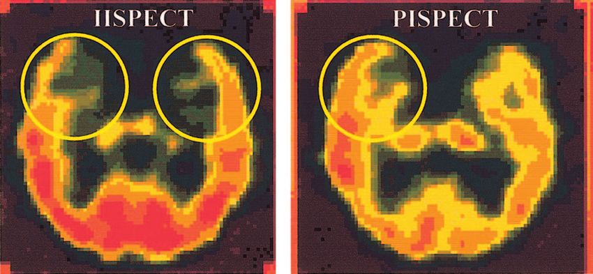
Downloaded from jnm.snmjournals.org by on February 17, 2020. For personal use only.

interictal and the immediate postictal periods. The IISPECT and   tain, with the study available in only 29 of the 42 patients
PISPECT were then compared to evaluate the preictal and imme-     (69%). This was because the other patients had all the
diate postictal perfusion patterns.                               required seizures before the next batch of isotope was
                                                                  stocked, because the second and third seizures occurred
RESULTS
                                                                  within the 24-h window when another injection was not
   Of the 42 patients, who were included in the study, 40         permitted and the patient was discharged, or because the
had IISPECT scans. Two patients did not have IISPECT due          seizures occurred over the weekend when scanning was not
to technical reasons. The relative concordance of the 2           available. In 2 cases, only PISPECT was available. This was
studies to the EEG is shown in Table 1. Of the 40 patients        done before IISPECT because the patients had the first
who had IISPECT, 36 (90%) showed evidence of focal                seizure before an interictal study could be performed and
temporal hypoperfusion. Four (10%) were nonlocalizing, 2          they had to be discharged before IISPECT could be done.
because they were normal and 2 because they had evidence          Interictal hypoperfusion was the most common finding seen
of bilateral temporal hypoperfusion. PISPECT was avail-           in 36 of 40 (90%) patients, with 32 of the 36 (88.9%) being
able for 29 of the 42 patients. Of the 29 who had PISPECT,
                                                                  concordant with the EEG focus. None had interictal hyper-
19 (65.5%) showed focal hyperperfusion. In 10 (34.5%)
                                                                  perfusion. PISPECT was available in 29 patients. Immedi-
patients, there was accentuation of the focal hypoperfusion
                                                                  ate postictal hyperperfusion was seen in 19 of 29 (65.5%)
in PISPECT, on the same side as the interictal hypoperfu-
                                                                  patients and was less frequent than the occurrence of inter-
sion. None had interictal hyperperfusion.
   The mean injection time was 1.86 ⫾ 0.88 min from onset         ictal hypoperfusion but 18 of 19 (94.7%) were concordant
of the seizure. In our experience, it took 15–20 s for the        with the EEG focus. This was slightly better than the rate of
bolus of 99mTc-ECD to be administered. Since it takes 20 s        concordance (88.9%) for interictal hypoperfusion but was
for 95% brain extraction of the isotope (37), only the            not significantly better (P ⱖ 0.5). Keeping this in mind, our
patients in whom the injection was started within 40 s of         data would indicate that PISPECT has a higher concordance
seizure onset would have had 95% extraction of the isotope        rate (94.7%) with the EEG focus than IISPECT (88.9%).
within 60 s. The timing of the injection is extremely impor-      The lower incidence of hyperperfusion in the immediate
tant if one is to obtain true ictal SPECT since the electrical    postictal studies, (65.5%) as opposed to hypoperfusion in
activity, to which the initial rise in blood flow is coupled,     interictal studies (90%), was due to the occurrence of the
lasts 60 –140 s (39) only. Because our mean injection time        phenomenon of accentuation of hypoperfusion in PISPECT,
was about 114 s in most cases, the brain extraction of the        compared with their own interictal studies, in 10 of 29
isotope occurred after the rhythmic electrical activity asso-     (34.5%) patients. In 9 (90%) of these 10 patients who had
ciated with the seizure had peaked or was on the decline          ipsilateral accentuation of hypoperfusion in PISPECT, it
when the injection was finished. Therefore, we considered         was concordant with the EEG focus, whereas in 1 (10%) the
that the term immediate postictal or periictal SPECT              accentuation of hypoperfusion was contralateral.
(PISPECT) was more appropriate than the term ictal
SPECT, even though the injections in our patients were
accomplished in a very short time to justify our use of the       DISCUSSION
term “ictal SPECT” as it has been used in the literature (13)        In 1993, Oommen et al., using long-term ECoG and
by other investigators.                                           cortical CBF monitoring with specially designed subdural
   In our series of 42 patients, we observed that IISPECT         strip electrodes capable of measuring CBF and ECoG at the
was easier to obtain, with the study being done in 40 of 42,      same time, showed that the ictal hyperperfusion was cou-
or 95.2%, of patients. PISPECT was more difficult to ob-          pled to the ictal spike train. It was also shown that that the
                                                                  rhythmic electrical discharge in the temporal lobe during the
                                                                  seizure is short-lived and that the CBF after the cessation of
                          TABLE 1                                 the rhythmic electrical activity or spike train may show 3
                        SPECT Findings                            different phenomena: The blood flow may immediately
                        Total   Ipsilateral to    Contralateral   revert to the preictal level, change to a hyperperfusional
   SPECT finding         (n)     EEG focus*      to EEG focus*    state, or change to a hypoperfusional state. These latter
                                                                  phenomena lasted up to 3 h in that particular study (39).
 IISPECT
    Hypoperfusion        36       32 (88.9)         4 (11.1)      Thus, the dynamic nature of blood flow in the periictal
    Hyperperfusion        0        0                0             period systematically studied by Penfield (23) and Penfield
 PISPECT                                                          et al. (24) during surgery, and later demonstrated by Plum et
    Hyperperfusion       19       18 (94.7)         1 (5.3)
                                                                  al. (25) experimentally, was confirmed by Oommen et al. by
    Hypoperfusion        10        9 (90)           1 (10)
                                                                  in vivo CBF monitoring in the human brain using this novel
                                                                  technology. This observation became the basis of their
  *Data are presented as number (%).
                                                                  combined interpretation of IISPECT and immediate post-

                                                  WHEN    TO   DO SPECT   TO   LOCALIZE EPILEPSY? • Oommen et al.         2023
Downloaded from jnm.snmjournals.org by on February 17, 2020. For personal use only.

FIGURE 1. Pattern 1: Focal interictal hypoperfusion (left) and   FIGURE 3. Pattern 3: Bilateral interictal hypoperfusion and
immediate postictal hyperperfusion in same temporal lobe.        unilateral (right) immediate postictal accentuation of hypoperfu-
                                                                 sion in temporal lobe.

ictal SPECT studies published subsequently (40). As our          CONCLUSION
experience evolved, we divided these into patterns 1–3. In
                                                                    Interictal hypoperfusion in spontaneous seizures of tem-
pattern 1 (Fig. 1), there is hypoperfusion in IISPECT and
                                                                 poral lobe origin can be demonstrated with SPECT and the
hyperperfusion ipsilaterally in PISPECT. In pattern 2 (Fig.
                                                                 pattern of periictal and postictal CBF is useful in predicting
2), the IISPECT is either normal with unilateral hyperper-
                                                                 the seizure focus. True ictal hyperperfusion is practically
fusion in PISPECT or, if there is bilateral hypoperfusion in
                                                                 difficult to obtain in epilepsy-monitoring units because of
IISPECT, it is normalized unilaterally or is focally hyper-
                                                                 the transient nature of seizures and the dynamics of CBF
perfused on one side in PISPECT. In pattern 3 (Fig. 3), there
                                                                 during seizures. Except for very few instances in which
is focal or bilateral hypoperfusion in IISPECT and im-
                                                                 injection of the isotope is made at the onset of seizures,
mediate postictal accentuation of the hypoperfusion in
                                                                 using isotopes that are rapidly (within seconds) taken up by
PISPECT.
                                                                 the brain tissue, most ictal studies reported in the literature
   In the combined analysis of the IISPECT and PISPECT
                                                                 have been periictal or, in some cases, postictal studies. This
data, pattern 1 was concordant with the EEG focus in 13 of
                                                                 is one reason for the controversy regarding the usefulness of
14 (92.9%) patients. When the accentuation of hypoperfu-
                                                                 SPECT findings in the “so-called” ictal studies reported in
sion occurred on the same side as the interictal hypoperfu-
                                                                 the literature. The paradoxical increase in flow in interictal
sion, (pattern 3), it also significantly (90% concordance)
correlated with the EEG focus. Pattern 2 was noted in only       studies reported in the literature was in some cases due to
1 patient in the current series and had 100% correlation with    the presence of unrecognized structural lesions and in others
                                                                 due to interictal epileptiform activity. We did not see this
the EEG focus (Table 2). Thus, bitemporal hypoperfusion,
                                                                 phenomenon in any of our patients, all of whom had a
which usually does not help in localization, became useful
                                                                 demonstrated seizure-free period of about 24 h before their
in localization, when combined with the hyperperfusion in
PISPECT. Many previous studies have noted focal hypo-            IISPECT studies. SPECT studies should therefore be done
perfusion in the ictal studies, and others have looked at its    under controlled circumstances with video-EEG monitoring
                                                                 to define the relationship of the scans to interictal EEG
usefulness in localization. Recognition of this phenomenon
                                                                 activity and seizures. When performed in this way, both
is critical in avoiding false localization, because an area of
                                                                 IISPECT and PISPECT have their place in the localization
hypoperfusion in SPECT may result in the homologous area
                                                                 of seizure foci and, when both are available, they should be
on the other side appearing to be hyperperfused, because of
                                                                 interpreted keeping the dynamic nature of blood flow during
the inherent qualitative nature of most SPECT studies.
                                                                 seizures in mind. The recognition and use of patterns 1–3
                                                                 using IISPECT and PISPECT will improve the value of

                                                                                          TABLE 2
                                                                   Relationship of Perfusion Patterns to EEG Localization

                                                                                                Localization by SPECT pattern
                                                                                      Total     Ipsilateral to    Contralateral
                                                                   SPECT pattern       (n)       EEG focus*      to EEG focus*

                                                                      Pattern 1        14         13 (92.9)          1 (7.1)
                                                                      Pattern 2         1          1 (100)           0
                                                                      Pattern 3        10          9 (90)            1 (10)
FIGURE 2. Pattern 2: Symmetric bilateral interictal hypoper-
fusion and asymmetric unilateral (right) immediate postictal
                                                                   *Data are presented as number (%).
hyperperfusion.

2024     THE JOURNAL    OF   NUCLEAR MEDICINE • Vol. 45 • No. 12 • December 2004
Downloaded from jnm.snmjournals.org by on February 17, 2020. For personal use only.
SPECT in localization of epileptogenic foci, and the com- 21. Engel J Jr, Kuhl D, Phelps M, et al. Interictal cerebral glucose metabolism in
partial epilepsy and its relation to EEG changes. Ann Neurol. 1982;12:510 –517.
bined interpretation can improve the usefulness of the pro- 22. Theodore W, Brooks R, Sato S, et al. The role of positron emission tomography
cedure, particularly in patients in whom the IISPECT is in the evaluation of seizure disorders. Ann Neurol. 1984;15:S176 –S179.
normal or shows bilateral hypoperfusion. 23. Penfield W. The evidence for a cerebral vascular mechanism in epilepsy. Ann
Intern Med. 1933;7:303–310.
24. Penfield W, von Santha K, Cipriani A. Cerebral blood flow during induced
REFERENCES epileptiform seizures in animals and man. J Neurophysiol. 1939;2:257–267.
25. Plum F, Posner J, Troy B. Cerebral metabolic and circulatory responses to
1. Gloor P. Contributions of electroencephalography and electrocortico-graphy to
induced convulsion in animals. Arch Neurol. 1968;18:1–13.
the neurosurgical treatment of the epilepsies. Adv Neurol. 1975;8:59 –106.
26. Engel J Jr, Henry T, Risinger M, et al. Pre-surgical evaluation of partial epilepsy:
2. Walter RD. Principles of clinical investigation of surgical candidates. Adv Neu-
relative contributions of chronic depth electrode recording vs. FDG-PET and
rol. 1975;8:49 –58.
scalp sphenoidal ictal EEG. Neurology. 1990;40:1670 –1677.
3. Crandall PH. Prospective management and criteria for evaluation. Adv Neurol.
27. Weinand M, Carter L, Patton D, Oommen K, Labiner D, Talwar D. Long term
1975;8:265–280.
surface cortical cerebral blood flow monitoring in temporal lobe epilepsy. Neu-
4. Jack CR Jr, Sharbrough FW, Twomey CK, et al. Temporal lobe seizures:
rosurgery. 1994;35:657– 664.
lateralization with MR volume measurements of hippocampal formation. Radi-
28. Weinand M, Carter L. Surface cortical cerebral blood flow monitoring and single
ology. 1990;175:423– 429.
photon emission computed tomography: prognostic factors for selecting temporal
5. Bronen RA, Fulbright RK, Spencer DD, Spencer SS, Kim JH, Lance RC. MR
lobectomy candidates. Seizure. 1994;3:55–59.
characteristics of neoplasms and vascular malformations associated with epi-
29. LaManna M, Sussman N, Harner R, et al. Initial experience with SPECT imaging
lepsy. Magn Reson Imaging. 1995;13:1153–1162.
of the brain using I-123 p-iodoamphetamine in focal epilepsy. Clin Nucl Med.
6. Spencer SS. The relative contributions of MRI, SPECT and PET imaging in
1989;14:428 – 430.
epilepsy. Epilepsia. 1994;35(suppl 6):S72–S89.
30. Devous M Sr, Thisted R, Morgan G, Leroy R, Rowe C. SPECT brain imaging in
7. Ingvar DH. Regional cerebral blood flow in focal cortical epilepsy. Stroke.
epilepsy: a meta analysis. J Nucl Med. 1998;39:285–293.
1973;4:359 –360.
31. Oliveira A, Costa J, Hilario L, Anselmi O, Palmini A. Localization of epilepto-
8. Lavy S, Melamed E, Portnoy Z, et al. Interictal regional cerebral blood flow in
genic zone by ictal and interictal SPECT with Tc-ethyl cysteinate dimer in
patients with partial seizures. Neurology. 1976;26:418 – 422. patients with medically refractory epilepsy. Epilepsia. 1999;40:693–702.
9. Kuhl DE, Engel J Jr, Phelps ME, Selin C. Epileptic patterns of local cerebral 32. Rowe C, Berkovic S, Austin M, McKay W, Bladin P. Patterns of postictal
metabolism and perfusion in humans determined by emission computed tomog- cerebral blood flow in temporal lobe epilepsy: qualitative and quantitative anal-
raphy of 18 FDG and 13 NH3. Ann Neurol. 1980;8:348 –360. ysis. Neurology. 1991;41:1096 –1103.
10. Magistretti P, Uren R, Blume H, Schomer D, Royal H. Delineation of epileptic 33. Juni JE, Waxman AD, Devous MD Sr, et al. Brain perfusion single photon
focus by single photon emission tomography. Eur J Nucl Med. 1982;7:484 – 485. emission computed tomography (SPECT) using Tc-99m radiopharmaceuticals.
11. Hougaard K, Oikawa T, Sveindottir E, et al. Regional cerebral blood flow in focal Procedure Guidelines. 1999:113–118. Society of Nuclear Medicine Web site.
cortical epilepsy. Arch Neurol. 1976;33:527–535. Available at: http://interactive.snm.org/index.cfm?PageID⫽800. Accessed Octo-
12. Sakai F, Meyer JS, Naritomi H, et al. Regional cerebral blood flow and EEG in ber 31, 2004.
patients with epilepsy. Arch Neurol. 1978;35:648 – 657. 34. Duncan R, Patterson J, Roberts R, Hadley D, Bone I. Ictal/postictal SPECT in
13. Weis M, Feistel H, Stefan H. Utility of ictal SPECT: peri-ictal, postictal. Acta presurgical localization of complex partial seizures. J Neurol Neurosurg Psychi-
Neurol Scand. 1994;152(suppl):145–147. atry. 1993;56:141–148.
14. Kuikka JT, Mervaala E, Vanninen E, Kalviainen E. Does technetium-99m bici- 35. Jibiki I, Kubota T, Fujimoto K, et al. Reexamination of I231-IMP-SPECT at
sate image local brain metabolism in late ictal temporal lobe epilepsy? Eur J Nucl interictal stages in adult partial epilepsy. Jpn J Psychiatry Neurol. 1989;43:385–
Med. 1994;21:1247–1251. 388.
15. Devous M, Leroy R, Homan R. Single photon emission computed tomography in 36. Jibiki I, Yamaguchi N, Matsuda H, Hisada K. Fluctuations of interictal brain
epilepsy. Semin Nucl Med. 1990;20:325–341. imaging in repeated I231-IMP-SPECT scans in an epileptic patient. J Neurol.
16. Alavi A, Hirsch L. Studies of central nervous system disorders with single photon 1990;237:372–375.
emission computed tomography: evolution over the past two decades. Semin Nucl 37. Walovitch R, Hill T, Garrity S, et al. Characterization of technetium-99-m-
Med. 1991;21:58 – 81. 1,1ECD for brain perfusion imaging. Part I. Pharmacology of technetium-99-m
17. Rowe C, Berkovic S, Austin M, Saling M, Kalnins R. Visual and quantitative ECD in nonhuman primates. J Nucl Med. 1989;30:1892–1901.
analysis of interictal SPECT with technetium-99m-HMPAO in temporal lobe 38. Leveille J, Demonceau G, Walovitch R. Intrasubject comparison between tech-
epilepsy. J Nucl Med. 1991;32:1688 –1694. netium 99m-ECD and technetium 99m-HMPAO in healthy human subjects.
18. Newton M, Berkovic S, Austin M, Rowe C, McKay W, Bladin P. Ictal postictal J Nucl Med. 1992;33:480 – 484.
and interictal single-photon emission tomography in the lateralization of temporal 39. Oommen K, Carter L, Weinand M. Cerebral blood-flow over epileptogenic
lobe epilepsy. Eur J Nucl Med. 1994;21:1067–1071. temporal cortex before, during and after seizures. Epilepsia. 1993;34(suppl
19. Berkovic S, Newton M, Chiron C, Dulac O. Single photon emission tomography. 6):127–128.
In: Engel J Jr, ed. Surgical Treatment of the Epilepsies. 2nd ed. New York, NY: 40. Oommen K, King J, Carter LP, Chacko G, Wilson D. Utility of combined
Raven Press; 1993:233–243. inter-ictal and immediate post-ictal SPECT (IISPECT and PISPECT) in local-
20. Chugani H. Functional brain imaging in pediatrics. Pediatr Clin North Am. ization of epileptogenic foci by using 99mTc ECD (Neurolite) [abstract]. Epilep-
1992;39:777–799. sia. 1997;38(suppl 8):146.
WHEN TO DO SPECT TO LOCALIZE EPILEPSY? • Oommen et al. 2025Downloaded from jnm.snmjournals.org by on February 17, 2020. For personal use only. The Relative Localizing Value of Interictal and Immediate Postictal SPECT in Seizures of Temporal Lobe Origin Kalarickal J. Oommen, Sadia Saba, Joseph A. Oommen, Paul C. Francel, Charles D. Arnold and Don A. Wilson J Nucl Med. 2004;45:2021-2025. This article and updated information are available at: http://jnm.snmjournals.org/content/45/12/2021 Information about reproducing figures, tables, or other portions of this article can be found online at: http://jnm.snmjournals.org/site/misc/permission.xhtml Information about subscriptions to JNM can be found at: http://jnm.snmjournals.org/site/subscriptions/online.xhtml The Journal of Nuclear Medicine is published monthly. SNMMI | Society of Nuclear Medicine and Molecular Imaging 1850 Samuel Morse Drive, Reston, VA 20190. (Print ISSN: 0161-5505, Online ISSN: 2159-662X) © Copyright 2004 SNMMI; all rights reserved.
You can also read