Technical feasibility of radiomics signature analyses for improving detection of occult tonsillar cancer
←
→
Page content transcription
If your browser does not render page correctly, please read the page content below
www.nature.com/scientificreports
OPEN Technical feasibility of radiomics
signature analyses for improving
detection of occult tonsillar cancer
Jeong Hoon Lee1, Eun Ju Ha2*, Jin Roh3, Su Jin Lee4 & Jeon Yeob Jang5
Diagnosis of occult palatine tonsil squamous cell carcinoma (SCC) using conventional magnetic
resonance imaging (MRI) is difficult in patients with cervical nodal metastasis from an unknown
primary site at presentation. We aimed to establish a radiomics approach based on MRI features
extracted from the volume of interest in these patients. An Elastic Net model was developed
to differentiate between normal palatine tonsils and occult palatine tonsil SCC. The diagnostic
performances of the model with radiomics features extracted from T1-weighted image (WI), T2WI,
contrast-enhanced T1WI, and an apparent diffusion coefficient (ADC) map had area under the receiver
operating characteristic (AUROC) curve values of 0.831, 0.840, 0.781, and 0.807, respectively, for
differential diagnosis. The model with features from the ADC alone showed the highest sensitivity of
90.0%, while the model with features from T1WI + T2WI + contrast-enhanced T1WI showed the highest
AUROC of 0.853. The added sensitivity of the radiomics feature analysis were 34.6% over that of
conventional MRI to detect occult palatine tonsil SCC. Therefore, we concluded that adding radiomics
feature analysis to MRI may improve the detection sensitivity for occult palatine tonsil SCC in patients
with a cervical nodal metastasis from cancer of an unknown primary site.
The palatine tonsil is the most common site of squamous cell carcinoma (SCC) in the o ropharynx1,2. However,
because tonsillar fossae are anatomically complex and many patients commonly have enlarged tonsils due to
underlying chronic tonsillitis, identification of SCC of the palatine tonsil using physical and endoscopic examina-
tion, or conventional imaging such as computed tomography (CT) or magnetic resonance imaging (MRI) can be
complicated3–5. Since occult palatine tonsil SCC patients commonly show a palpable neck lymphadenopathy at
the initial presentation, it is difficult to differentiate them from carcinoma of unknown primary sites (CUP) of
the head and neck, which makes decisions for appropriate treatment difficult for physicians as well as p atients3–5.
Currently, because more than 90% of CUPs of the head and neck reflect human papillomavirus (HPV)-associated
cancers, the palatine tonsils are expected to be the most likely source of the primary cancer, and randomly
directed biopsies of the tonsil and/or diagnostic tonsillectomy are commonly performed for a more focused
therapy in these patients6–9.
Recently, several papers have reported that radiomics features derived from the volumetric analyses of a whole
tumor in CT or MRI scans have shown potential for tumor detection, grading, and predicting the recurrence of
head and neck c ancer10–13. In oropharyngeal cancer, a few papers have reported the potential of radiomic features
analysis (RFA) for discrimination of HPV status, tumor grading, and detection of local recurrence11–13. However,
to date, no information exists on the potential for the early detection of occult tonsillar cancer from normal or
hyperplastic lymphoid tissue. Given the histologic heterogeneity of tumor-harboring tonsillar tissue compared
to normal palatine tonsils, RFA has the potential to demonstrate occult palatine tonsil SCC in patients with a
neck lymph node metastasis from an unknown primary s ite3.
This retrospective study aimed to establish a technical feasibility of radiomics approach based on MRI features
extracted from the volume of interest (VOI) to detect occult palatine tonsil SCC in patients with cervical nodal
metastasis from a cancer of an unknown primary site.
1
Division of Biomedical Informatics, Seoul National University Biomedical Informatics (SNUBI), Seoul National
University College of Medicine, Seoul 110799, Republic of Korea. 2Department of Radiology, Ajou University
School of Medicine, Wonchon‑Dong, Yeongtong‑Gu, Suwon 443‑380, Korea. 3Department of Pathology,
Ajou University School of Medicine, Wonchon‑Dong, Yeongtong‑Gu, Suwon 443‑380, Korea. 4Department
of Nuclear Medicine, Ajou University School of Medicine, Wonchon‑Dong, Yeongtong‑Gu, Suwon 443‑380,
Korea. 5Department of Otolaryngology, Ajou University School of Medicine, Wonchon‑Dong, Yeongtong‑Gu,
Suwon 443‑380, Korea. *email: radhej@naver.com
Scientific Reports | (2021) 11:192 | https://doi.org/10.1038/s41598-020-80597-3 1
Vol.:(0123456789)
www.nature.com/scientificreports/
Occult palatine tonsil SCC
Overt palatine tonsil SCC (n = 49) (n = 29) Normal palatine tonsils (n = 94) P-value
Age (years) 56.3 ± 8.0 63.4 ± 9.5 61.1 ± 9.4 0.006
Sex (M:F) 21:8 43:6 78:16 0.226
Tonsil size (mm)
Axial 38.0 ± 8.8 31.3 ± 6.3 24.0 ± 6.2 < 0.001
Sagittal 35.3 ± 9.0 28.6 ± 6.2 21.6 ± 6.3 < 0.001
Coronal 35.3 ± 9.0 28.3 ± 6.5 21.1 ± 6.7 < 0.001
HPV status (%) 71.1 (27/38) 95.9 (25/26) 71.4 (5/7)* 0.038
Table 1. Patient demographic characteristics. Numbers for age and tonsil size are the means ± standard
deviation. HPV human papillomavirus; SCC squamous cell carcinoma. *The percentage indicates values for a
limited number of patients with a carcinoma of an unknown primary site.
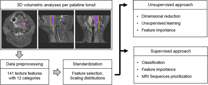
Figure 1. Box and whisker plots of the distribution of the representative values of shape features and fractal
analyses in patients with overt palatine tonsil SCC, occult palatine tonsil SCC, and normal palatine tonsils on
T2-weighted images.
Results
Comparison of radiomics features. Table 1 summarizes demographic data for overt palatine tonsil SCC,
occult palatine tonsil SCC, and normal palatine tonsils in our study cohort. The mean size of the palatine tonsils
in the axial, sagittal, and coronal images differed significantly among the three groups, and the values increased
from normal palatine tonsils to occult palatine tonsil SCC, and to overt palatine tonsil SCC. The ANOVA analy-
ses showed that age was significantly different (P = 0.006). There were no significant differences in sex among the
groups (P = 0.226), but there was a significant difference in the status of HPV (P = 0.038).
Figure 1 shows the representative values of the radiomics features obtained through UMAP dimensional
reduction. The representative values of the shape features, 3D fractal analyses, and moment features were sig-
nificantly different factors among the three groups, regardless of the MRI sequences (all P < 0.001, respectively).
The representative values of shape features, fractal analyses and moment features increased or decreased, in the
order of normal palatine tonsils, occult palatine tonsil SCC, and overt palatine tonsil SCC. The UMAP dimen-
sionality reduction results for all 12 categories are shown in the Fig. 2. This tendency was also seen in the UMAP
visualization of the shape features (Fig. 3). The AUROC for the 12 feature categories to discern occult palatine
tonsil SCC from normal palatine tonsils according to the MRI sequences are shown in Supplemental Table S1.
Value of RFA in occult palatine tonsil SCC detection. The diagnostic performance of the model with
whole radiomics features extracted from T1WI, T2WI, contrast-enhanced T1WI, and ADC had AUROCs of
0.831, 0.840, 0.781, and 0.807, respectively, for the differential diagnosis of occult palatine tonsil SCC from
normal palatine tonsils (Table 2, Supplemental Table S2). In terms of sensitivity, the best performing model
with features from ADC alone showed the highest sensitivity of up to 90.0%. The model with features from
T1WI + T2WI + contrast-enhanced T1WI had the highest AUROC of up to 0.853 (Table 3, Supplemental
Table S2).
Scientific Reports | (2021) 11:192 | https://doi.org/10.1038/s41598-020-80597-3 2
Vol:.(1234567890)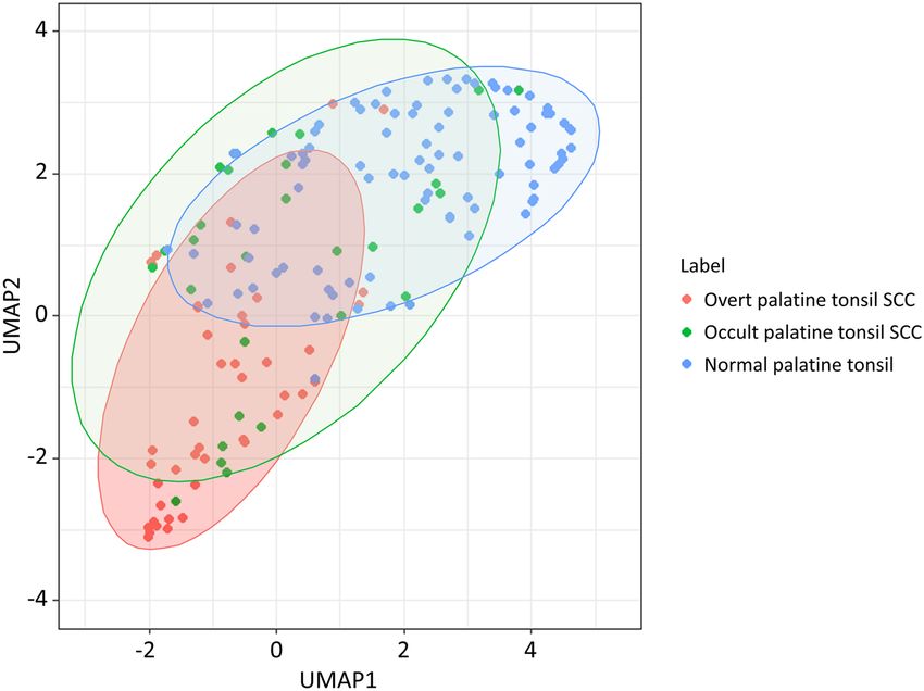
www.nature.com/scientificreports/
Figure 2. Box and whisker plots of the distribution of the representative values of radiomics features in patients
with overt palatine tonsil SCC, occult palatine tonsil SCC, and normal palatine tonsils on T2-weighted images.
Figure 3. Dimensionality reduction of shape features through the Uniform Manifold Approximation and
Projection algorithm20 (https://github.com/ropenscilabs/umapr, v0.0.0.9001).
Additional value of radiomics over conventional MRI or 18F‑FDG PET/CT. Table 4 shows the diag-
nostic performance of the different imaging modalities to detect occult palatine tonsil SCC among patients with
cervical lymph node metastasis from CUP at presentation. The sensitivity and accuracy of conventional MRI
alone were 41.4% and 51.4%, respectively, and those of 18F-FDG PET/CT were 79.3% and 75.7%, respectively.
The SUVmax of the occult palatine tonsil SCC was 9.3 ± 3.4 (range 2.5–16.2), and that of the contralateral ton-
sil was 5.5 ± 1.7 (range 2.4–10.6). The mean difference in the SUVmax between the two (occult palatine tonsil
SCC—contralateral normal palatine tonsil) was 3.8 ± 3.5 (range − 4.5 to 11.0). The added value of the RFA of
Scientific Reports | (2021) 11:192 | https://doi.org/10.1038/s41598-020-80597-3 3
Vol.:(0123456789)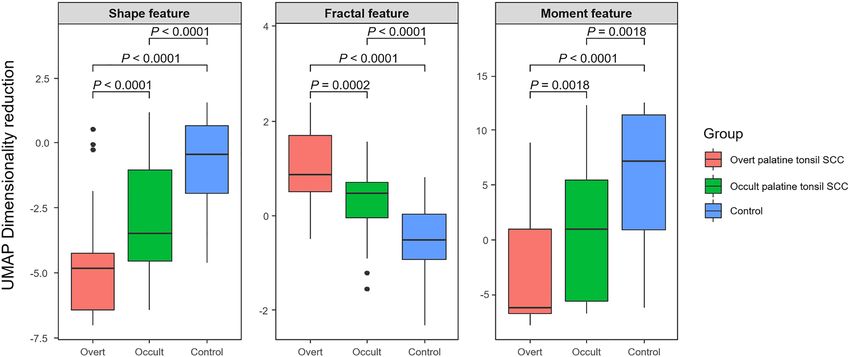
www.nature.com/scientificreports/
MRI sequences AUROC Sensitivity (%) Specificity (%)
T1WI 0.831 81.4 89.5
T2WI 0.840 86.5 82.4
Contrast-enhanced T1WI 0.781 83.2 80.2
ADC 0.807 90.0 77.9
Table 2. Diagnostic performance of RFA to discern occult palatine tonsil SCC from normal palatine tonsils
according to the MRI sequences. ADC apparent diffusion coefficient; AUROC area under the receiver operating
curve; T1WI T1-weighted image; T2WI T2-weighted image.
MRI sequence AUROC Sensitivity (%) Specificity (%)
ADC 0.807 90.0 77.9
T1WI + T2WI 0.810 89.7 75.4
T1WI + contrast-enhanced T1WI 0.765 81.4 79.8
T2WI + contrast-enhanced T1WI 0.843 84.8 85.3
T1WI + T2WI + contrast-enhanced T1WI 0.853 81.4 87.4
Table 3. Diagnostic performance of radiomics features to discern occult palatine tonsil SCC from normal
palatine tonsils according to a combination of MRI sequences. ADC apparent diffusion coefficient; AUROC
area under the receiver operating characteristics; T1WI T1-weighted image; T2WI T2-weighted image.
Modality Sensitivity Specificity Accuracy PPV NPV
Radiomics feature (ADC) 76.0 80.0 80.3 94.1 57.7
MRI 41.4 87.5 51.4 92.3 29.2
18
F-FDG PET/CT 79.3 62.5 75.7 88.4 53.8
Table 4. Diagnostic performance to detect occult palatine tonsil SCC using different imaging modalities.
Numbers for diagnostic performance are percentages. ADC apparent diffusion coefficient; PPV positive
predictive value; NPV negative predictive value.
ADC was 34.6% for sensitivity and 28.9% for accuracy over conventional MRI alone. This indicates comparable
sensitivity and accuracy compared to 18F-FDG PET/CT.
Discussion
Our study demonstrated that a radiomics approach based on MRI features extracted from a VOI of the palatine
tonsil has the potential to differentiate occult palatine tonsil SCC from normal palatine tonsils in patients with
cervical nodal metastasis from a CUP. The added sensitivity of RFA for detecting occult palatine tonsil SCC was
34.6% over conventional MRI and it was comparable to 18F-FDG PET/CT.
Despite recent advances in diagnostic tools for use in head and neck cancers, it is common to encounter
patients who present with cervical lymph node metastasis without an apparent primary site following thor-
ough clinical and radiological e xaminations6,14. In previous studies, SCC was the most common histologic type,
accounting for 78% of cases. However, the treatment strategies have been controversial, until now6,8,9. Although
radiation therapy with or without neck dissection, including irradiation of all potential mucosal disease sites,
has been effective, it causes substantial morbidity and side e ffects6,14. Therefore, efforts should be made to find
the primary tumor, as this allows a more focused therapy, with less morbidity, and possibly a better o utcome9.
In this respect, a randomly directed biopsy and/or diagnostic tonsillectomy is common, as up to 25% of primary
tumors can be detected in this way whereas some have a delayed diagnosis of the primary tumor during the
follow-up period9.
More recently, research based on medical imaging informatics has greatly improved. Radiomics, a data mining
approach that extracts high-dimensional data in the form of a multitude of features from clinical images to build
machine-learning or statistical models, has been applied to various imaging modalities to answer relevant clinical
questions10. In the field of head and neck cancer radiomics, classification and survival regression models have
been applied to predict molecular markers and identify genomic signatures for the diagnostic differentiation of
suspected tissues, survival prognostication, and to predict treatment responses11–13. In our study, we developed
models using radiological images and integrated quantitative radiomics features to arrive at a better clinical
decision and treatment planning for head and neck cancer. We focused on the potential of a radiomics approach
to differentiate occult palatine tonsil SCC from normal palatine tonsil, based on the histologic heterogeneity of
Scientific Reports | (2021) 11:192 | https://doi.org/10.1038/s41598-020-80597-3 4
Vol:.(1234567890)www.nature.com/scientificreports/
tumor-harboring tonsillar tissue compared to normal palatine tonsils. Representative values of shape features,
3D fractal analyses, and moment features showed significant differences among normal palatine tonsils and
occult and overt palatine tonsil SCC, regardless of the MRI sequences. Based on these results, the model with
the radiomics features extracted from an ADC map showed the highest sensitivity of up to 90.0%. These results
are similar to those of Choi et al., which showed the potential for histogram analyses of ADC for differential
diagnosis of occult palatine tonsil SCC. Adding histogram analyses of ADC to conventional MRI showed the
potential to improve the detection sensitivity up to 52.6% (from 26.3% to 78.9%) in their s tudy3.
The application of advanced diagnostic methods such as 18F-FDG PET-CT can be useful for differential
diagnosis in patients with C UP15,16. A previous study that included data from 302 patients with occult primary
head and neck tumors found that FDG PET detected 24.5% more histologically proven primary lesions com-
pared to conventional assessment methods17. However, because the range of physiological FDG uptake in nor-
mal palatine tonsil varies considerably, establishing a cut-off threshold to distinguish between normal palatine
tonsil uptakes from occult palatine tonsil SCC is not easy15,18. In addition, FDG PET had a false positive rate of
39.3% for detecting occult palatine tonsil primary cancers17. In our study, the mean differences in the SUVmax
between the occult palatine tonsil SCC and the contralateral normal palatine tonsil also varied from − 4.5 to
11.0. Because a subtle asymmetric difference between the bilateral tonsils without tonsillar malignancy is also
common in practice, clinical decisions in patients with CUP are sometimes very difficult. In this study, adding
the RFA over conventional MRI alone showed the potential for improving sensitivity, which was comparable to
18
F-FDG PET-CT. Further research in a larger population sample is needed based on these results.
There were some limitations to our study. First, we only included 29 patients with occult palatine tonsil SCC
and six patients who had an MRI in another institution were excluded. Since an external validation cohort is
lacking in this study, further prospective studies with a larger population are necessary to validate of our study
results and to achieve reproducibility and generalizability. Second, there may be a technical challenge in delin-
eating tonsil boundaries when drawing the regions of interest and it may produce segmentation errors in tumor
volumes and their associated radiomic features3. Stability analysis of MRI radiomic features in palatine tonsils
are required in the future.
In conclusion, occult palatine tonsil SCC may be differentiated from normal palatine tonsils using RFA in
patients with cervical nodal metastases from CUP. A radiomics approach has the potential to improve targeted
treatment and reduce morbidity.
Methods
Study population. Our study protocol was reviewed and approved by the Ajou University hospital Insti-
tutional Review Board. Ethics committees/Institutional Review Board (AJIRB-MED-MDB-18-508) waived the
need for you to obtain informed consent because of the retrospective nature of the study. All methods were per-
formed in accordance with the relevant guidelines and regulations. We reviewed the medical records of patients
at our institution between May 2010 and April 2020 that had cervical nodal metastasis from CUP at presentation
and which was finally confirmed as palatine tonsillar SCC. We enrolled the patients who met the following cri-
teria: a histologically proven metastatic SCC in cervical lymph node at clinical presentation, negative or equivo-
cal findings of a contrast-enhanced neck CT scan, a pretreatment neck MRI examination, and a pathologically
proven palatine tonsil SCC after a tonsillectomy or biopsy. A total of 39 patients with occult palatine tonsil SCC
that had cervical nodal metastasis at presentation were identified. After the exclusion of 10 patients who did not
undergo pretreatment neck MRI or an MRI outside of the hospital (n = 6), or who had a severe dental or motion
artifact on the MRI (n = 4), 29 patients were finally enrolled (mean age, 56.3 years; age range, 32–74 years).
Tumor grades were determined by an experienced pathologist (J. R.: 10 years of clinical experience) to be well-
differentiated (n = 2), moderately differentiated (n = 18), or poorly differentiated (n = 9).
For comparison, we enrolled 49 patients with an overt palatine tonsil SCC (mean age, 63.4 years; age range,
44–82 years) as positive control subjects as follows: pathologically proven palatine tonsil SCC, evidence of an
overt palatine tonsil SCC on a contrast-enhanced neck CT, and a pretreatment neck MRI at our hospital. Bilateral
palatine tonsils from eight patients with CUP of head and neck (mean age, 54.5 years; age range, 36–74 years)
and the contralateral palatine tonsils of 78 patients with an occult or overt palatine tonsil SCC were included as
negative control subjects with a histopathologic c onfirmation3.
Imaging Techniques. All MR examinations were performed with 1.5 or 3T MRI units (Signa HDxt or
Discovery MR 750; GE Healthcare Systems, Illinois, USA). In all of the patients, the protocol included axial
T2-weighted fast spin-echo images with fat suppression (4 mm slice thickness with 0.4 mm slice spacing), axial
T1-weighted fast spin-echo images (4 mm slice thickness with 0.4 mm slice spacing), and contrast-enhanced
axial T1-weighted images (WI) following a bolus injection of 0.1 mmol per kilogram of body weight of gadoteri-
dol (ProHance; Bracco, Milan, Italy). All T1WI and T2WI were acquired with 30 imaging sections and a field
of view of 200 (anterior to posterior) × 200 (right to left) × 120 (feet to head) mm. Diffusion-weighted imaging
was performed before the contrast-enhanced T1WI; 30 fat-suppressed diffusion-weighted images of the head
and neck were acquired in the axial plane using a spin-echo single-shot echo-planar image sequence (section
thickness 4 mm/gap 0.4 mm; field of view, 230 (anterior to posterior) × 230 (right to left) × 120 (feet to head) mm;
acquisition matrix 112 × 112; reconstruction matrix 256 × 256; b values of 0 and 1000 s/mm2) in the axial plane
from the skull base to the hyoid bone level. The diffusion gradients were applied in three orthogonal directions
(x, y, and z). Axial trace apparent diffusion coefficient (ADC) maps were generated for all of the obtained images
with the manufacturer’s software included in the MR unit using the b values of 0 and 1000 s/mm23.
Scientific Reports | (2021) 11:192 | https://doi.org/10.1038/s41598-020-80597-3 5
Vol.:(0123456789)www.nature.com/scientificreports/
Figure 4. Workflow scheme of this study. Flow diagrams show the process from a VOI segmentation to model
evaluation.
Imaging processing and analyses. RFA was performed using Pyradiomics in A-VIEW Research 1.0v
(Coreline Soft; Seoul, Korea; https://www.corelinesoft.com/aview-research-2/)19. The T1WI, T2WI, contrast-
enhanced T1WI, and ADC maps of all patients were exported to the software. Three-dimensional VOIs per
palatine tonsil were semi-automatically created by the software, and an experienced radiologist (E.J.H.: 15 years
of clinical experience) modified the VOIs on each axial images. For each VOI, 141 texture features with 12
categories were computed, including shape features (26 features), first-order statistics features (19 features),
and histogram/percentile/gradient features (17 features). Second-order statistics features were derived from the
gray-level co-occurrence matrix (GLCM, 24 features), gray-level dependence matrix (GLDM, 14 features), gray-
level run length matrix (GRLM, 16 features), gray-level size zone matrix (GSZM, 16 features), and neighborhood
gray-tone difference matrix (NGTDM, 5 features). Higher-order features included fractal analyses (1 feature)
and moment features (3 features). All features were transformed to the same scale through Z-score normaliza-
tion. There was no further manipulation in a pre-processing step before the radiomics feature extraction. The
study workflow is detailed in Fig. 4.
To assess the value of adding RFA to conventional MRI and 18F-fluorodeoxyglucose (FDG) positron emis-
sion tomography (PET)/CT for differentiating normal palatine tonsils from occult palatine tonsil SCC, one
radiologist (E.J.H.) reviewed conventional MRIs of 29 patients with occult palatine tonsil SCC and 8 patients
with CUP, including T1WI, T2WI, and contrast-enhanced T1WI, and the radiologist was blinded to all other
data including the final histologic diagnoses. In the MRIs, SCC of the palatine tonsil was defined as a mass that
could be discriminated from the surrounding tonsillar tissue by its signal intensity or the degree of enhance-
ment. Tonsillar asymmetry was also considered positive for the presence of palatine tonsil S CC3. One nuclear
medicine physician (S.J.L.: 16 years of clinical experience) reviewed 18F-FDG PET/CT images, and asymmetric
hypermetabolism on the 18F-FDG PET/CT images was considered to indicate a positive palatine tonsil SCC
finding. The maximum standardized uptake values (SUVmax) of the bilateral palatine tonsils were recorded as
reference values.
Statistical analyses. Statistical analyses were performed using R v. 3.6.3. (R Foundation for Statistical
Computing, Vienna, Austria). One-way analysis of variance (ANOVA) was used to compare the demographic
characteristics among the groups. A Wilcoxon test was used to compare the quantitative texture feature catego-
ries among the groups. To obtain representative values of the feature categories, a nonlinear dimension reduc-
tion algorithm, Uniform Manifold Approximation and Projection (UMAP), was applied, except for the fractal
analyses, which comprised only one f eature20. The discriminative power of the representative value of each cat-
egory was evaluated through the geometric means of the p-value through group-wise comparisons.
Elastic Net regularization for generalized linear models is the linear combination of lasso and ridge regu-
larization methods21. An Elastic Net model was developed to differentiate between normal palatine tonsils and
occult palatine tonsil SCC. We constructed area under the receiver operating characteristic (AUROC) curves
to determine the best predictive model and threshold values from the radiomics features. The best predictive
model was selected based on an AUROC of fivefold cross-validation, and the alpha parameter of the Elastic Net
was set to 0.5. In each fivefold cross-validation, the test set was predicted through the penalty parameter with the
minimum mean cross-validated error in the training set. A P value < 0.05 was considered statistically significant.
Received: 22 September 2020; Accepted: 21 December 2020
Scientific Reports | (2021) 11:192 | https://doi.org/10.1038/s41598-020-80597-3 6
Vol:.(1234567890)www.nature.com/scientificreports/
References
1. Chi, A. C., Day, T. A. & Neville, B. W. Oral cavity and oropharyngeal squamous cell carcinoma: An update. CA Cancer J. Clin. 65,
401–421. https://doi.org/10.3322/caac.21293 (2015).
2. Stambuk, H. E., Karimi, S., Lee, N. & Patel, S. G. Oral cavity and oropharynx tumors. Radiol. Clin. North Am. 45, 1–20. https://
doi.org/10.1016/j.rcl.2006.10.010 (2007).
3. Choi, Y. J. et al. Histogram analysis of apparent diffusion coefficients for occult tonsil cancer in patients with cervical nodal metas-
tasis from an unknown primary site at presentation. Radiology 278, 146–155. https://doi.org/10.1148/radiol.2015141727 (2016).
4. Cinar, F. Significance of asymptomatic tonsil asymmetry. Otolaryngol. Head Neck Surg. 131, 101–103. https://doi.org/10.1016/j.
otohns.2004.02.004 (2004).
5. Jumper, J. R., Fischbein, N. J., Kaplan, M. J., Klein, H. Z. & Dillon, W. P. The, “small, dark tonsil” in patients presenting with meta-
static cervical lymphadenopathy from an unknown primary. AJNR Am. J. Neuroradiol. 26, 411–413 (2005).
6. Piazza, C., Incandela, F. & Giannini, L. Unknown primary of the head and neck: A new entry in the TNM staging system with old
dilemmas for everyday practice. Curr. Opin. Otolaryngol. Head Neck Surg. 27, 73–79. https://doi.org/10.1097/MOO.0000000000
000528 (2019).
7. Tribius, S. et al. HPV status in patients with head and neck of carcinoma of unknown primary site: HPV, tobacco smoking, and
outcome. Oral Oncol. 48, 1178–1184. https://doi.org/10.1016/j.oraloncology.2012.05.022 (2012).
8. Rassy, E., Nicolai, P. & Pavlidis, N. Comprehensive management of HPV-related squamous cell carcinoma of the head and neck
of unknown primary. Head Neck 41, 3700–3711. https://doi.org/10.1002/hed.25858 (2019).
9. Ryan, J. F. et al. The impact of a stepwise approach to primary tumor detection in squamous cell carcinoma of the neck with
unknown primary. Laryngoscope 129, 1610–1616. https://doi.org/10.1002/lary.27625 (2019).
10. Haider, S. P., Burtness, B., Yarbrough, W. G. & Payabvash, S. Applications of radiomics in precision diagnosis, prognostication and
treatment planning of head and neck squamous cell carcinomas. Cancers Head Neck 5, 6. https: //doi.org/10.1186/s41199 -020-00053
-7 (2020).
11. Head, M. D. A. C. C. & Neck Quantitative Imaging Working, G. Investigation of radiomic signatures for local recurrence using
primary tumor texture analysis in oropharyngeal head and neck cancer patients. Sci Rep 8, 1524. https://doi.org/10.1038/s4159
8-017-14687-0 (2018).
12. Leijenaar, R. T. et al. Development and validation of a radiomic signature to predict HPV (p16) status from standard CT imaging:
A multicenter study. Br. J. Radiol. 91, 20170498. https://doi.org/10.1259/bjr.20170498 (2018).
13. Yu, K. et al. Radiomic analysis in prediction of human papilloma virus status. Clin. Transl. Radiat. Oncol. 7, 49–54. https://doi.
org/10.1016/j.ctro.2017.10.001 (2017).
14. Wang, Y. et al. Cervical lymph node carcinoma metastasis from unknown primary site: A retrospective analysis of 154 patients.
Cancer Med. 7, 1852–1859. https://doi.org/10.1002/cam4.1458 (2018).
15. Wong, W. L., Gibson, D., Sanghera, B., Goodchild, K. & Saunders, M. Evaluation of normal FDG uptake in palatine tonsil and
its potential value for detecting occult head and neck cancers: A PET CT study. Nucl. Med. Commun. 28, 675–680. https://doi.
org/10.1097/MNM.0b013e32829152b1 (2007).
16. Davison, J. M., Ozonoff, A., Imsande, H. M., Grillone, G. A. & Subramaniam, R. M. Squamous cell carcinoma of the palatine tonsils:
FDG standardized uptake value ratio as a biomarker to differentiate tonsillar carcinoma from physiologic uptake. Radiology 255,
578–585. https://doi.org/10.1148/radiol.10091479 (2010).
17. Rusthoven, K. E., Koshy, M. & Paulino, A. C. The role of fluorodeoxyglucose positron emission tomography in cervical lymph
node metastases from an unknown primary tumor. Cancer 101, 2641–2649. https://doi.org/10.1002/cncr.20687 (2004).
18. Kawabe, J. et al. Physiological FDG uptake in the palatine tonsils. Ann. Nucl. Med. 15, 297–300. https://doi.org/10.1007/BF029
87850(2001).
19. CorelineSoft. https://www.corelinesoft.com/wp-content/uploads/2018/11/AVIEW_Research.pdf (2018).
20. Becht, E. et al. Dimensionality reduction for visualizing single-cell data using UMAP. Nat. Biotechnol. https://doi.org/10.1038/
nbt.4314 (2018).
21. Zou, H. & Hastie, T. Regularization and variable selection via the elastic net. J. R. Stat. Soc. B 67, 301–320 (2005).
Acknowledgements
This work was supported by the BRACCO Research Foundation. This work was supported by the 2018 intramural
research fund of Ajou University Medical Center.
Author contributions
J.H.L. data collection and manuscript writing, E.J.H. acquisition of imaging data statistical analysis and manu-
script review, J.R., S.J.L. and J.Y.J. manuscript review. All authors read and approved the manuscript.
Competing interests
The authors declare no competing interests.
Additional information
Supplementary Information The online version contains supplementary material available at https://doi.
org/10.1038/s41598-020-80597-3.
Correspondence and requests for materials should be addressed to E.J.H.
Reprints and permissions information is available at www.nature.com/reprints.
Publisher’s note Springer Nature remains neutral with regard to jurisdictional claims in published maps and
institutional affiliations.
Scientific Reports | (2021) 11:192 | https://doi.org/10.1038/s41598-020-80597-3 7
Vol.:(0123456789)www.nature.com/scientificreports/
Open Access This article is licensed under a Creative Commons Attribution 4.0 International
License, which permits use, sharing, adaptation, distribution and reproduction in any medium or
format, as long as you give appropriate credit to the original author(s) and the source, provide a link to the
Creative Commons licence, and indicate if changes were made. The images or other third party material in this
article are included in the article’s Creative Commons licence, unless indicated otherwise in a credit line to the
material. If material is not included in the article’s Creative Commons licence and your intended use is not
permitted by statutory regulation or exceeds the permitted use, you will need to obtain permission directly from
the copyright holder. To view a copy of this licence, visit http://creativecommons.org/licenses/by/4.0/.
© The Author(s) 2021
Scientific Reports | (2021) 11:192 | https://doi.org/10.1038/s41598-020-80597-3 8
Vol:.(1234567890)You can also read