Is ultrasonography mandatory in all children at their first febrile urinary tract infection?
←
→
Page content transcription
If your browser does not render page correctly, please read the page content below
Pediatric Nephrology
https://doi.org/10.1007/s00467-020-04909-5

    ORIGINAL ARTICLE

Is ultrasonography mandatory in all children at their first
febrile urinary tract infection?
Marco Pennesi 1 & Stefano Amoroso 2 & Giulia Pennesi 3 & Manuela Giangreco 1 & Elisabetta Cattaruzzi 1 & Marco Pesce 2 &
Marina Busetti 4 & Egidio Barbi 1,2 & Ester Conversano 2

Received: 26 September 2020 / Revised: 29 November 2020 / Accepted: 22 December 2020
# IPNA 2021

Abstract
Background This study investigated whether performing kidney ultrasound (KUS) only in children presenting either a pathogen
other than E. coli at their first febrile urinary tract infection (fUTI) or experiencing fUTI recurrence would increase missed
diagnoses of kidney anomalies.
Methods Patients aged 2–36 months with fUTI who underwent KUS evaluation from 2 January 2013 to 31 June 2018 were
enrolled. Cystourethrography was performed after pathological KUS or recurring fUTIs. Thereafter, we retrospectively assessed
the detection rate of kidney anomalies through performing KUS only in patients with atypical pathogen at first fUTI or with
recurring fUTIs.
Results In 263 patients included, the isolated pathogen was E. coli in 223 cases (84.8%) and atypical in 40 cases (15.2%). KUS
detected kidney anomalies in 14/223 (6.3%) of fUTIs caused by E. coli and in 11/40 (27.5%) of fUTIs caused by an atypical
pathogen (OR 5.5, 95%CI 2.5–14.5). Cystourethrography was performed in 40 patients and vesicoureteral reflux (VUR) found in
20 cases. None of the high grade VUR diagnoses or other kidney anomalies would have been lost through a different diagnostic
protocol that required the presence of an atypical pathogen at the first fUTI or a fUTI recurrence to perform the KUS.
Conclusions A diagnostic protocol that requires presence of an atypical pathogen at the first fUTI or a second episode of fUTI to
perform the KUS would allow a reduction in the number of negative ultrasounds with a negligible risk of missed diagnoses of
kidney anomalies.

Keywords Children . Febrile urinary tract infections . Recurrence . Kidney ultrasound . VCUG . CAKUT

Abbreviations                                                             VCUG         Voiding cystourethrography
CAKUT Congenital anomalies of the kidney and urinary                      VUR          Vesicoureteral reflux
           tract
fUTI       Febrile urinary tract infection
KUS         Kidney ultrasound
SINEPE Italian Society of Paediatric Nephrology                           Introduction

                                                                          In recent decades, different tests have been recommended by
                                                                          guidelines in the management of first febrile urinary tract in-
                                                                          fection (fUTI) in children, including kidney ultrasound
* Stefano Amoroso
  stefanoamoroso1234@gmail.com                                            (KUS), cystography and renal scintigraphy [1–6]. In particu-
                                                                          lar, KUS plays a pivotal role in decision-making when choos-
                                                                          ing which patients require further testing in order to exclude
1
     Institute for Maternal and Child Health IRCCS “Burlo Garofolo”,
     Trieste, Italy
                                                                          underlying kidney anomalies. The majority of guidelines,
2
                                                                          such as the Italian Society of Paediatric Nephrology
     University of Trieste, Piazzale Europa 1, 34127 Trieste, Italy
                                                                          (SINEPE) guidelines [1], American Academy of Pediatrics
3
     Edinburgh Napier University, Edinburgh, UK                           guidelines [2], EAU/ESPU guidelines [3] and Canadian
4
     Microbiology Unit, University Hospital ASUITS, Trieste, Italy        Paediatric Society guidelines [4], continue to recommend a
Pediatr Nephrol
routine KUS for all children at the first fUTI, likely in part The aim of this study was to investigate whether
given the non-invasive nature of the investigation. On the performing KUS only in patients presenting either a pathogen
other hand, due to the fact that this approach is not based on other than E. coli at their first fUTI or experiencing a second
robust evidence or a convincing cost-benefit ratio, other fUTI episode would result in a significant number of missed
guidelines such as NICE [5] and KHA-CARI [6] suggest that kidney anomalies, challenging the Italian guidelines in a ret-
KUS should only be performed on a number of selected pa- rospective simulation. Furthermore, we determined the benefit
tients according to specific risks. of this approach in terms of number of KUS and VCUGs that
Despite being a non-invasive and radiation-free method, would have been avoided and the relative cost saving.
KUS tests negative in 83% of cases of fUTIs and possesses
low specificity for low grade vesicoureteral reflux (VUR) [7].
This lack of specificity, together with the strikingly high num-
ber of normal KUS performed on all children in adherence to Methods
the current guidelines [1–4], often results in a waste of re-
sources and time [4–6]. We conducted a retrospective monocentric study enrolling all
Although current guidelines have led to a remarkable re- patients aged 2 to 36 months diagnosed with first fUTI, in
duction in the number of voiding cystourethrographies accordance with SINEPE guidelines, who subsequently
(VCUGs) performed, the large number of negative ultra- underwent US evaluation for study of the kidneys and urinary
sounds remains an issue [1–9] and prompts investigations tract (Fig. 1) [1, 10]. Patients with fUTI were recruited from 2
about the real usefulness of KUS. From this perspective, stud- January 2013 to 31 June 2018. Urine cultures were carried out
ies aimed at identifying patients at higher risk may limit the in the microbiology laboratories of the Integrated University
universal use of KUS in children at their first fUTI. Recent Healthcare Hospital of Trieste and the Institute for Maternal
evidence regarding VUR suggests that the presence of patho- and Child Health Burlo Garofalo. Patients with a minimum
gens different from E. coli may help to identify children who follow-up of 24 months from the first episode of fUTI were
necessitate further investigations [8–12]. included. Urine samples were collected following SINEPE
recommendations according to the child’s clinical condition:
Fig. 1 Suggested imaging approach after a febrile UTI in children aged 2 months to 3 years of age (modified from SINEPE guidelines [1, 9])Pediatr Nephrol in febrile children in good clinical condition, urine samples guidelines [1, 10] (Fig. 1). The secondary outcome was to were collected by clean voided urine or by bladder catheter- see how many KUS and VCUGs would have been avoided isation if clean catch was not possible. In febrile children in following this approach and the relative cost saving. poor general clinical condition, urine samples were collected Categorical variables are described as absolute frequency by transurethral bladder catheterisation or suprapubic aspira- and percentage, while median and interquartile range were tion [1, 10]. Patients with urine culture who tested negative or used for quantitative variables. The difference in the continu- with the presence of polymicrobial flora, patients with positive ous variables between the groups of a dichotomous variable urine culture and prenatal diagnosis of congenital anomalies was tested by non-parametric Wilcoxon Mann-Whitney test. of the kidney and urinary tract (CAKUT) and those who had Association between categorical variables was evaluated by not performed the follow-up with KUS after the first episode Chi-square test. To evaluate the probability of obtaining a of fUTI were excluded. VCUG was performed after a patho- positive KUS, a logistic regression model was constructed. logical KUS or after the recurrence of fUTI even after a neg- Type of bacterium at the first urine culture was the indepen- ative KUS, in adherence to SINEPE guidelines [1, 10]. None dent variable, and the model was adjusted for age class and of our patients was on antibiotic prophylaxis. sex. A p value < 0.05 was considered as statistically signifi- The following data were collected: demographic patient cant. All statistical analysis was conducted using SAS soft- data (date of birth, gender) and type of pathogen in urine ware, Version 9.4 (SAS Institute Inc., Cary, NC, USA). The cultures classified as “E. coli” and “other than E. coli”, which study was approved by the Institutional Review Board of the was defined as “atypical”. The follow-up was performed at the Institute for Maternal and Child Health Burlo Garofalo (ID paediatric nephrology department of the Institute for Maternal 2018-120). and Child Health Burlo Garofalo. KUS and VCUG were per- formed, in adherence with SINEPE guidelines [1, 10], at the radiology department of the Institute for Maternal and Child Health Burlo Garofolo. Results The primary outcome of the study was to assess the rate of lost kidney anomalies through a different type of follow-up Four hundred and five patients with fUTI and positive urine consisting of performing KUS only in patients with an atypi- cultures aged 2–36 months were enrolled. Of these, 38 pa- cal pathogen at the first fUTI or with recurring fUTIs, regard- tients with a prenatal diagnosis of urinary tract malformations less of the type of causal pathogen (Fig. 2), compared with the and 104 patients in which the KUS was either not performed standard nephrological follow-up according to SINEPE or unavailable after a first diagnosis of fUTI were excluded. Fig. 2 The study diagnostic protocol
Pediatr Nephrol

Fig. 3 Study flowchart with the                                                           405
“standard” protocol, according to
SINEPE guidelines                                                                 Posive urine culture

                                                                                                                         142 excluded
                                                                                                              -     38 CAKUT (26.8%)
                                                                                                              -     104 US not performed

                                                                                           263

                                                                                   Enrolled in the study

                                    14 E.coli               25 (9.5%)                                              238 (90.5%)             209 E.coli
                                    11 Non E.coli        Pathological US                                           Normal US               29 Non E.coli

                                                                  VCUG                                                                       8
                                                                                                                                    Lost at follow-up

                                                        10 VUR
                                                        1 urethral valves
                                                                                                                         15
                                                        14 negave VCUG
                                                                                                                  fUTI recurrence

                                                                                                                            VCUG

                                                                                                                  10 VUR
                                                                                                                  5 negave VCUG

   Of the 263 patients included in the study (Fig. 3), 96 were              KUS detected kidney anomalies in 14 out of 223 cases (6.3%)
males (36.5%) and 167 were females (63.5%). The median                      in UTIs caused by E. coli and in 11 out of 40 cases (27.5%) in
age at the first episode of fUTI was 8.5 months (IQR 3.9;                   fUTIs caused by an atypical pathogen (OR 5.5, 95%CI: 2.5–
12.9); the median age was 5.2 months (IQR 2.5; 9.5) in males                14.5, p < 0.001).
and 10.4 months (IQR 6.2; 16.0) in females. The median
follow-up was 3.6 years (IQR 2.0; 5.1). KUS was normal in                   Table 2   Isolated pathogens in patients with positive urine culture
238 cases (90.5%) and pathological in 25 cases (9.5%)                       Germ detected in the urine culture           Number             Percentage
(Table 1). The isolated pathogen was “E. coli” in 223 cases
(84.8%) and “non-E. coli” in 40 cases (15.2%) (Table 2). The                E. coli                                      223                84.8
                                                                            Atypical pathogen                            40                 15.4
                                                                            Klebsiella spp.                              12                 4.5
Table 1   Anomalies found on pathological ultrasounds                       Proteus                                      11                 4.1
                                                                            Enterococcus faecalis                        7                  2.6
Pathological KUS                      Number              Percentage
                                                                            Enterobacter spp.                            2                  0.7
Hydronephrosis                        14                  56                Citrobacter                                  2                  0.7
Hydroureteronephrosis                 7                   28                Pseudomonas                                  2                  0.7
Renal hypoplasia                      1                   4                 Serratia species                             1                  0.4
Duplicated collecting system          1                   4                 Sphingomonas paucimobilis                    1                  0.4
Ureterocele                           1                   4                 Staphylococcus warneri                       1                  0.4
Bladder diverticula                   1                   4                 Streptococcus agalactiae                     1                  0.4
Total                                 25                  100%              Total                                        263                100%
Pediatr Nephrol
Therefore, in the “new” protocol that requires the presence Table 3 Isolated bacteria in the 30 patients with a second episode of
fUTI
of an atypical pathogen to perform the KUS, 40 out of 263
patients would have undergone the KUS, and of those, 11 Isolated bacteria in recurring fUTIs Number
(27.5%) would have had a pathological KUS. Of the 223
patients presenting E. coli at the first fUTI, KUS would have E. coli 18
been performed on 30 (13.5%) of them due to a second epi- Atypical pathogen 12
sode of fUTI. Of these 30 ultrasounds, 10 (33.3%) would have Proteus 5
tested positive (Fig. 4). Bacteria isolated in the 30 patients Klebsiella spp. 3
with a fUTI recurrence are shown in Table 3. Enterococcus faecalis 2
VCUG was performed on a total of 40 patients, due to Pseudomonas 1
pathological KUS in 25 cases and fUTI recurrence in the Enterobacter spp. 1
remaining 15. VUR was found in 20 out of 40 patients Total 30
(50%), and anterior urethral valves were found in one case
(2.5%). In 19 out of 40 cases (47.5%), VCUG tested negative
(Table 4). None of the VUR diagnoses found with the “stan-
dard” protocol according to SINEPE guidelines [1, 10] would patients with normal KUS after a fUTI recurrence in the ret-
have been missed with this “new” protocol (Fig. 4). rospective simulation (Fig. 4), in reality (Fig. 3), 19 had nor-
Furthermore, following the “new” protocol would have result- mal VCUGs (no VUR missed), while 1 patient was lost at
ed in a 47.5% decrease in VCUGs performed. Of the 20 follow-up and did not undergo VCUG.
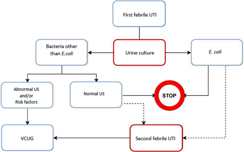
Fig. 4 Study flowchart with the 405
“new” protocol
Posive urine culture
142 excluded
- 38 CAKUT (26.8%)
- 104 US not performed (73.2%)
263
Enrolled in the study
40 (15.2%) 223 (84.8%)
Non E.coli E.coli
US No US
4
Missed diagnosis of pathological
11 (27.5%) 29 (72.5%) US with no UTI recurrence
Pathological US Normal US
30
Febrile UTI recurrence
US
21 VCUG
10 (33.3%) 20 (66.7%)
20 VUR
Pathological US Normal US
1 anterior urethral valvesPediatr Nephrol
Table 4 Voiding cystourethrography caused by episodes of fUTI as was previously believed
VCUG Number Percentage [10–16]. This is further supported by the analysis of data ob-
tained from the dialysis and transplant registries, which con-
Pathological 21 52.5% firms that aggressive protocols and surgical correction man-
VUR 20 agement or antibiotic prophylaxis of VUR, performed on the
Grade I 2 grounds of the assumption that kidney damage was acquired
Grade II 2 rather than congenital, did not reduce the number of paediatric
Grade III 10 patients requiring dialysis or transplant [17–20].
Grade IV 6 Numerous prospective randomised controlled trials
Grade V 0 assessing the efficacy of prophylaxis versus the treatment of
Anterior urethral valve 1 single fUTI relapses have been conducted on these findings
Negative 19 47.5% over the past 10 years. These studies have shown little or no
benefit in the prevention of fUTIs [21–28]. These findings
have led to a re-evaluation of the imaging protocols following
Therefore, when compared with the “standard” protocol, a first fUTI, and today, all major international guidelines [1–4]
the “new” protocol would have failed to detect four kidney recommend, albeit with some differences, the execution of
anomalies, all grade 1 hydronephrosis (Table 5), with non- KUS to select those children who will need to undergo more
evolutive disease at long-term follow-up. Considering a pop- invasive investigations such as VCUG and renal scintigraphy.
ulation of 1,628,739 children between 2 and 36 months in This new approach has led to an important reduction in the
Italy [13] and considering that about 5% of these children will number of VCUGs performed (up to 88% less) but the price to
experience a fUTI [14], 81,436 KUS would be performed be paid remains the high number of KUS which test negative
according to the majority of current guidelines [1–4]. (around 80%) [10, 29]. The top-down approach showed the
According to our data, 73% fewer KUS would be performed highest sensitivity in detecting kidney damage but also the
with the new protocol requiring the presence of an atypical highest economic and radiation costs. There is no ideal diag-
pathogen or a fUTI recurrence, with no lost diagnoses of se- nostic protocol following a first fUTI. An aggressive protocol
vere kidney anomalies and a total of 59,448 KUS avoided. has a high sensitivity for detecting VUR and scarring but
Assuming an average cost across the various Italian regions of carries high financial and radiation costs with questionable
€ 82 per KUS [9], the total yearly savings for the National benefit [9].
Healthcare System would amount to € 4,874,736, plus € The goal of this study was to create novel selection criteria
304,942 considering a reduction of 47.5% VCUGs assuming in order to reduce the number of negative ultrasounds and, if
an average cost of € 83 per VCUG [9]. possible, also that of VCUGs, and to investigate the impact
this novel protocol would have on the number of missed di-
agnoses of kidney anomalies other than VUR. These data
confirm the high percentage of negative KUS in patients with
Discussion
fUTI, with only 25 out of 263 (9.5%) ultrasounds performed
showing some anomaly (Fig. 3). If we had performed the KUS
This study showed that a fUTI caused by an atypical pathogen
only in patients with fUTIs caused by a pathogen other than
and recurring fUTIs appear to be targetable risk factors to
E. coli (40) or with fUTIs recurrence (30), a total of 70 KUS
detect kidney anomalies and could both be used to identify
would have been performed (Fig. 4), of which 20/70 positive
patients for whom a KUS would likely be positive and useful.
(28.6%), compared with the 25/263 (9.5%) obtained follow-
Over the past few years, the improvement in prenatal ultra-
ing the “standard” protocol, therefore resulting in a significant
sound technique has clarified how the anomalies of the kidney
reduction in KUS performed (73% less, 70 instead of 263). As
and urinary tract, especially dysplastic kidney, often highlight-
a matter of fact, the probability of having a positive KUS was
ed in association with VUR, are mainly congenital rather than
five times higher for those infected by an atypical agent rather
Table 5 Patients with positive ultrasound who would be missed with than E. coli. This correlation could be explained on the basis
the new protocol that pathogens lacking the adhesive capacity to the
urothelium, such as those which we classified as “other than
Patient N Age (months) Sex RUS VCUG
E. coli", can cause infection more frequently in children with
1 2.9 Male Grade 1 hydronephrosis Negative urinary tract anomalies, due to lacking of the protective mech-
2 1.6 Female Grade 1 hydronephrosis Negative anism provided by a normal urinary stream [15]. Alberici et al.
3 3.5 Male Grade 1 hydronephrosis Negative assessed the effectiveness of the risk factors proposed by the
4 23.4 Female Grade 1 hydronephrosis Negative Italian guidelines, highlighting that the only factor with a sta-
tistically significant correlation with the presence of a high-Pediatr Nephrol
grade VUR was the presence of a pathogen other than E. coli Conclusions
[10, 11]. Pauchard et al. found the same correlation with path-
ogens other than E. coli in 0–3-month-old babies, with the In conclusion, this study showed that a fUTI caused by an
26% chance of having a high-grade VUR rising to 55% if atypical pathogen appears to be a specific and targetable risk
associated with an abnormal US [8, 12]. factor to detect kidney anomalies that could be used as a se-
Of the patients with E. coli infection who would not have lection criterion to identify patients at the first episode of fUTI
undergone the KUS after the first episode of fUTI with no for whom a KUS would be useful. In children with E. coli
recurring fUTIs, we would have had 4 missed diagnoses of infection, the execution of an US after a fUTI recurrence could
pathological KUS, all first grade hydronephrosis with non- identify a further minority of patients at risk of malformations.
evolutive disease at long-term follow-up. The role of prenatal This approach would allow a significant reduction in the num-
US for mass screening for CAKUT in this setting appears ber of negative tests, saving time and reducing costs, with a
crucial, considering the low number of urinary anomalies negligible risk of missed diagnoses of kidney anomalies.
found in the post-natal evaluation of the cohort of our study Since children with abnormal prenatal KUS were excluded,
[14–16]. We tested the Italian guidelines considering the high these conclusions apply to children with normal prenatal
level of prenatal ultrasound screening in Italy where most KUS.
cases of significant CAKUT are detected. Prenatal US is rou- Further prospective multicentre randomised controlled tri-
tinely performed in the 20° and 30° week in all pregnancies as als will be needed to confirm our data, together with a better
a screening guaranteed by our National Health Care System. analysis of recurrence of fUTIs, as a tool for recognising non-
According to our data, 73% fewer KUS and 47.5% fewer previously intercepted patients to better evaluate a more spe-
VCUGs would be performed with the new protocol, with no cific target in the follow-up protocol of patients with fUTIs.
lost diagnoses of severe kidney anomalies and a cost-benefit
analysis predicting an almost 5 million Euro yearly saving for
the Italian National Health Care System.
Furthermore, considering that a minimum of 1-h time of What is known on this subject
engagement per family is required to perform a KUS, the
savings in terms of hours of work and transportation costs The kidney ultrasound is currently recommended in children
would be relevant as well. at their first febrile urinary tract infection by the majority of
Limitations of this study include the retrospective nature of current guidelines, but the large number of negative tests re-
the data employed. A further limitation is found in the fact that mains an issue.
we did not perform VCUGs in all the patients with atypical Despite being a non-invasive and radiation-free method,
pathogens and negative KUS, potentially losing some diagno- kidney ultrasound possesses low specificity for vesicoureteral
ses of VUR. However, according to the data collected, almost reflux.
none of those patients had a recurrence of fUTIs, making it
unlikely for them to have an underlying kidney abnormality.
On the other hand, our goal was not to identify low grade
VUR, which is generally not indirectly detectable via KUS, What this study adds
which is not associated with significant kidney damage and
does not require any treatment [24]. However, it is also no- A diagnostic protocol that required the presence of an atypical
ticed that KUS could have up to 79% sensitivity to detect high pathogen at the first febrile urinary tract infection or a second
grade VUR. On the contrary, we were interested in under- febrile urinary tract infection to perform the kidney ultrasound
standing what kind of kidney diseases other than VUR would would reduce negative tests with a negligible risk of missed
have gone undiagnosed by not performing a KUS. This choice kidney anomaly diagnoses.
is supported by the fact that VUR does not appear to be an
important and modifiable risk factor for recurrent fUTIs, new Authors’ contributions Dr Amoroso, Drs Conversano, and Drs Busetti
conceptualized and designed the study, drafted the initial manuscript, and
kidney scarring or CKD stage 5. Since new kidney scarring
reviewed and revised the manuscript. Drs Giangreco, Dr Pesce, Pennesi
develops only after recurrent fUTIs, the focus should be on the G., and Drs Cattaruzzi designed the data collection instruments, collected
prompt diagnosis and treatment of fUTIs [29]. Lastly, as the data, carried out the initial analyses, and reviewed and revised the man-
study was conducted in a third level-centre which provides uscript. Dr Pennesi M. and Prof Barbi conceptualized and designed the
study, coordinated and supervised data collection, and critically reviewed
excellent prenatal screening, the low number of kidney anom-
the manuscript for important intellectual content. All authors approved
alies detected during our post-natal evaluation could be due to the final manuscript as submitted and agree to be accountable for all
this factor. aspects of the work.Pediatr Nephrol
Compliance with ethical standards 15. Venhola M, Uhari M (2009) Vesicoureteral reflux, a benign condi-
tion. Pediatr Nephrol 24:223–226
Conflict of interest The authors declare that they have no conflict of 16. Hodson J, Maling TM, McManamon PJ, Lewis MG (1975) Reflux
interest. nephropathy. Kidney Int Suppl 4:S50–S58
17. Hewitt I, Montini G (2020) Vesicoureteral reflux is it important to
find? Pediatr Nephrol. https://doi.org/10.1007/s00467-020-04573-
9
References 18. Harambat J, van Stralen KJ, Kim JJ, Tizard EJ (2012)
Epidemiology of chronic kidney disease in children. Pediatr
1. Ammenti A, Alberici I, Brugnara M, Chimenz R, Guarino S, La Nephrol 27:363–373
Manna A, La Scola C, Maringhini S, Marra G, Materassi M, Morello 19. Craig JC, Irwig LM, Knight JF, Roy LP (2000) Does treatment of
W, Nicolini G, Pennesi M, Pisanello L, Pugliese F, Scozzola F, Sica F, vesicoureteric reflux in childhood prevent end-stage renal disease
Toffolo A, Montini G (2020) Updated Italian recommendations for the attributable to reflux nephropathy? Pediatrics 105:1236–1241
diagnosis, treatment and follow-up of the first febrile urinary tract in- 20. Brakeman P (2008) Vesicoureteral reflux, reflux nephropathy, and
fection in young children. Acta Paediatr 109:236–247 end-stage renal disease. Adv Urol 2008:508949
2. Subcommittee on urinary tract infection (2016) Reaffirmation of 21. Garin EH, Olavarria F, Garcia Nieto V, Valenciano B, Campos A,
AAP Clinical Practice Guideline: the diagnosis and management Young L (2006) Clinical significance of primary vesicoureteral re-
of the initial urinary tract infection in febrile infants and young flux and urinary antibiotic prophylaxis after acute pyelonephritis: a
children 2-24 months of age. Pediatrics 138:e20163026 multicenter, randomized, controlled study. Pediatrics 117:626–632
3. Stein R, Dogan HS, Hoebeke P, Kočvara R, Nijman RJ, Radmayr 22. Brandström P, Esbjörner E, Herthelius M, Holmdahl G, Läckgren
C, Tekgül S, European Association of Urology; European Society G, Nevéus T, Sillén U, Sixt R, Sjöberg I, Stokland E, Jodal U,
for Pediatric Urology (2015) Urinary tract infections in children: Hansson S (2010) The Swedish reflux trial in children: I. Study
EAU/ESPU guidelines. Eur Urol 67:546–558 design and study population characteristics. J Urol 184:274–279
4. Robinson JL, Finlay JC, Lang ME, Bortolussi R, Canadian 23. Pennesi M, Travan L, Peratoner L, Bordugo A, Cattaneo A, Ronfani
Paediatric Society, Infectious Diseases and Immunization L, Minisini S, Ventura A, North East Italy Prophylaxis in VUR
Committee, Community Paediatrics Committee (2014) Urinary study group (2008) Is antibiotic prophylaxis in children with
tract infections in infants and children: diagnosis and management. vesicoureteral reflux effective in preventing pyelonephritis and renal
Paediatr Child Health 19:315–325 scars? A randomized, controlled trial. Pediatrics 121:e1489–e1494
5. NICE: National Institute for Health and Clinical Excellence. 24. Montini G, Rigon L, Zucchetta P, Fregonese F, Toffolo A, Gobber
Urinary tract infection in children: diagnosis, treatment and long D, Cecchin D, Pavanello L, Molinari PP, Maschio F, Zanchetta S,
term management. Available at: www.nice.org.uk/nicemedia/pdf/ Cassar W, Casadio L, Crivellaro C, Fortunati P, Corsini A,
CG54fullguideline.pdf. Accessed October 15, 2020 Calderan A, Comacchio S, Tommasi L, Hewitt IK, Da Dalt L,
6. McTaggart S, Danchin M, Ditchfield M, Hewitt I, Kausman J, Zacchello G, Dall'Amico R, IRIS Group (2008) Prophylaxis after
Kennedy S, Trnka P, Williams G, Kidney Health Australia - first febrile urinary tract infection in children? A multicenter, ran-
Caring for Australasians with Renal Impairment (2015) KHA- domized, controlled, noninferiority trial. Pediatrics 122:1064–1071
CARI guideline: diagnosis and treatment of urinary tract infection 25. Roussey-Kesler G, Gadjos V, Idres N, Horen B, Ichay L, Leclair
in children. Nephrology (Carlton) 20:55–60 MD, Raymond F, Grellier A, Hazart I, de Parscau L, Salomon R,
7. Abdelhalim A, Khoury AE (2017) Critical appraisal of the top-down Champion G, Leroy V, Guigonis V, Siret D, Palcoux JB, Taque S,
approach for vesicoureteral reflux. Investig Clin Urol 58:S14–S22 Lemoigne A, Nguyen JM, Guyot C (2008) Antibiotic prophylaxis
8. Pauchard JY, Chehade H, Kies CZ, Girardin E, Cachat F, Gehri M for the prevention of recurrent urinary tract infection in children
(2017) Avoidance of voiding cystourethrography in infants youn- with low grade vesicoureteral reflux: results from a prospective
ger than 3 months with Escherichia coli urinary tract infection and randomized study. J Urol 179:674–679
normal renal ultrasound. Arch Dis Child 102:804–808
26. Craig JC, Simpson JM, Williams GJ, Lowe A, Reynolds GJ, SJ
9. La Scola C, De Mutiis C, Hewitt IK, Puccio G, Toffolo A,
MT, Hodson EM, Carapetis JR, Cranswick NE, Smith G, Irwig
Zucchetta P, Mencarelli F, Marsciani M, Dall'Amico R, Montini
LM, Caldwell PH, Hamilton S, Roy LP, Prevention of Recurrent
G (2013) Different guidelines for imaging after first UTI in febrile
Urinary Tract Infection in Children with Vesicoureteric Reflux and
infants: yield, cost, and radiation. Pediatrics 131:e665–e671
Normal Renal Tracts (PRIVENT) Investigators (2009) Antibiotic
10. Ammenti A, Cataldi L, Chimenz R, Fanos V, La Manna A, Marra
prophylaxis and recurrent urinary tract infection in children. N Engl
G, Materassi M, Pecile P, Pennesi M, Pisanello L, Sica F, Toffolo A,
J Med 361:1748–1759
Montini G, Italian Society of Pediatric Nephrology (2012) Febrile
27. Trial Investigators RIVUR, Hoberman A, Greenfield SP, Mattoo
urinary tract infections in young children: recommendations for the
TK, Keren R, Mathews R, Pohl HG, Kropp BP, Skoog SJ, Nelson
diagnosis, treatment and follow-up. Acta Paediatr 101:451–457
CP, Moxey-Mims M, Chesney RW, Carpenter MA (2014)
11. Alberici I, La Manna A, Pennesi M, Starc M, Scozzola F, Nicolini
Antimicrobial prophylaxis for children with vesicoureteral reflux.
G, Toffolo A, Marra G, Chimenz R, Sica F, Maringhini S, Monasta
N Engl J Med 370:2367–2376
L, Montini G (2019) First urinary tract infections in children: the
role of the risk factors proposed by the Italian recommendations. 28. Hewitt IK, Pennesi M, Morello W, Ronfani L, Montini G (2017)
Acta Paediatr 108:544–550 Antibiotic prophylaxis for urinary tract infection-related renal scar-
12. Hewitt IK, Montini G (2017) Re-evaluating the use of ultrasound to ring: a systematic review. Pediatrics 139:e20163145
investigate first febrile urinary tract infections in childhood. Acta 29. Pennesi M, L'erario I, Travan L, Ventura A (2012) Managing chil-
Paediatr 106:1727–1728 dren under 36 months of age with febrile urinary tract infection: a
13. http://dati.istat.it/ Accessed on 1 May 2020 new approach. Pediatr Nephrol 27:611–615
14. Shaikh N, Morone NE, Bost JE, Farrell MH (2008) Prevalence of
urinary tract infection in childhood: a meta-analysis. Pediatr Infect Publisher’s note Springer Nature remains neutral with regard to jurisdic-
Dis J 27:302–308 tional claims in published maps and institutional affiliations.You can also read