Moderate Versus Low Intensity Aerobic Exercise on Bone Mineral Density in Patients on Hemodialysis
←
→
Page content transcription
If your browser does not render page correctly, please read the page content below

Moderate Versus Low Intensity Aerobic Exercise on Bone
           Mineral Density in Patients on Hemodialysis
      Nesreen G. El-Nahas* , Heba A. Bahey, ** Shimaa N.Aboelazm***
 * Department of physical therapy for Cardiovascular \Respiratory Disorder and Geriatrics, Faculty of
 Physical Therapy, Cairo University. ** Lecturer of Physical Therapy for Surgery, Faculty of Physical
 Therapy, Misr University for Sciences and Technology ,.*** Department of Basic Sciences, Faculty of
 Physical Therapy, Misr University for sciences and technology.

                                           Abstract
        Chronic kidney disease (CKD) is recognized as a major health problem reflecting
the growing elderly population and increasing numbers of patients with diabetes and
hypertension. Medical researches confronted with management of complex medical
problems that are unique to patients with chronic renal impairment and renal dialysis where
patients suffer from hypocalcemia that subjected them to osteoporosis. Objective: The aim
of this study was to compare the effect of two different intensities of aerobic exercises on
bone mass density in patients on haemodialysis Subjects: Thirty male patients underwent
renal haemodialysis for 2 years ago with mean age (52.75±4.51)were recruited from Police
hospital . Methods: They were assigned randomly into two groups, 15 patients in each
group. Group (A) attended a program of moderate intensity aerobic treadmill exercise (60-
70%MHR), where Group (B) attended a program of light intensity aerobic treadmill
exercise (40-60%MHR), both for 6 months (3 sessions of exercise per week) prior to the
dialysis session. Laboratory investigations for serum calcium and phosphorus level in
addition to a dual X ray absorpimetry (DXA) were applied at baseline and after 6 months
of training for both groups. Results: The study revealed a significant difference in bone
mineral density in favor of group A with P- value 0.01 as well as a significant increase in
serum calcium by 12.29 %, 4.23 % and significant decrease in serum phosphorus with
21.67 %, 6.52 % for group A and B respectively. Conclusion: Moderate intensity aerobic
exercise is more effective than light intensity aerobic exercise in modulating serum calcium
and phosphorus and thus improving BMD in patients with hemodialysis.
Key words: Bone Mass Density /Osteoporosis/ Renal Haemodialysis/ Aerobic
Exercises.

INTRODUCTION
        Chronic kidney disease (CKD) is a progressive condition that often
 comes with other multiple complications, such as diabetes, hypertension,
 renal osteodystrophy, anemia, cardiovascular disease, and malnutrition. The
 earlier the recognition of CKD and treatment of its complications the better
 the long-term outcomes (1). kidneys have many important roles, such as
 regulating fluid and minerals in the body, they stimulate bone marrow to
 make red blood cells, synthesize vitamin D, regulate blood pressure, excrete
 waste chemicals in the urine and regulate acid-base levels. In kidney failure,

                                                  1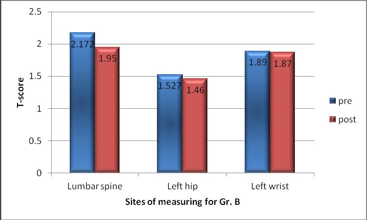
the blood concentrations of calcium and phosphorus become abnormal.
Calcium level drop is a condition called hypocalcaemia that can cause
muscle weakness and nerve problems. In contrast, phosphorus levels rise.
This is a condition called hyperphosphatemia, which can cause bone
problems and itching. (2).

      Hypocalcaemia occurs in kidney failure for at least two reasons. First,
kidneys cannot synthesize vitamin D which normally raises the level of
calcium in the body. Without vitamin D, calcium is not absorbed from the
diet. Second, high levels of phosphate that could not bind to calcium deposit
in the tissues as the diseased kidney could not excrete it. Low calcium levels
encourage the release of parathyroid hormone ( PTH). This hormone
increases blood calcium by reabsorbing calcium from the bones. This can
lead to a condition called renal osteodystrophy(ROD) .(3) . The syndrome
known as chronic kidney disease–mineral and bone disorder (CKD-MBD) is
composed of clinical, biochemical and radiological abnormalities where
progressive bone loss and muscle cramping frequently occur (4).

   Bone strength reflects the integration of two main features: bone density
and bone quality. Bone density is expressed as grams of mineral per area or
volume, and in any given individual is determined by peak bone mass and
amount of bone loss. Bone quality refers to architecture, turnover, damage
accumulation (e.g.,microfractures) and mineralization (5) .Normal bone
density is defined as being (-1 standard deviation) or greater than the mean
at 30-40 years (peak bone mass). Bone density between -1 SD and -2.5 SD
of peak bone mass (T score between −1.5 and −2.5) has been defined by the
WHO as osteopenia, and equal or below 2.5 SD of peak bone mass (a T
score ≤ − 2.5), as osteoporosis (6) .However, not every person diagnosed with
osteopenia will develop osteoporosis (7).

      Renal osteodystrophy (ROD) is a spectrum of bone mineral changes
that could range from the high-turnover lesions of secondary
hyperparathyroidism to the low-turnover lesions of a dynamic bone disease
.The impact of different types of ROD on bone density in patients with CKD
remains undefined. Dual energy X-ray absorptiometry (DXA) is the
commonest method used to screen for osteoporosis in adults due to its
precision and accuracy, short scan time and low radiation (3).

                                      2
Aerobic exercise increases bone mass by using body weight as the
resistance. Walking and running are great ways to increase or maintain bone
mass while increasing cardiovascular fitness. (8)
Exercise training in adults with CKD can affect the following factors:
Muscular hypotrophy, strength, endurance & physical functioning (9,10).The
structure and number of capillaries and mitochondria (11). Glucose metabolism
(12)
.Aerobic capacity (13).Blood pressure (14)and Cardiac performance (11).
It is known that inactivity, muscle wasting and reduced physical
functioning especially for those on long-term dialysis are associated with
increased mortality in CKD. Exercise in patients receiving regular dialysis
as a treatment for end-stage renal disease was first introduced 3 decades ago,
but is still only offered in a minority of renal units around the world, despite
a significant body of evidence to support its use. Work is needed to increase
awareness of the potential benefits of increased physical activity for patients
with advanced CKD (15).
This study was conducted to compare the effect of two different
intensities of aerobic exercises on bone mass density in patients with
haemodialysis.
SUBJECTS, MATERIALES AND METHODS
Subjects:
Thirty male patients with mean age (52.75±4.51) years were enrolled in
this study; they underwent renal haemodialysis for 2 years ago with rate of 3
times/week. They were randomly selected from Police hospital using one to
one base. All patients gave their written informed consent for the
participation in the study that had been preceded by the explanation of the
aim of the study and its course, their role in it with regard to time and
money, assurance of protection of the obtained data, and information about
free-willingness to participate in the study and the possibility to withdraw
from the study at any time.
Inclusion crieteria:
- body mass index ranged from 25 to 29 kg/m2.
- systolic blood pressure ranged between 130-190 mm Hg.
- diastolic blood pressure between 85-100 mm Hg).
3- T- score between -1.1and -2.4 SD according to DEXA measurements.
- Male subjects with age ranged from 45 to 55 years
Exclusion crieteria:
-chest ,cardiac, or hepatic diseases.
- severe life limiting illness (e.g. malignancy).
- marked anaemia(Ht1) AP view of the lumbar (L1–L4) spine.
2) AP view of the left hip providing information on the femur.
3) AP view of the left wrist with the subject supine.
While the scanner moved across the left hip, providing information on the
femur neck (whole hip), left wrist (33% of left radius), and measure lateral
view of the lumbar (L1–L4) spine. Regional and total body BMD
measurements with this technique are highly reliable when subject positioning
is carefully standardized (16)..
The test results included the following scores:
T score, Z score, Bone mineral density, Percentage, Age matched percentage.
Training program:
Patients were randomly assigned into two groups of equal number, Group A
and Group B (each group consists of fifteen patients) Patients were recruited
two hours early prior to dialysis session . electronic treadmill and pulsometer
were used to perform walking training program, with maximum Heart Rate
(MHR) calculated according to (220-Age) for men.
Group A received a program of moderate intensity aerobic exercise (60%-70%
MHR) with an exercise period of 40 minutes divided as warming up phase:3-5
min. with 30% MHR, actual phase:20-30 min. with 60%-70% MHR and cooling
down phase:3-5 min. with 30% MHR three times weekly for six months.
Group B received a program of light intensity aerobic exercise (40%-60%
MHR) with an exercise period of 40 minutes divided as warming up phase : 3-5
min. with 30% MHR, actual phase: 20-30 min. with 40%-60% MHR and cooling
down phase:3-5 min. with 30% MHR three times weekly for six month. The
training program was performed under careful supervision for both groups.
RESULTS
Table (1): Demographic characteristics of the patients in both groups
(A&B).
Items Group A Group B Comparison
Mean ±SD Mean ±SD t- P- S
value value
Age (yrs) 51.86 ±4.22 53.6 ±4.82 1.04 0.3 NS
Weight (Kg) 86.26 ±10.06 84.66 ±7.37 0.49 0.62 NS
Height (cm) 172.26 ±5.68 169.33 ±6.97 1.26 0.21 NS
2
BMI (Kg/m ) 29.02 ±2.61 29.56 ±2.53 0.57 0.57 NS
5Systolic blood 162.66 ±17.91 165.33 ±15.52 0.43 0.66 NS
pressure
(mmHg)
Diastolic blood 93.66 ±5.49 91.66 ±5.87 0.96 0.34 NS
pressure
(mmHg)
Yrs: years, Kg.: Kilograms, Cm. centimeters, Kg/m2: Kilogram per meter
square, mmHg: millimeters mercury
Table (2): Statistical Analysis of Calcium levels pre and post treatment
for both groups (A & B).
Calcium level Group A Group B Between both
groups
Pre Post Pre Post Post post
Mean ± SD 7.97±0.7 8.95±0.6 8.03±0. 8.38±0.52 0.06 0.57
2 1 5
t-value 7.5 3.02 0.26 2.75
P-value 0.0001* 0.009* 0.79 0.01*
Percentage of
12.29 %
improvement 4.23 %
SD: Standard Deviation, *: Significance
Group (A) Group (B)
10
8 8.95
8.38
7.97 8.03
Calcium level
6
4
2
0
Pre treatment Post treatment
Fig.(1): Mean and ±SD of Calcium level pre and post treatment of groups
(A,B).
Table (3): Statistical Analysis of Phosphorus levels pre and post
6treatment for both groups (A & B).
Phosphorus Group A Group B Between
level both groups
Pre Post Pre Post Post post
Mean ± SD 6.46±1.39 5.05±1.21 6.44±0.8 6.02±0.78 0.01 0.96
t-value 6.71 3.09 0.03 2.59
P-value 0.0001* 0.008* 0.97 0.01*
Percentage 6.52 %
of 21.67 %
improvement
SD: Standard Deviation, *: Significance
Group (A) Group (B)
10
8
Phosphorus level
6
6.46 6.44 6.02
4 5.05
2
0
Pre treatment Post treatment
Fig.(2): Mean and ±SD of Phosphorus level pre and post treatment of
groups (A,B).
Table(4): Mean values of T score pre and post treatment at lumbar
spine, left hip, left wrist for group A
Group A pre Post T- value P- Percentage
exercise exercise value of change
Lumbar 1.30 ± 1.10 ± 6.96 0.001* 18.2%
spine 0.747 0.759
Left hip 1.36 ± 1.20 ± 6.07 0.001* 13.3%
0.894 0.928
Left wrist 1.40 ± 1.30 ± 7.22 0.001* 7.69%
0.692 0.776
*: Significance
7Fig (3): The mean values of T score before and after exercise at lumbar
spine, left hip, left wrist in group A.
Table(5) : Mean values of T score pre and post treatment at lumbar
spine, left hip, left wrist for group B
Group B Pre Post T- P- Percentage
exercise exercise value value of change
Lumbar 2.17 ± 1.95 ± 0.90 6.96 0.001* 11.3%
spine 0.72
Left hip 1.53 ± 1.50 ± 0.95 6.07 0.001* 2%
0.91
Left wrist 1.89 ± 1.0 1.87 ± 0.97 7.23 0.345 ---------
*: Significance
Fig (4): The mean values of T score before and after exercise at
lumbar spine, left hip, and left wrist in group B.
Table (6): Comparison between post treatment between both groups
(A&B):-
8Group A Group B T- P- value
value
Lumbar spine 1.95±0.82 1.10±0.75 -2.779 0.010*
Left hip 1.50±0.95 1.20±0.92 -0.735 .467
Left wrist 1.90±0.96 1.40±0.77 1.51 0.14
DISCUSSION:
Changes in calcium metabolism during exercise are dependent on the
exercise intensity. Moderate endurance exercise increases serum calcium
level (17) but decreases serum PTH (18). In bone, endurance exercise increases
bone mineral density (BMD), bone strength (19) and bone formation rate (20).
Thus, moderate endurance exercise seems to induce positive calcium
balance, and has a beneficial effect on bone metabolism. In addition, a
combination of moderate-impact exercise and adequate calcium intake can
increase bone strength during childhood (21). Interestingly, modes of
exercise, such as running (weight-bearing exercise) and swimming (non-
weight-bearing exercise) can affect bone calcium metabolism in a different
way.
It is established that physical activity before dialysis treatment
increases urea Kt/V through improved perfusion of muscle, the main urea-
containing body compartment. Similar effects have been described for
phosphate removal with predialysis physical activity increasing phosphate
removal by 6% and intradialytic activity even by 9% (22).
Even moderate exercise is related to an enhanced bone mineral density
in peripubertal boys and also in young men compared to controls with a low
level of physical activity. Animal studies have demonstrated that such an
increase in bone mass is the result of an enhanced formation of organic bone
matrix and a higher apposition rate of minerals such as calcium (Ca). A
moderate level of physical exercise can already acutely influence various Ca
metabolic parameters in untrained human subjects: Alterations can include a
decrease in ionized serum Ca levels and an increase in serum parathyroid
hormone (PTH) levels (23).
In the present study there were significant difference between the two
9groups (group A &B) in serum blood sample of calcium and phosphorus, as
there were significant increase in groups (A& B) in serum calcium (12.29%,
4.23%) respectively, and significant decrease in serum phosphorus in group
A compared to group B (21.67%, 6.52%) respectively in response to the
designed aerobic exercise program.
As well as increased percent of improvement in T score for group A in
the measured sites lumbar spine, left hip and left wrist by 18.2%,13.3% and
7.69% respectively . Regarding to group B the percent of improvement was
less as shown in lumbar spine by 11.3% and left hip by 2% with no change
in the left wrist. That dragged the emphasis to the effect of the moderate
exercise applied to group A that gives significant increase in bone mineral
density for patients on renal hemodialysis.
The results of the study after the suggested period of treatment confirmed
the findings of John et al., 2007 (24) who stated that moderate exercise
intensity results in regional increase in bone mass.
This Coincided with Asadi et al., 2007 (25) who studied the effects of
exercise in reducing phosphorus levels and reported that although exercise
decreased the level of phosphorus, the significant effects and changes could
be observed in long-term and perhaps more intense exercise might be
required for some patients.
The rehabilitation of the hemodialysis patients is enhanced, most
likely because aerobic exercise induces elongation and an increase in the
diameter of the striated muscle fibre, improves their capillary vasculature, as
well as their aerobic capacity, and positively affects their blood pressure
measurements, their brain function and the lipid profile. The increased ionic
calcium of the cell sarcoplasma in the skeletal muscles, which is prevalent
during the muscle contractions (26) .
The results of numerous studies have shown that exercise training to be
of benefit for dialysis patients(27)during haemodialysis on physical
performance and nutrition assessment that agreed with results of this study.
In addition to its well-known beneficial effects on cardiovascular fitness and
mortality (28) exercise also has an anabolic effect and has been shown to
reduce muscular atrophy in dialysis patients (29).
10The results of the present study showed significant improvement in
calcium and phosphorus electrolytes with aerobic exercise during
hemodialysis that coincided with the data presented byVaithilingam et al.,
2004 (22), who suggest that an aerobic exercise movement’s regimen for 15
minutes during hemodialys is sessions improve serum phosphate and
calcium levels in a period of 8 weeks. This observation might be due to
direct beneficial effects of aerobic exercise or general effects of regular
intradialytic exercise.
These findings agree with Hagberg et al., 2001 (30) who stated that
prolonged low-to-moderate-intensity physical activity was associated with
higher BMD.
The results supports the findings of Vencint and Braith, 2002 (31),who
reported that, regional BMD can be increased via high-intensity resistance
exercise even in healthy elderly persons. The results also indicate that both
high- and low-intensity resistance exercises can change biochemical indices
of bone turnover. As evidenced by increased OC/PYD and BAP/PYD ratios,
these changes seemingly favor increased bone formation.
The results of this study are also consistent with that stated by Hurley
and Stephen ,2000 (32) who reported that strength training is considered a
promising intervention for reversing the loss of muscle function and the
deterioration of muscle structure that is associated with advanced age as well
as osteoporotic effects due to renal dialysis. This reversal is thought to result
in improvements in function abilities and health status in patients on dialysis
by increasing muscle mass, strength and power and by increasing bone
mineral density (BMD).
Conclusion
The results of this study supported the good effect of aerobic exercise
on serum calcium and phosphorus in patients under renal hemodialysis.
Aerobic exercise showed a significant increase serum calcium and
significant decrease serum phosphorus in both groups in addition to
increased BMD. The result of this study concluded that moderate intensity
aerobic exercise (60%-70%MHR) is beneficial than light intensity aerobic
exercise (40%-60%MHR) in modulating serum calcium and phosphorus in
hemodialytic patients reflected on BMD.
11References
1-U.S. Renal Data System, USRDS 2006 Annual Data Report: Atlas of End-Stage
Renal Disease in the United States, National Institutes of Health, National Institute of
Diabetes and Digestive and Kidney Diseases, Bethesda, MD, 2006. Available from:
http://www.usrds.org/adr.htm
2-Kumar V (2009); “Robbins and Cotran Pathologic Basis of Disease.” 8th Ed, 2
3-Zittermann A, Sabatschus O., Jantzen S., Platen P., Danz A. and P. Stehle P.
(2002): Evidence for an acute rise of intestinal calcium absorption in response to aerobic
exercise Eur J Nutr 41 : 189–196 (2002)
4-Fauci AS (2008); "Harrison's Principles of Internal Medicine" 2008; 17th Edition
268:1373, 1313.
5-Anthony D. Woolf, Kristina Akesson. Book of An Atlas of Investigation and
Manegement, Clinical Publishing, Oxford,2008. (book1)
6-WHO Scientific Group on the Prevention and Management of Osteoporosis
(2000 : Geneva, Switzerland) (2003). "Prevention and management of osteoporosis :
report of a WHO scientific group" (pdf). Retrieved 2007-05-31. (thesis 2)
7-Wluka, A.E., Wang, Y., Davis, S.R. and Cicuttini, F.M.: Tibial plateau size is
related to grade of joint space narrowing and osteophytes in healthy women and in
women with osteoarthritis: Ann Rheum Dis., 64: 1033 – 1037, 2005.
8-Beck BR and Snow CM(2003): Bone health across the lifespan exercising our
options. Exerc Sport Sci Rev.; 31:117-122.
9-Heiwe S, Clyne N, Tollback A, Borg K. (2005). “Effects of regular resistance training
on muscle histopathology and morphometry in elderly patients with chronic
kidney disease”. American journal of physical medicine & Rehabilitation
/Association of Academic Physiatrists.; 84(11):865-74.
10-Mcintyre CW, Selby NM, Sigrist M, Pearce LE, Mercer TH, Naish PF. (2006).
Patients receiving maintenance dialysis have more severe functionally
significant skeletal muscle wasting than patients with dialysis-independent
chronic kidney disease. Nephrol Dial Transplant.; 21(8):2210-6.
11-Cheema BS, Abas H, Smith B, O'Sullivan AJ, Chan M, Patwardhan A, Kelly J,
Gillin A, Pang G, Lloyd B, Berger K, Baune BT, Fiatarone Singh MA. (2010).
12“Effects of resistance training during hemodialysis on circulating cytokines”: a
randomised controlled trial. Eur J Appl Physiol Dec (Epub ahead of print).
12- Painter P, Moore G, Carlson L, Paul S, Myll J, Philips W and Haskell W. (2002).
Effect of exercise training plus normalization of hematocrit on exercise
capacity and health related quality of life. American Journal of Kidney
Disease 39:257-265.
13-DePaul V, Moreland J, Eager T, Clase CM. (2002). The effectiveness of aerobic
and muscle strength training in patients receiving hemodialysis and EPO: a
randomized controlled trial. Am J Kidney Dis. Dec; 40(6):1219-29.
14- Pechter U, Ots M, Mesikepp S, Zilmer K, Kullissaar T, Vihalemm T, et al.
(2003). Beneficial effects of water-based exercise in patients with chronic
kidney disease. International journal of rehabilitation research Internationale
Zeitschrift fur Rehabilitationsforschung. Jun; 26(2):153-6.
15- Kosmadakis GC., Bevington A., Smith AC., Clapp EL., Viana JL., Bishop
NC and Feehally J. (2010). Physical Exercise in Patients with Severe
Kidney Disease Nephron Clin Pract; 115:c7-c16
16-Carter D, Bouxsein M, and Marcus R: New approaches for interpreting projected
bone densitometry data. J Bone Miner Res 7:137-145. (thesis 1) (1992)
17- Yeh JK, Aloia JF (1990). “Effect of physical activity on calciotropic hormones and
calcium balance in rats”. Am J Physiol; 258: E263–E268.
18- Yasumura S. “Effect of physical activity on calcium and phosphorus metabolism in
the rat”. Am J Physiol 1989; 256: E1–E6.
19- Huang TH, Lin SC, Chang FL, Hsieh SS, Liu SH, Yang RS. (2003). “Effects of
different exercise modes on mineralization, structure, and biomechanical
properties of growing bone”. J Appl Physiol ; 95: 300–3
20- Hart KJ, Shaw JM, Vajda E, Hegsted M, Miller SC. (2001): “Swim-trained rats
have greater bones mass, density, strength, and dynamics”. J Appl Physiol;
91: 1663–1668.
21- Welch JM and Weaver CM (2005). “Calcium and exercise affect the growing
skeleton”. Nutr Rev; 63: 361–373.
22- Vaithilingam I, Polkinghorne KR, Atkins RC, Kerr PG (2004): “Time and
exercise improve phosphate removal in hemodialysis patients”. Am J Kidney
Dis; 43:85–89.
23- Suki WN. (2008). Effects of sevelamer and calcium-based phosphate binders on
mortality in hemodialysis patients: Results of a randomized clinical trial. J
Ren Nutr 18: 91–98.
1324- John C. Stevenson and Michael S. Marsh (2007): An Atlas of Osteoporosis, third
edition.
25- Asadi N A, Bassampour S H, Zolfaghari M (2007). Critical care nursing ICU,
CCU, dialysis. Salemi publication; 2th ed.; p. 448-53.
26- Ioannis Karamouzis, Dimitrios Grekas, Michael Karamouzis, Konstantinos
Kallaras, Vassiliki Stergiou-Michailidou, Evangelia Kouidi, Asterios Deligiannis,
Norma Vavatsi-Christaki :( 2009) Physical training in patients on hemodialysis has a
beneficial effect on the levels of eicosanoid hormone-like substances. HORMONES,
8(2):129-137
27- Konstantinidou E, Koukouvou G, Kouidi E, Deligiannis A, Tourkantonis A,:
2002 Exercise training in patients with end-stage renal disease on hemodialysis:
comparison of three rehabilitation programs. J Rehabil Med 34: 40- 45.
28- Deligiannis A, 2004: Cardiac adaptations following exercise training in
hemodialysis patients. Clin Nephrol 61: 539-545.
29- Kouidi A, Albani M, Natsis K. (1998). The effects of exercise training on muscle
atrophy in haemodialysis patients. Nephrol Dial Transplant; 13: 685–699)
30-Hagberg JM, Zmuda JM, McCole SD, Rodgers KS, Ferrell RE, Wilund KR,
Moore GE.(2001): Moderate physical activity is associated with higher bone mineral
density in postmenopausal women. J Am Geriatr Soc. Nov;49(11):1411-7.
31- Vincent R K and Braith W R (2002): Resistance exercise and bone turnover in
elderly men and women. Medicine and science in sports and exercise: 17-23.
32- Hurley FB and Stephen MR (2000): Strength Training in the Elderly. Effects on
Risk Factors for Age-Related Diseases. Sports Med Oct; 30 (4): 249-268.
Role of funding source
No benefits or funds were received in support of this study. None of the
authors has received or will receive benefits for personal or professional use
from a commercial party related directly or indirectly to the subject of this
article.
Acknowledgements
We would like to express a great gratitude to Dr. Nivin A. Abdel-Kawy
physical therapist at Police Hospital for her help and support during and
after conduction of the study.
Conflict of interest :- Author has not declared any conflict of interest.
1415
You can also read