Complement and tissue factor-enriched neutrophil extracellular traps are key drivers in COVID-19 immunothrombosis
←
→
Page content transcription
If your browser does not render page correctly, please read the page content below
Complement and tissue factor-enriched neutrophil extracellular
traps are key drivers in COVID-19 immunothrombosis
Panagiotis Skendros, … , John D. Lambris, Konstantinos Ritis
J Clin Invest. 2020. https://doi.org/10.1172/JCI141374.

 Concise Communication      In-Press Preview COVID-19     Immunology

Emerging data indicate that complement and neutrophils contribute to the maladaptive immune response that fuels
hyper-inflammation and thrombotic microangiopathy, thereby increasing COVID-19 mortality. Here, we investigated how
complement interacts with the platelet/neutrophil extracellular traps (NETs)/thrombin axis, using COVID-19 specimens,
cell-based inhibition studies and NETs/human aortic endothelial cell (HAEC) co-cultures. Increased plasma levels of
NETs, tissue factor (TF) activity and sC5b-9 were detected in patients. Neutrophils of patients yielded high TF expression
and released NETs carrying active TF. Treatment of control neutrophils with COVID-19 platelet-rich plasma generated TF-
bearing NETs that induced thrombotic activity of HAEC. Thrombin or NETosis inhibition or C5aR1 blockade attenuated
platelet-mediated NET-driven thrombogenicity. COVID-19 serum induced complement activation in vitro, consistent with
high complement activity in clinical samples. Complement C3 inhibition with compstatin Cp40 disrupted TF expression in
neutrophils. In conclusion, we provide a mechanistic basis for a pivotal role of complement and NETs in COVID-19
immunothrombosis. This study supports strategies against SARS-CoV-2 that exploit complement or NETosis inhibition.

Find the latest version:
https://jci.me/141374/pdf
Complement and tissue factor-enriched neutrophil extracellular traps are key

drivers in COVID-19 immunothrombosis

Panagiotis Skendros1,2*, Alexandros Mitsios2*, Akrivi Chrysanthopoulou2*, Dimitrios

C. Mastellos3, Simeon Metallidis4, Petros Rafailidis5, Maria Ntinopoulou2, Eleni

Sertaridou6, Victoria Tsironidou2, Christina Tsigalou7, Maria Tektonidou8, Theocharis

Konstantinidis2,     Charalampos     Papagoras1,2,   Ioannis   Mitroulis1,2,   Georgios

Germanidis4, John D. Lambris9#, Konstantinos Ritis1,2#.

* These authors contributed to this work equally
#
    These authors co-supervised this work.

1
    First Department of Internal Medicine, University Hospital of Alexandroupolis,

Democritus University of Thrace, Alexandroupolis, Greece.
2
    Laboratory of Molecular Hematology, Department of Medicine, Democritus

University of Thrace, Alexandroupolis, Greece.
3
    National Center for Scientific Research „Demokritos‟, Aghia Paraskevi, Athens,

Greece.
4
    First Department of Internal Medicine, AHEPA University Hospital, Aristotle

University of Thessaloniki, Thessaloniki, Greece.
5
    Second Department of Internal Medicine, University Hospital of Alexandroupolis,

Democritus University of Thrace, Alexandroupolis, Greece.
6
    Intensive Care Unit, University Hospital of Alexandroupolis, Alexandroupolis,

Greece.
7
    Laboratory of Microbiology, University Hospital of Alexandroupolis, Democritus

University of Thrace, Alexandroupolis, Greece.

                                             1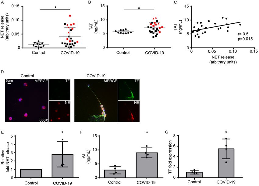
8
    First Department of Propaedeutic Internal Medicine, National and Kapodistrian

University of Athens, Greece
9
    Department of Pathology & Laboratory Medicine, Perelman School of Medicine,

University of Pennsylvania, Philadelphia, PA, USA.

Correspondence to:

Konstantinos Ritis, MD, PhD
First Department of Internal Medicine,
University Hospital of Alexandroupolis,
Democritus University of Thrace,
Alexandoupolis, 68100, Greece
Tel. +00302551351103
e-mail: kritis@med.duth.gr

Conflict of interest

JDL is the founder of Amyndas Pharmaceuticals which develops complement

inhibitors for therapeutic purposes, and inventor of patents that describe the

therapeutic use of complement inhibitors, some of which are developed by Amyndas.

JDL is also the inventor of the compstatin technology licensed to Apellis

Pharmaceuticals (i.e., 4(1MeW)7W/POT-4/APL-1 and PEGylated derivatives such as

pegcetacoplan and APL-9).

The remaining authors have no conflict of interest to declare.

.

                                           2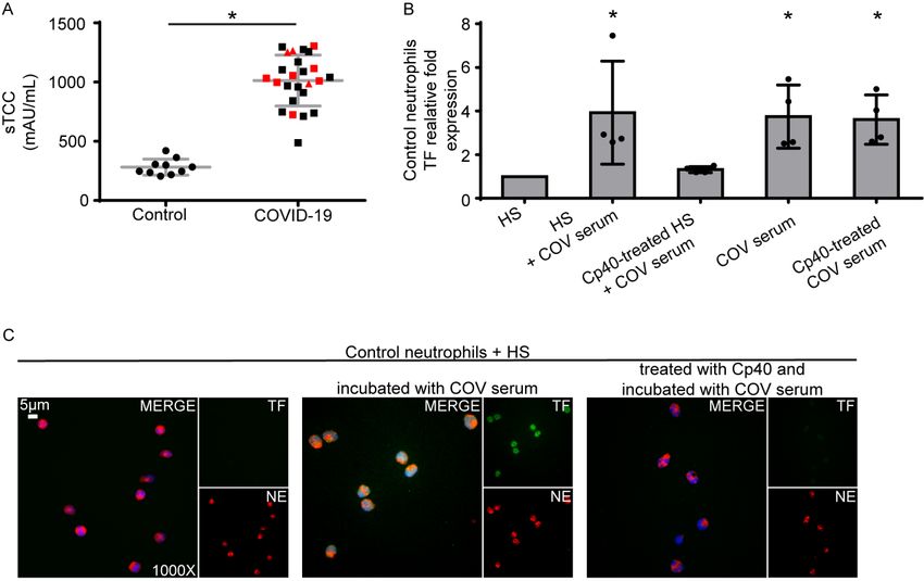
Abstract

Emerging data indicate that complement and neutrophils contribute to the maladaptive

immune response that fuels hyper-inflammation and thrombotic microangiopathy,

thereby increasing COVID-19 mortality. Here, we investigated how complement

interacts with the platelet/neutrophil extracellular traps (NETs)/thrombin axis, using

COVID-19 specimens, cell-based inhibition studies and NETs/human aortic

endothelial cell (HAEC) co-cultures. Increased plasma levels of NETs, tissue factor

(TF) activity and sC5b-9 were detected in patients. Neutrophils of patients yielded

high TF expression and released NETs carrying active TF. Treatment of control

neutrophils with COVID-19 platelet-rich plasma generated TF-bearing NETs that

induced thrombotic activity of HAEC. Thrombin or NETosis inhibition or C5aR1

blockade attenuated platelet-mediated NET-driven thrombogenicity. COVID-19

serum induced complement activation in vitro, consistent with high complement

activity in clinical samples. Complement C3 inhibition with compstatin Cp40

disrupted TF expression in neutrophils. In conclusion, we provide a mechanistic basis

for a pivotal role of complement and NETs in COVID-19 immunothrombosis. This

study supports strategies against SARS-CoV-2 that exploit complement or NETosis

inhibition.

Keywords: Complement, compstatin Cp40, platelets, neutrophil extracellular traps,

tissue factor, immunothrombosis, COVID-19.

                                          3
Introduction
Accumulated clinical evidence during the evolving COVID-19 pandemic indicates
that severe acute respiratory syndrome coronavirus 2 (SARS-CoV-2) infection
triggers thrombotic complications that affect multiple vital organs, increasing the
mortality burden in COVID-19 patients (1, 2). The pathological process of
coagulopathy in COVID-19 is commonly characterized as an immunothrombosis,
since it is related to a maladaptive host immune response fueled by excessive
activation of innate immune pathways, deregulated thromboinflammation and
endothelial dysfunction (1, 2). Understanding the immunothrombotic mechanisms in
COVID-19 constitutes a significant medical challenge.
Increased neutrophil counts have been associated with disease severity and poor
prognosis in COVID-19, and extensive neutrophil infiltration of pulmonary capillaries
has been described in autopsy specimens (3–7). In several inflammatory disorders (8),
neutrophil extracellular traps (NETs) have been shown to exert thrombogenic activity
through the expression of functionally active tissue factor (TF). NETs appear to be
involved in COVID-19 as well (9).
Complement activation has been implicated as driver of the maladaptive
inflammatory response in COVID-19. Complement can enhance neutrophil/monocyte
activation and recruitment to the infected lungs, and several complement effectors,
acting in concert with platelets, can fuel thromboinflammation, microvascular
thrombosis and endothelial dysfunction (thrombotic microangiopathy) (7, 10, 11).
Therefore, early clinical data have prompted the initiation of trials to evaluate various
complement therapeutics in COVID-19 patients (12).
The well-established cross-talk between complement and neutrophils in human
immunothrombosis (13–15) led us to hypothesize that the collaboration of these
4innate immune systems may mediate early events leading to coagulopathy in COVID-
19. Here, we have investigated the role of neutrophils in COVID-19
thromboinflammation and provide evidence that complement activation potentiates
the platelet/NETs/TF/thrombin axis during SARS-CoV-2 infection.
Results and discussion
NETs are associated with activation of the TF/thrombin axis in COVID-19
We first used ELISA to measure the levels of myeloperoxidase (MPO)/DNA
complexes (Figure 1A), a well-defined surrogate marker of NETosis (8), in the
plasma of control and COVID-19 patients and detected significantly increased levels
in COVID-19 patients. The levels of these complexes were positively correlated with
thrombin-antithrombin (TAT) activity, indicating activation of the TF/thrombin axis
(Figure 1B,C). These findings were further confirmed by confocal
immunofluorescence microscopy in neutrophils collected from four patients with
severe COVID-19 (Supplemental Table S1). We found spontaneous formation of
NETs expressing TF (Figure 1D,E) that were functional, as indicated by TAT assay
(Figure 1F). Moreover, the concomitant increase in TF mRNA levels (Figure 1G)
further confirmed previous studies indicating that neutrophils constitute source of TF
in thromboinflammation. Although detection of NETosis markers in plasma by
similar laboratory techniques has already been reported (9), we now show that NET
release is positively correlated with in vivo thrombotic potency in COVID-19 (Figure
1C).
In an effort to corroborate our ex vivo findings, and because platelet-neutrophil
interactions are necessary for NET formation in several thomboinflammatory
disorders (8, 16), neutrophils isolated from healthy individuals (control neutrophils)
5were stimulated with PRP from COVID-19 patients. We found that these PRP-
stimulated control neutrophils had increased levels of TF mRNA (Figure 2A) and
efficiently generated TF-bearing NETs (Figure 2B,D,E); the corresponding NETs
showed high TAT activity (Figure 2C), supporting the important role of platelets in
NET-mediated COVID-19 immunothrombosis. On the other hand, although COVID-
19 serum or plasma led to intracellular TF expression in control neutrophils, they
were not able to trigger efficient NET formation (Supplemental Figure S1A-C),
further supporting previous studies indicating that the release of TF-expressing NETs
is a “double-hit phenomenon” (8, 16). That is, PRP includes both the first hit, which
induces TF expression (plasma), and the second hit, which enables NET formation
and extracellular exposure of TF via NETs (platelets).
Considering the proposed role of endothelial cells in the thrombotic microangiopathy
of COVID-19 (2, 17), we next undertook an in vitro investigation of the interplay
between HAEC and the platelet/neutrophil/TF axis. Only NETs generated in vitro by
control neutrophils that had been exposed to PRP from COVID-19 patients were able
to induce TF expression in HAEC that led to thrombotic potential, as indicated by TF
qPCR (Supplemental Figure S2A), and TAT assay in cell culture supernatants
(Supplemental Figure S2B). In contrast, although HAEC treated with COVID-19-
derived PRP alone expressed endothelial activation markers (Supplemental Figure
S2C), the COVID-19 PRP was not able to stimulate TF activity in HAEC
(Supplemental Figure S2A,B). Moreover, treatment with PMA-induced NETs or
PMA-induced NETs mixed with COVID-19 PRP had no effect on HAEC-derived TF
expression and TAT activity (Supplemental Figure S2A,B). In parallel, endothelial
activation markers were not up-regulated upon treatment of HAEC with PMA-
induced NETs (Supplemental Figure S2C).
6Collectively, these results suggest that COVID-19 PRP-induced NETs may amplify
the TF/thrombin axis by activating endothelial cells to express TF.
Inhibition of NET-driven immunothrombosis
We next investigated the possible mechanisms governing platelet/NET/TF cross-talk.
Given that activated platelets provide a catalytic surface for thrombin generation (18),
we examined the role of thrombin in platelet/neutrophil interactions. Thrombin
inhibition with dabigatran mitigated TF expression (Figure 2A,F) and activity (Figure
2C), as well as NET release (Figure 2B,F) in COVID-19 PRP-stimulated control
neutrophils, indicating that thrombin contributes to platelet/neutrophil-mediated
thrombogenicity. Similar effects were also obtained by blocking thrombin signaling
with the FLLRN peptide against the PAR1 receptor (Figure 2G). These findings are in
line with current guidelines recommending the use of low molecular weight heparin
(LMWH) in COVID-19 patients (1). Moreover, the intracellular signaling of thrombin
through PAR1 implies a potential link with inflammatory pathways (8).
Several reports have demonstrated that autophagy is tightly associated with NET
formation (19). Pretreatment of control neutrophils with the autophagy inhibitor
hydroxychloroquine (HCQ), which is currently being tested in COVID-19 patients,
abolished NET formation in PRP-stimulated control neutrophils (Figure 2B,H),
leading to a reduction in NET-bound active TF, as assessed by TAT assay (Figure
2C). This finding suggests that autophagy may be involved in platelet-induced NET
formation. It also suggests additional action by HCQ in COVID-19 against
immunothrombosis and further supports data indicating a promising effect for HCQ in
NET-related thromboinflammatory disorders (20, 21).
Since C5a is a key mediator of neutrophil TF expression (13–15), we selectively
inhibited its signaling in control neutrophils stimulated with PRP obtained from
7COVID-19 patients. C5a receptor (C5aR1) blockade attenuated TF expression both at
the mRNA and protein levels (Figure 2A and I, respectively); C5aRI blockade also
inhibited NET release, as indicated by ELISA of MPO/DNA complex levels and
immunostaining (Figure 2B and I, respectively). Consequently, we also saw
diminished TF functionality as assessed by TAT assay (Figure 2C). These findings
are consistent with previous studies indicating a dual role for complement activation
in both TF expression by neutrophils (13–15) and mechanisms related to NET
formation (22, 23). In addition, the COVID-19 patients exhibited considerably
increased plasma levels of sC5b-9 (terminal complement complex/TCC) (Figure 3A).
Our findings are in agreement with recent evidence demonstrating significantly higher
plasma levels of sC5b-9 and C5a in severely affected COVID-19 patients and
widespread complement deposition along with microvascular thrombosis in lung and
skin tissue samples (7, 24, 25).
Compstatin inhibits complement activation, disrupting neutrophil-mediated TF
expression
Emerging evidence from COVID-19 patients indicates that complement activation
predominantly involves the lectin and alternative complement pathways. However,
the possible contribution of the classical pathway, due to immune complexes formed
by natural autoantibodies or antibodies against SARS-CoV-2, cannot be ruled out (10,
25).
Given the central role of C3 as a point of convergence of all complement pathways,
we hypothesized that C3 activation is a crucial upstream mechanism driving
C5a/C5aR1-mediated responses in COVID-19.
Prompted by our findings regarding the high levels of complement activation in
COVID-19 patients (increased sC5b-9), we next examined whether COVID-19
8patient serum, which represents the disease‟s inflammatory environment, is able to
induce complement activation upstream of C5, leading to TF expression in control
neutrophils. In this context, to address the role of C3 activation we developed a serum
co-incubation system (for details see Supplemental Methods, section 1.4.1). Serum
from healthy individuals (control serum), serving as a source of non-activated
complement, was pre-treated or not treated with the compstatin analog Cp40 (which
blocks all complement pathways by targeting the central protein C3) (10, 15), and
then co-incubated with COVID-19 serum. Cp40-mediated inhibition of C3
significantly decreased the capacity of our serum co-incubation system to induce TF
expression in control neutrophils, at both the mRNA (Figure 3B) and protein levels
(Figure 3C). These results indicate that C3 inhibition disrupts the source that triggers
TF release from neutrophils, broadly preventing complement activation and impairing
the thrombogenicity of the TF/thrombin axis in COVID-19.
COVID-19 serum contains a plurality of plausible factors that could trigger de novo
C3 activation in the control serum; these include immune complexes due to the
presence of natural autoantibodies, or cross-reactive IgM recognizing conserved
epitopes of „common cold‟ coronavirus strains, or SARS-CoV-2-induced specific IgG
antibodies. Triggers of C3 activation could also include virus-released pathogen-
associated molecular patterns (PAMPs) such as the nucleocapsid (N) protein that has
been linked to mannan-binding lectin serine protease-2 (MASP-2)-dependent lectin
pathway activation (10, 25).
The above finding is further supported by recent, promising clinical data from the
compassionate use of the C3-targeted therapeutic AMY-101 (Cp40-based drug
candidate, clinically developed by Amyndas Pharmaceuticals) in severely ill COVID-
19 patients with ARDS (12). AMY-101 treatment resulted in rapid normalization of
9inflammatory markers and improvement of respiratory function, indicating a
pronounced effect of C3 inhibition on COVID-19 thromboinflammation (12). In this
regard, the impact of C3 inhibition on neutrophil-derived TF expression may provide
important mechanistic insights into how AMY-101 can broadly suppress the
thomboinflammatory response that culminates in pronounced microvascular injury
and thrombosis in COVID-19 patients. A phase II clinical trial has been launched to
assess the impact of AMY-101 in patients with severe COVID-19 (NCT04395456).
Many pieces of the immunothrombosis puzzle have been identified as components of
the complement/neutrophil/TF axis in various disorders (13–15, 22). In addition, it
has recently been demonstrated that C3aR signaling can mediate platelet activation in
coronary artery disease (26). The ability of Cp40 to attenuate neutrophil-driven
thromboinflammation in our study could reflect a broader impact of C3 activation on
neutrophil-platelet interactions that promote thrombogenic responses. In fact, C3
inhibition by compstatin disrupts CR3-dependent platelet-neutrophil complex
formation which could likely enhance thrombotic reactions relevant to COVID-19
pathology (27). Moreover, enhanced C3 expression is part of the unique host
transcriptional signature in response to SARS-CoV-2 infection (28) and C3aR
upregulation in the lung microvascular endothelium was recently correlated with
disease severity (29), increased thrombosis and aberrant angiogenesis in COVID-19
lung biopsies (2). Collectively, these studies support a key role for C3 activation in
driving the platelet/NETs coagulopathy axis. Here, we report that this axis is
substantially involved in the pathophysiology of COVID-19 (Figure 4). We propose
that COVID-19 constitutes an emerging clinical model of immunothrombosis that
may help to shed light on the dark side of complement-related disorders (11). Our
findings argue for the timely application of therapeutic strategies that can disrupt the
10vicious cycle of COVID-19 immunothrombosis by targeting complement activation
or/and NET formation (8, 10–12, 19, 24, 25) (Figure 4).
Methods
The subjects were 25 patients hospitalized at University Hospital of Alexandroupolis
or AHEPA University Hospital of Thessaloniki, with moderate (n=15), severe (n=7)
or critical (n=3) COVID-19 (12 males, 13 females; mean age, 62.1±13.8 years;
Supplemental Table S1); 10 healthy age- and sex-matched individuals served as
controls. Neutrophils, sera, citrated plasma and platelet-rich plasma (PRP) were
isolated from COVID-19 patients and controls. For in vitro stimulation, appropriate
cultures of human aortic endothelial cell (HAEC) were also used.
Statistics
Statistics were performed using GraphPad Prism 6 and SPSS 26. Comparisons
between two groups were performed using Student‟s t-test (2-tailed) and Levene‟s test
for equality of variances. For comparisons among more groups, the Friedman's Two-
Way Analysis of Variance by Rank was used. Bivariate correlation analysis was
performed using Spearman‟s correlation test (at 95% confidence intervals). P value
was set to 0.05. Data are presented as means ± standard error of the mean.
Study approval
The study protocol was reviewed and approved by the Ethics Review Board of
Alexandroupolis and AHEPA/Thessaloniki university hospitals. Subjects or their
relatives provided informed consent prior to study participation.
Detailed information for all methods is provided in the Supplemental Methods.
11Author contributions
PS wrote the manuscript, conceived and designed experiments, acquired and analyzed
clinical data; AM, AC, designed and conducted experiments, analyzed data and
contributed to writing; DCM contributed to data analysis and writing; MN, VT, CT,
TC, conducted in vitro experiments; SM, PR, ES, MT, CP, IM, GG provided clinical
samples and analyzed data; JDL, KR contributed to writing, conceived, designed and
co-supervised the study. All authors have read and approved the final manuscript.
Acknowledgments
We thank Dr. Vasileios Papadopoulos for his advice in statistical analysis and Dr. D.
McClellan for editorial assistance.
This study was supported by GSRT, EYDE-ETAK Research & Innovation
Programme CYTONET, Grant no: Τ1ΕDK-00617.
AM is co-financed by Greece and the European Union (European Social Fund-ESF),
(MIS-5000432) program, implemented by the State Scholarships Foundation (ΙΚΥ).
12References
1. Connors JM, Levy JH. COVID-19 and its implications for thrombosis and anticoagulation.
Blood 2020;135(23):2033–2040.
2. Ackermann M et al. Pulmonary Vascular Endothelialitis, Thrombosis, and Angiogenesis in
Covid-19. N. Engl. J. Med. [published online ahead of print: May 21, 2020];
doi:10.1056/NEJMoa2015432
3. Wang D et al. Clinical Characteristics of 138 Hospitalized Patients With 2019 Novel
Coronavirus–Infected Pneumonia in Wuhan, China. JAMA 2020;323(11):1061–1069.
4. Liu X, Zhang R, He G. Hematological findings in coronavirus disease 2019: indications of
progression of disease. Ann Hematol 2020;1–8.
5. Barnes BJ et al. Targeting potential drivers of COVID-19: Neutrophil extracellular traps. J.
Exp. Med. 2020;217(6). doi:10.1084/jem.20200652
6. Fox SE et al. Pulmonary and cardiac pathology in African American patients with COVID-
19: an autopsy series from New Orleans. Lancet Respir Med [published online ahead of print:
May 27, 2020]; doi:10.1016/S2213-2600(20)30243-5
7. Magro C et al. Complement associated microvascular injury and thrombosis in the
pathogenesis of severe COVID-19 infection: a report of five cases. Transl Res [published
online ahead of print: April 15, 2020]; doi:10.1016/j.trsl.2020.04.007
8. Stakos D, Skendros P, Konstantinides S, Ritis K. Traps N‟ Clots: NET-Mediated
Thrombosis and Related Diseases. Thromb. Haemost. 2020;120(3):373–383.
9. Zuo Y et al. Neutrophil extracellular traps in COVID-19 [Internet]. JCI Insight 2020;5(11).
doi:10.1172/jci.insight.138999
1310. Risitano AM et al. Complement as a target in COVID-19?. Nat. Rev. Immunol.
2020;20(6):343–344.
11. Java A et al. The complement system in COVID-19: friend and foe?. JCI Insight
[published online ahead of print: June 18, 2020]; doi:10.1172/jci.insight.140711
12. Mastaglio S et al. The first case of COVID-19 treated with the complement C3 inhibitor
AMY-101. Clin. Immunol. 2020;215:108450.
13. Ritis K et al. A novel C5a receptor-tissue factor cross-talk in neutrophils links innate
immunity to coagulation pathways. J. Immunol. 2006;177(7):4794–4802.
14. Redecha P et al. Tissue factor: a link between C5a and neutrophil activation in
antiphospholipid antibody induced fetal injury. Blood 2007;110(7):2423–2431.
15. Kourtzelis I et al. Complement anaphylatoxin C5a contributes to hemodialysis-associated
thrombosis. Blood 2010;116(4):631–639.
16. Stakos DA et al. Expression of functional tissue factor by neutrophil extracellular traps in
culprit artery of acute myocardial infarction. Eur. Heart J. 2015;36(22):1405–1414.
17. Varga Z et al. Endothelial cell infection and endotheliitis in COVID-19. Lancet
2020;395(10234):1417–1418.
18. Monroe DM, Hoffman M, Roberts HR. Platelets and thrombin generation. Arterioscler.
Thromb. Vasc. Biol. 2002;22(9):1381–1389.
19. Skendros P, Mitroulis I, Ritis K. Autophagy in Neutrophils: From Granulopoiesis to
Neutrophil Extracellular Traps. Front Cell Dev Biol 2018;6:109.
20. Jung H et al. The protective effect of antimalarial drugs on thrombovascular events in
systemic lupus erythematosus. Arthritis Rheum. 2010;62(3):863–868.
1421. Bertolaccini ML et al. Complement inhibition by hydroxychloroquine prevents placental
and fetal brain abnormalities in antiphospholipid syndrome. J. Autoimmun. 2016;75:30–38.
22. Huang Y-M, Wang H, Wang C, Chen M, Zhao M-H. Promotion of hypercoagulability in
antineutrophil cytoplasmic antibody-associated vasculitis by C5a-induced tissue factor-
expressing microparticles and neutrophil extracellular traps. Arthritis & Rheumatology
(Hoboken, N.J.) 2015;67(10):2780–2790.
23. Guglietta S et al. Coagulation induced by C3aR-dependent NETosis drives
protumorigenic neutrophils during small intestinal tumorigenesis [Internet]. Nat Commun
2016;7. doi:10.1038/ncomms11037
24. Cugno M et al. Complement activation in patients with COVID-19: A novel therapeutic
target. J. Allergy Clin. Immunol. [published online ahead of print: May 14, 2020];
doi:10.1016/j.jaci.2020.05.006
25. Gao T et al. Highly pathogenic coronavirus N protein aggravates lung injury by MASP-2-
mediated complement over-activation. medRxiv 2020;2020.03.29.20041962.
26. Sauter RJ et al. Functional Relevance of the Anaphylatoxin Receptor C3aR for Platelet
Function and Arterial Thrombus Formation Marks an Intersection Point Between Innate
Immunity and Thrombosis. Circulation 2018;138(16):1720–1735.
27. Hamad OA et al. Contact activation of C3 enables tethering between activated platelets
and polymorphonuclear leukocytes via CD11b/CD18. Thromb Haemost 2015;114(6):1207–
1217.
28. Blanco-Melo D et al. Imbalanced Host Response to SARS-CoV-2 Drives Development of
COVID-19. Cell 2020;181(5):1036-1045.e9.
29. SARS-CoV2 drives JAK1/2-dependent local and systemic complement hyper-activation
[Internet][published online ahead of print: June 9, 2020]; doi:10.21203/rs.3.rs-33390/v1
15Figures
Figure 1: Neutrophil extracellular traps (NETs) in the coagulopathy of COVID-
19.
A) Myeloperoxidase (MPO)-DNA complex levels representing NET release in
plasma from healthy individuals (controls, n=10) and COVID-19 patients (n=25) B)
Thrombin-antithrombin (TAT) complex levels in plasma from controls and COVID-
19 patients. In both A and B, red squares: severe patients, red triangles: critical
patients. C) Correlation between MPO-DNA levels and TAT levels in COVID-19
patients. D) Confocal fluorescence microscopy showing tissue factor (TF)/neutrophil
elastase (NE) staining in control and COVID-19 neutrophils. A representative
example of four independent experiments is shown. Original magnification: x600,
scale bar: 5 μm. Blue: DAPI, green: TF, red: NE. E) MPO-DNA levels in NETs
isolated from controls and COVID-19 patients. Data are from four independent
experiments (mean ± SD). F) TAT levels in plasma from controls treated with NETs
isolated from controls and COVID-19 patients. Data are from four independent
experiments (mean ± SD). G) Fold expression of TF mRNA in neutrophils isolated
from controls and COVID-19 patients. Data are from four independent experiments
(mean ± SD). D-G were performed in the same four patients (identified by * in
Supplementary Table S1). All conditions were compared to controls, and statistical
significance at p< 0.05 is indicated by *. A, B: Student‟s t-test, C: Spearman
correlation test, E-G: Friedman's test.
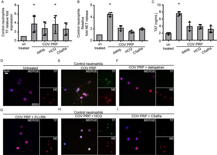
16Figure 2. Inhibition of platelet-rich plasma (PRP)-neutrophil interactions in
COVID-19.
A) Relative fold expression of tissue factor (TF) mRNA in control neutrophils treated
with COVID-19-derived PRP (“COV PRP”) and inhibited with dabigatran (thrombin
inhibitor), hydroxychloroquine (HCQ, autophagy inhibitor) or C5aR1 antagonist
(C5aRa/PMX-53). Data are from four independent experiments (mean ± SD). B)
Myeloperoxidase (MPO)-DNA complex levels in NET isolated from control
neutrophils treated with COV PRP and inhibited with dabigatran, HCQ or
C5aRa/PMX-53. Data are from four independent experiments (mean ± SD). C)
Thrombin-antithrombin (TAT) complex levels in control plasma stimulated with NET
structures isolated from control neutrophils treated with COV PRP and inhibited with
dabigatran, HCQ or C5aR. Data are from four independent experiments (mean ± SD).
D, E) Confocal fluorescence microscopy showing TF/neutrophil elastase (NE)
staining in control neutrophils treated with COV PRP and inhibited with F)
dabigatran, G) FLLRN (PAR1 receptor inhibitor), H) HCQ or I) C5aRa/PMX-53. A
representative example of four independent experiments is shown. Original
magnification: x600, scale bar: 5 μm. Blue: DAPI, green: TF, red: NE. All conditions
were compared to control/untreated; statistical significance at p< 0.05 is indicated by
*. A-C: Friedman's test. The mentioned above in vitro experiments are also
summarized in Supplemental Table S2.
17Figure 3. C3 inhibition disrupts neutrophil-driven thromboinflammation in
COVID-19.
A) Soluble terminal complement complex (sTCC) levels in plasma from controls
(n=10) and COVID-19 patients (n=25). Red squares: severe patients, red triangles:
critical patients. B) Relative fold expression of tissue factor (TF) mRNA in control
neutrophils stimulated with: serum from healthy individuals (HS), HS incubated with
COVID-19 serum (“COV serum”), HS treated with compstatin analog Cp40 and then
COV serum, COV serum alone, or COV serum treated with Cp40. Data are from four
independent experiments (mean ± SD). C) Fluorescence microscopy showing
TF/neutrophil elastase (NE) staining in control neutrophils stimulated with: HS, HS
incubated with COV serum, or HS treated with Cp40 and then COV serum. A
representative example of four independent experiments is shown. Original
magnification: x1000, scale bar: 5 μm. Blue: DAPI, green: TF, red: NE. All
conditions were compared to HS alone (control), and statistical significance at p<
0.05 is indicated by *. A: Student‟s t-test, B: Friedman's test. These in vitro
experiments are also listed in Supplemental Table S2.
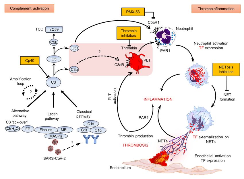
18Figure 4. Proposed mechanism of COVID-19 immunothrombosis.
During COVID-19, SARS-CoV-2 triggers complement activation by interacting with
mannan-binding lectin (MBL) serine proteases (MASPs) or possibly through
(auto)antibodies or/and immunocomplexes. C3 activation, as a point of convergence
of all complement pathways, leads to C3a, C5a and sC5b-9 (terminal complement
complex/TCC) generation. Subsequently, C3a might activate platelets (PLT), while
C5a and PLT-derived thrombin induce both neutrophil tissue factor (TF) expression
and neutrophil extracellular traps (NETs) carrying active TF. These thrombogenic
NETs may induce endothelial cell activation towards TF expression thus increasing
their procoagulant activity. This may further amplify (e.g. via PAR1), inflammation
and PLT activation, thereby fueling a complement/NETs-driven vicious cycle of
immunothrombosis. Complement, thrombin and NETosis represent promising
therapeutic targets. The central pink box includes components of COVID-19
thromboinflammatory environment. Question marks & dotted lines indicate
provisional pathways/connections that have not yet been investigated in COVID-19.
Cp40, compstatin analog; FP, properdin; PAR1, protease-activated receptor 1; PMX-
53, specific C5a receptor antagonist; SARS-CoV-2, severe acute respiratory
syndrome coronavirus 2.
19You can also read