Lower local recurrence rate after robot assisted thoracoscopic esophagectomy than conventional thoracoscopic surgery for esophageal cancer
←
→
Page content transcription
If your browser does not render page correctly, please read the page content below
www.nature.com/scientificreports
OPEN Lower local recurrence rate
after robot‑assisted thoracoscopic
esophagectomy than conventional
thoracoscopic surgery
for esophageal cancer
Satoru Motoyama1,2,3*, Yusuke Sato1,3, Akiyuki Wakita1,3, Yushi Nagaki1,3, Hiromu Fujita1,3,
Ryohei Sasamori1,3, Kohei Kemuriyama1,3, Shinogu Takashima3, Kazuhiro Imai3 &
Yoshihiro Minamiya3
The oncological advantages of robot-assisted thoracoscopic esophagectomy (RATE) over conventional
thoracoscopic esophagectomy (TE) for thoracic esophageal cancer have yet to be verified. In this
study, we retrospectively analyzed clinical data to compare the incidences of recurrence within the
surgical field after RATE and TE as an indicator of local oncological control. Among 121 consecutive
patients with thoracic esophageal or esophagogastric junction cancers for which thoracoscopic
surgery was indicated, 51 were treated with RATE while 70 received TE. The number of lymph nodes
dissected from the mediastinum, duration of the thoracic portion of the surgery, and morbidity due
to postoperative complications did not differ between the two groups. However, the rate of overall
local recurrence within the surgical field was significantly (P = 0.039) higher in the TE (9%) than the
RATE (0%) group. Lymph node recurrence within the surgical field occurred in left recurrent nerve, left
tracheobronchial, left main bronchus and thoracic paraaortic lymph nodes, which were all difficult
to approach to dissect. The other two local failures occurred around the anastomotic site. This study
indicates that using RATE enabled the incidence of recurrence within the surgical field to be reduced,
though there were some limitations.
About 20 years have passed since the first experiences with the innovative transthoracic robot-assisted thoraco-
scopic esophagectomy (RATE) were reported1–3. Theoretically, the four arms employed with RATE have sufficient
dexterity to increase operative precision and maneuverability within the narrow space of the mediastinum. In
addition, it was reported that RATE was feasible and safe, and surgeons could learn to use it within a relatively
short period4–7. Investigators then began comparing the intraoperative and short-term outcomes between their
early cases with RATE and those with conventional thoracoscopic esophagectomy (TE)8–11. RATE reportedly
reduced blood loss, the incidence of vocal cord palsy, and hospital stay duration as compared to TE, though the
operations took longer and had a significantly higher financial cost in several cohorts. Regarding intraoperative
oncological factors, RATE enabled dissection of a higher number of lymph nodes along the left recurrent laryn-
geal nerve (RLN) without increasing morbidity10,11. We also reported that the extent of lymph node dissection
around the left RLN in the left lateral decubitus position was more powerful with RATE than TE12. On the other
hand, there have been few reports demonstrating definitive a mid- or long-term oncological benefit with RATE.
Thus, further evidence showing an oncological benefit of RATE over TE is urgently needed. To add to the avail-
able data, in the present study we assessed recurrence within the surgical field with RATE in our early cases as
an indicator of the ability to maintain local control for mid-term oncological benefit.
1
Esophageal Surgery, Akita University Hospital, Akita University School of Medicine, 1‑1‑1 Hondo, Akita 010‑8543,
Japan. 2Comprehensive Cancer Control, Akita University Graduate School of Medicine, Akita, Japan. 3Thoracic
Surgery, Akita University Graduate School of Medicine, Akita, Japan. *email: motoyama@doc.med.akita-u.ac.jp
Scientific Reports | (2021) 11:6774 | https://doi.org/10.1038/s41598-021-86420-x 1
Vol.:(0123456789)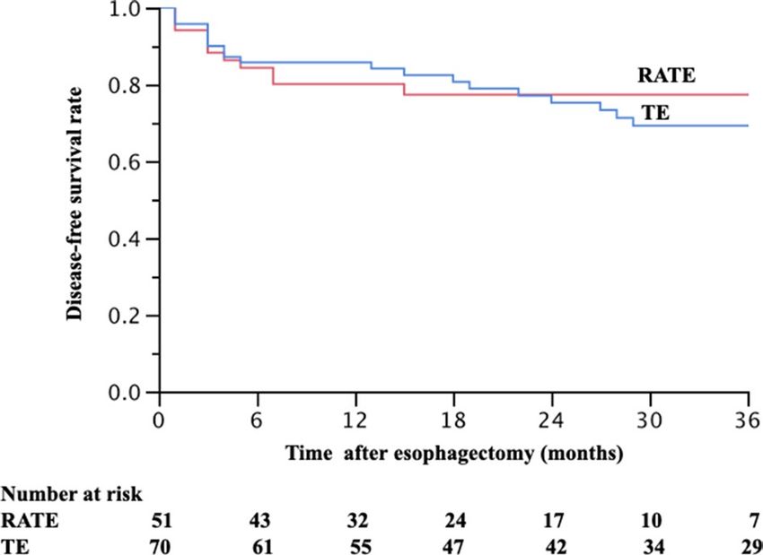
www.nature.com/scientificreports/

                                                                                        RATE (N = 51)   TE (N = 70)   P
                                             Age, years, median (range)                 65 (44–80)      67 (41–85)    0.212
                                             Sex, n (%)
                                             Male                                       45 (88%)        60 (86%)
                                                                                                                      0.790
                                             Female                                     6 (12%)         10 (14%)
                                             Tumor location, n (%)
                                             Upper                                      8 (16%)         11 (16%)
                                             Middle                                     20 (39%)        30 (43%)
                                                                                                                      0.839
                                             Lower                                      16 (31%)        23 (33%)
                                             Esophagogastric junction                   7 (14%)         6 ( 9%)
                                             cT, n(%)
                                             T1                                         18 (35%)        23 (37%)
                                             T2                                         9 (18%)         6 (9%)
                                                                                                                      0.494
                                             T3                                         23 (45%)        37 (53%)
                                             T4                                         1 ( 2%)         1 (1%)
                                             cN, n (%)
                                             0                                          23 (45%)        34 (49%)
                                                                                                                      0.717
                                             1–2                                        28 (55%)        36 (51%)
                                             Number of involved nodes, median (range)   1 ( 0–6)        1 ( 0–4)      0.560
                                             cStage, n (%)
                                             IA                                         13 (25%)        23 (33%)
                                             IB                                         5 (10%)         3 ( 4%)
                                             IIA                                        5 (10%)         8 (11%)
                                             IIB                                        9 (18%)         4 ( 6%)
                                                                                                                      0.448
                                             IIIA                                       9 (18%)         18 (26%)
                                             IIIB                                       6 (12%)         9 (13%)
                                             IIIC                                       1 ( 2%)         1 ( 1%)
                                             IV                                         3 ( 6%)         4 ( 6%)
                                             Histological type of cancer
                                             Squamous cell carcinoma                    43 (84%)        64 (91%)
                                                                                                                      0.200
                                             Adenocarcinoma                             6 (12%)         6 (9%)
                                             Other                                      2 (4%)          0 (0%)
                                             Neoadjuvant therapy, n (%)
                                             Chemoradiotherapy                          27 (53%)        31 (44%)
                                             Chemotherapy                               2 (4%)          7 (10%)
                                                                                                                      0.311
                                             Endoscopic resection                       1 (2%)          5 (7%)
                                             None                                       21 (41%)        27 (39%)

                                            Table 1.  Characteristics of the patients in the RATE and TE groups.

                                            Results
                                            Between December 2014 and April 2020, 51 consecutive patients who had undergone RATE with R0 resection
                                            using the da Vinci S, Si or Xi Surgical System were enrolled in this retrospective study of our early experience.
                                            All operations using the da Vinci Surgical System were performed by a consultant surgeon (SM). During the
                                            same period, 70 patients underwent conventional TE with R0 resection in a left lateral position. The operations
                                            were performed by five surgeons, including two certified esophageal surgeons and three noncertified surgeons.
                                            Thus, the early experiences of three noncertified surgeons are included. Patients were observed for a median of
                                            20 months (range, 5–77 months) after esophagectomy in the RATE group and 41 months (range, 6–72 months)
                                            in the TE group.
                                                The characteristics of the patients and clinical stages, histological type of the cancers, and preoperative treat-
                                            ments in the RATE and TE groups are summarized in Table 1. There were no significant differences between the
                                            two groups with respect to age, sex, tumor location, tumor depth, lymph node metastasis, number of involved
                                            nodes, distant metastasis, clinical stage or histological type. The percentages of patients who received neoadjuvant
                                            therapy also did not differ.
                                                The operation fields and times for the thoracic portion of the surgery were nearly the same in the two groups;
                                            however, blood loss during the thoracic surgery was significantly lower in the RATE group than the TE group
                                            (Table 2). The number of dissected lymph nodes did not differ between the two groups. Morbidity due to post-
                                            operative complications, such as pneumonia (Uniform Pneumonia Score (UPS) ≥ 2, with at least 1 point being
                                            assigned due to infiltrative findings on pulmonary radiography), anastomotic leakage (Type ≥ I in Esophageal

          Scientific Reports |   (2021) 11:6774 |                    https://doi.org/10.1038/s41598-021-86420-x                                                 2

Vol:.(1234567890)
www.nature.com/scientificreports/
RATE (N = 51) TE (N = 70) P
Area of lymph node dissection, n (%)
Mediastinum and upper abdomen (2-field) 4 (8%) 6 (9%)
Bilateral neck, mediastinum and upper abdomen (3-field) 40 (78%) 58 (83%) 0.664
Lower mediastinum and upper abdomen 7 (14%) 6 ( 9%)
Reconstruction
Gastric tube 49 (96%) 67 (96%)
0.921
Pedicled colon 2 (4%) 3 (4%)
Approach for the abdominal portion
Open 27 (53%) 47 (67%)
Hand-assisted laparoscopic 14 (27%) 19 (27%)
0.003*
Total laparoscopic 1 (2%) 4 (6%)
Robot-assisted laparoscipic 9 (18%) 0 ( 0%)
Operation time (min), median (range)
All 646 (485–852) 606 (410–975) 0.201
Thoracic portion 297 (188–457) 298 (144–580) 0.753
Blood-loss (ml), median (range)
All 407 (43–2355) 417 (129–3366) 0.684
Thoracic portion 91 (0–623) 148 (12–1858) 0.004*
Number of dissected lymph nodes, median (range)
All 52 (14–104) 54 (7–97) 0.325
Mediastinal 21 (0–45) 19 (0–68) 0.741
Pneumonia (UPS), n (%)
Negative 43 (84%) 54 (77%) 0.365
Positive 8 (16%) 16 (23%)
Anastomotic leak (Type ≧ I), n (%) 1.000
Negative 46 (90%) 63 (90%)
Positive 5 (10%) 7 (10%)
Recurrent laryngeal nerve palsy (Type ≧ I), n (%) 0.414
Negative 39 (76%) 48 (67%)
Positive 12 (24%) 22 (33%)
Death in hospital 0 (0%) 0 (0%) 1.000
Death within 90 days after esophagectomy 0 (0%) 0 (0%) 1.000
Table 2. Surgical outcomes of patients in the RATE and TE groups. UPS uniform pneumonia score.
*Statistically significant difference.
Complications Consensus Group (ECCG) standardized definitions), and recurrent nerve palsy (Type ≥ I in
ECCG standardized definitions) also did not differ between the two groups (Table 2 and Supplement Table)13,14.
During the observational period, recurrence rates were 22% and 27% in the RATE and TE group, respectively.
Interestingly, the lymph node recurrence rate within the surgical field was 6% (4 patients) in the TE group, but
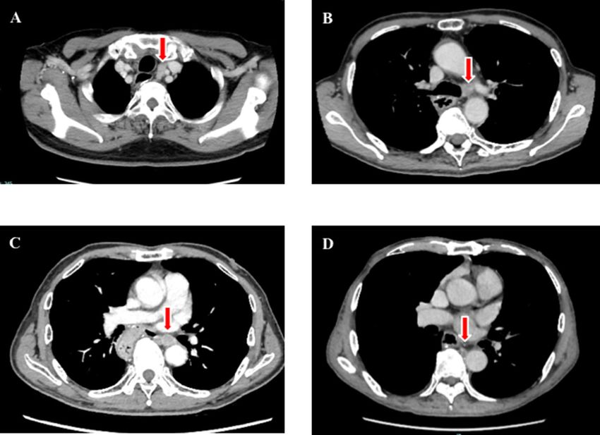
was 0% in the RATE group. Lymph node recurrences involved a left recurrent nerve lymph node (106recL: Lymph
node number according to the Japanese Classification of Esophageal Cancer, 11th edition), a left tracheobron-
chial lymph node (106tbL), a left main bronchus lymph node (109L), and a thoracic paraaortic lymph node
(112ao), which were difficult to approach for lymph node dissection (Fig. 1)15,16]. In addition, two patients had
recurrences around the anastomotic site. Overall, the local recurrence rate was 9% (6 patients) in the TE group,
which was significantly (P = 0.039) higher than the 0% recurrence rate in the RATE group (Table 3). Among
patients showing recurrence, 3 received neoadjuvant chemoradiotherapy, while the other 3 received up-front
surgery. In the RATE group, all recurrence sites were distant fields (distant organ or distant lymph node). On
the other hand, the ratio of surviving patients and the disease-free survival (DFS) rates did not differ between
the two groups (Table 3 and Fig. 2).
Discussion
This study revealed several interesting results. First, the local recurrence rate was 9% in the TE group, which was
significantly (P = 0.039) higher than the 0% recurrence rate in the RATE group. Second, in the TE group recur-
rence in mediastinal lymph nodes was in a region that was difficult to approach for lymph node dissection. By
contrast, in the RATE group all recurrence sites were in distant fields (a distant organ or distant lymph node).
Thoracic esophageal cancer is one of the most aggressive cancers and is characterized by rapid clinical pro-
gression and a poor prognosis. Consequently, neoadjuvant chemo- or chemoradiation therapy is usually indi-
cated, even in patients who will then undergo esophagectomy17. Indeed, the current international guidelines
Scientific Reports | (2021) 11:6774 | https://doi.org/10.1038/s41598-021-86420-x 3
Vol.:(0123456789)www.nature.com/scientificreports/
Figure 1. Contrast-enhanced computed tomography images of recurrent lymph nodes: (a) left recurrent
laryngeal nerve lymph node; (b) left tracheobronchial lymph node; (c) left main bronchus lymph node; (d)
thoracic paraaortic lymph node.
Robot (N = 51) Thoracoscopy (N = 70) P
Recurrence
None 40 (78%) 51 (73%) 0.393
Recurred 11 (22%) 19 (27%)
Lymph node recurrence within the surgical field
None 51 (100%) 66 (94%) 0.137
Recurred within the surgical field 0 (0%) 4 (6%)
Overall local recurrence (including anastomotic site)
None 51 (100%) 64 (91%) 0.039*
Recurred within a local field 0 (0%) 6 (9%)
Recurrence pattern
None 40 (78%) 51 (73%)
Local field 0 (0%) 6 (9%) 0.189
Distant lymph node 4 (8%) 5 (7%)
Distant organs 7 (14%) 8 (11%)
Survival
Alive 40 (78%) 55 (79%)
0.947
Esophageal cancer-specific death 8 (16%) 10 (14%)
Death from other diseases 3 (6%) 5 (7%)
Disease-free survival rate
1-year survival rate 76.5% 81.4%
0.697#
2-year survival rate 74.1% 71.8%
3-year survival rate 74.1% 64.7%
Table 3. Recurrence and survival among patients in the RATE and TE groups. *Statistically significant
difference. # Log-rank test.
Scientific Reports | (2021) 11:6774 | https://doi.org/10.1038/s41598-021-86420-x 4
Vol:.(1234567890)www.nature.com/scientificreports/
Figure 2. Disease-free survival curve in the patients received robot-assisted thoracoscopic esophagectomy
and conventional thoracoscopic esophagectomy. RATE robot-assisted thoracoscopic esophagectomy, TE
thoracoscopic esophagectomy.
recommend combined treatments consisting of chemotherapy, radiotherapy and surgery for patients with local-
ized advanced esophageal or esophagogastric cancer18–21. After neoadjuvant treatments, esophagectomy with
extended lymph adenectomy in the neck, mediastinum and abdomen was performed as the main component of
this curative and radical treatment strategy. Unfortunately, patients with esophageal cancer often have comor-
bidities and are in poor clinical condition as a result of advanced age, body weight loss, habitual alcohol use,
smoking, poor respiratory function (chronic obstructive pulmonary disease), hypertension, and/or a history of
prior cancer. As a result, postoperative complications are more frequent in these patients, even now. To decrease
the invasiveness of the operation and postoperative complications while providing a survival benefit to these
patients, surgeons have been applying minimally invasive techniques since the 1990’s. However, as Straatman et al.
reported, the randomized controlled TIME Trial revealed that there were no differences in 3-year disease-free or
overall survival between open transthoracic esophagectomy and minimally invasive T E22. With the rapid shift
from conventional TE to RATE, we must rapidly produce oncological benefit with RATE as compared to TE or
open transthoracic esophagectomy. To determine the long-term oncological benefit of RATE, a randomized trial
testing whether RATE is superior to conventional TE is currently in progress and is targeted to 5-year overall
survival as a primary endpoint23]. It will be several years before a conclusion is reached.
Regarding the intraoperative or short-term surgical outcomes with RATE, which will influence the oncological
benefit, several reports, including ours, have demonstrated radical lymph node dissection around the left RLN,
which is known to be a difficult field for lymph node dissection, without increasing left recurrent nerve palsy10–12.
Moreover, several other powerful findings were also recently added. Yong et al. reported in 2019 that their RATE
group (n = 280) yielded more lymph nodes along the RLN (4.8 vs. 4.1) with a shorter surgical duration for the
thoracic portion than their TE (n = 372) group (85.0 vs. 102.9 min), but the incidence of RLN injury was higher
in the RATE group (29.2% vs. 15.1%)24. Tagkalos et al. used propensity-matched analysis to assess consecutive
patients with esophageal cancer undergoing modified Ivor Lewis esophagectomy. They reported that there was
a trend toward improved lymphadenectomy with a shorter stay in the intensive care unit with RATE (n = 50)
than TE (n = 50)25. In addition, Harbison et al. conducted a retrospective analysis with risk-adjustment using a
nationally-validated database: the American College of Surgeons National Surgical Quality Improvement Pro-
gram (ACS-NSQIP). They found that surgical outcomes did not significantly differ between the RATE (n = 100)
and TE (n = 625) groups with respect to the incidence of 30-day postoperative mortality or overall m orbidity26.
However, van der Horst et al. reported that among patients with lymph node-positive thoracic esophageal cancer
in the superior mediastinum, RATE was associated with higher mortality and morbidity27.
Regarding the mid-term oncological benefits of RATE, Yong et al. reported that RATE was associated with
a lower rate of mediastinal lymph node recurrence (2.0% vs. 5.3%) (P = 0.044), but overall and disease-free
survival did not differ between the two cohorts24. Our present study strengthens and clarifies the observational
findings on the mid-term oncological benefits of RATE. Although the extent of the dissection and number of
lymph nodes dissected from the mediastinum did not differ between the RATE and TE groups, local recurrence,
including lymph node recurrence within the surgical fields, was significantly higher with conventional TE than
RATE. RATE enables surgeons to precisely maneuver and produce good oncological outcomes. This appears to
reflect the advantages of RATE, which include a 3D self-controlled magnified view enabling better visualization
of this narrow area and the ability to offer adequate depth perception. In addition, use of a self-controlled third
arm and a tremor filtering function enabled us to achieve fine tension and countertraction during dissection in
this narrow area. However, such recurrences are not always due to surgical failure, as these recurrences occurred
a considerable time (3–24 months) after esophagectomy. Although the rate of local failure was higher in the TE
group than the RATE group, the ratios of surviving patients and the DFS rates did not differ between the two
groups. One reason for that is the majority of recurrences were in distant fields in both groups, and the rates
of local recurrence were relatively low. In addition, local failures were treatable and had the possibility of cure.
Scientific Reports | (2021) 11:6774 | https://doi.org/10.1038/s41598-021-86420-x 5
Vol.:(0123456789)www.nature.com/scientificreports/
Indeed, we treated the six affected patients with additional surgical resection of the recurrent lymph node fol-
lowed by chemoradiotherapy or definitive chemoradiotherapy, and three patients were completely cured with
no further recurrence.
This study has several limitations. First, the study population was heterogeneous, the number of patients
in the cohort was small, and the number of events was limited. Second, it was a nonrandomized comparative
analysis, and there was considerable bias in the selection of RATE or TE as the surgical approach. Third, only
short- and mid-term results were determined. To assess overall oncological benefit, we will need to follow these
patients for a longer time. The presented result is extremely important, but it is only a preliminary report using
our first case series with RATE. Further analysis is therefore necessarily.
In summary, our findings indicate that RATE enables a reduction in the incidence of local recurrence within
the surgical field. However, this should be interpreted with caution due to the limitations of this study mentioned
above.
Methods
This study was approved by the Ethics Committee of Akita University Graduate School of Medicine (No. 1222).
All methods were performed in accordance with the relevant guidelines and regulations. All participants provided
informed consent and signed a human subject institutional review board consent form.
Selection of approach of surgery. We began using RATE for patients with thoracic esophageal and
esophagogastric junction cancer in December 2014. The procedure was indicated for all patients with thoraco-
scopically resectable cancers and with Eastern Cooperative Oncology Group performance status 0. Since April
2018, RATE has been covered by the health insurance system in Japan, which covers all of Japan’s citizens. It
enables us to perform RATE for all patients; however, there are several licenses that the surgeon must obtain.
Although in the present study all surgeries were performed by one team, the selection of approach (RATE vs.
TE) for the thoracic part depended upon the operating surgeon. Between December 2014 and March 2018,
RATE was selected by patients who desired to receive RATE, despite the lack of health insurance coverage; for
the other patients, TE was performed with insurance coverage. The extent and fundamental technique used for
dissection of the tumor and lymph nodes is same for both approaches. The abdominal surgeries were performed
using an open, hand-assisted laparoscopic or a total laparoscopic approach in the TE group. In the RATE group,
one surgeon added a robot-assisted laparoscopic approach to the other approaches for the abdominal surgery.
The selection of the abdominal approach was also decided by the operating surgeon.
Operative procedure. The patients were placed in the left lateral position under a combination of inhaled
and intravenous anesthesia, and a double-lumen endotracheal tube was used for single-lung ventilation during
the thoracic part of the surgery. With RATE, the right arm was raised 60° cranially to expose the right axillar
fossa, then tilted 20° cranially and 15° ventrally. The assistant surgeons stood on the left side of the patient. The
da Vinci trocars (8 mm) were inserted into the 2nd or 3rd intercostal space (ICS) on the anterior axillary line
(AL), the 4th or 5th ICS on the middle AL, the 6th or 7th ICS on the middle AL, and the 9th or 10th ICS on the
posterior AL. Generally, an additional trocar was inserted into the 4th ICS on the anterior AL for an assistant and
insufflation of the thoracic cavity with CO2 (8 mmHg). We mainly used a forward-oblique viewing endoscope
during the thoracic portion of the surgery. In TE, a 25-mm mini-thoracotomy or a 12-mm trocar insertion was
performed in the 4th ICS on the anterior AL for an assistant. Four 10.5-mm trocars were inserted into the 4th
ICS on the posterior AL for the operator’s left arm, the 5th ICS on the middle AL for the scope (for an assistant
surgeon), the 7th ICS on the posterior AL for the operator’s right arm, and the 8th ICS on the anterior AL for
an assistant. A forward-oblique viewing endoscope was used during the thoracic portion of the surgery. The
thoracic portion of the operation was nearly the same in the RATE and TE groups.
For thoracic esophageal cancer, the operation was begun with the thoracic portion and incising of the medi-
astinal pleura on the dissected line. The arch of the azygos vein was divided and then ligated. The lymph nodes
around the right RLN were dissected below the right subclavian artery. To dissect along the left RLN, trachea
and main bronchus were displaced to the ventral side to enlarge the limited space to increase the range of move-
ment of the surgical instrument. The thoracic duct was carefully preserved in T1-2 cancers, but was resected
together with the tumor in T3 cancers. The bilateral pulmonary branches of the vagal nerves were preserved. The
lower posterior mediastinal lymph nodes were dissected from the pericardium, left pleura, descending aorta,
and diaphragm. For middle and lower thoracic esophageal cancers, the esophagus was divided at the level of
the upper edge of the aortic arch by linear stapling; for upper thoracic esophageal cancers, it was divided in the
supradiaphragmatic area. Next, the abdominal portion of the surgery and bilateral neck lymph node dissection
were performed concurrently. Our standard strategy for lymph node dissection was 3-field lymphadenectomy
(bilateral neck, mediastinal and upper abdominal lymph nodes) for thoracic esophageal cancer. This was followed
by reconstruction using the stomach or pedicled colon and handsewn layer-to-layer anastomosis.
For esophagogastric junction cancers, the surgery was begun with the abdominal portion. This consisted of
abdominal lymph node dissection in the perigastric region and areas around the celiac axis, common hepatic
artery and splenic artery, and making a gastric roll using an open or a total laparoscopic approach, including a
robot-assisted approach. The thoracic portion of this surgery began with incision of the mediastinal pleura on
the dissected line. The arch of the azygos vein and thoracic duct were usually preserved. The lower and middle
posterior mediastinal lymph nodes were dissected from the pericardium, left pleura, descending aorta, and dia-
phragm. The lymph nodes around the main bronchus were prophylactically dissected in nearly all patients. Using
linear stapling, the esophagus was divided at the middle thoracic esophagus. The gastric tube was then pulled
Scientific Reports | (2021) 11:6774 | https://doi.org/10.1038/s41598-021-86420-x 6
Vol:.(1234567890)www.nature.com/scientificreports/
up into the intrathoracic space and reconstructed by making an anastomosis between the remaining esophagus
and the gastric tube using linear or circular stapling.
Clinical staging. The clinical staging, including diagnosis of lymph node metastasis, was defined at a con-
ference attended by radiologists, physicians and surgeons according to the International Union Against Cancer
tumor-node-metastasis (TNM) Classification of Malignant Tumors (seventh edition) based on findings from
endoscopy, esophagography, contrast-enhanced computed tomography (CE-CT), and systematic [ 18F] fluoro-
deoxyglucose-positron emission tomography/computed tomography (FDG-PET/CT)15,16,20,21. Regional nodes
were considered positive for malignancy when they were positive in FDG-PET/CT (the maximum standardized
uptake value; SUVmax ≥ 2.5) and/or round or ovoid shaped with short axes ≥ 8 mm in thin-sliced CE-CT.
Follow‑up program. The post-surgical follow-up program consisted of blood tests, including those for
tumor makers (squamous cell carcinoma antigen and carcinoembryonic antigen), and neck/chest/abdominal
CE-CT every 4 months for up to 3 years, then every 6 months for up to 5 years. Upper gastrointestinal endoscopy
was done yearly. FDG-PET/CT was used when recurrence was suspected. As a general rule, the patients visited
the hospital once every 2 months for at least 5 years after their surgery.
Outcomes. The intraoperative oncological surgical outcomes (operation time, estimated blood loss, the
number of lymph nodes dissected from the mediastinum) and short- and midterm outcomes (incidence rate of
postoperative complications, rate of local recurrence within surgical fields, and its site, patterns, and 3-year DFS
rates) were compared between the two groups. The operating time for the thoracic portion of the surgery was
defined as the time from the start of chest incision through closure of the trocar sites in the chest. Blood loss was
estimated by weighing the suctioned blood and gauze pieces with absorbed blood. Surgical complications were
evaluated using the ECCG standardized d efinitions14. Anastomotic leakage was observed using esophagography
on postoperative day 8 and counted when it was Type ≥ I according to the ECCG definitions. Recurrent nerve
palsy was observed using bronchoscopy on postoperative day 2 and counted when it was Type ≥ I according to
the ECCG definitions. Postoperative pneumonia was scored according to the UPS and was evaluated as positive
when UPS ≥ 2 with at least 1 point being assigned due to infiltrative findings on pulmonary r adiography13. The
other surgical complications were evaluated using the Clavien–Dindo c lassification28.
Local recurrences were confirmed using both CE-CT and FDG-PET/CT without pathological diagnosis,
excluding patients who received surgical resection. Regarding local recurrence around the anastomotic site, we
cannot confirm whether it was due to infiltration from a lymph node recurrence to the anastomotic site or from
expansion of intramural metastasis in the esophagus or from the stomach, or to invasion of the anastomosis
after regrowth of residual cancer cells around the anastomotic site. This is because intraoperative frozen sections
showed these patients to be cancer free at the margin, and a clearly epithelial lesion was not seen at the time of
recurrence. DFS was measured as the period from esophagectomy to the date of confirmed recurrence, death
(whichever happened first), or the date of the investigators’ last note of disease-free status on November 21, 2020.
Data analysis. We retrospectively analyzed clinical data. Continuous variables are presented as medians
(minimum–maximum), and differences between the two groups were analyzed using the Mann–Whitney-U
test. Categorical data were analyzed using Pearson’s Chi square test or Fisher’s exact probability test. Overall
survival was characterized using Kaplan–Meier curves. DFS curve was compared between the two groups using
the log-rank test. All statistical analyses were performed using JMP15 (SAS Institute Inc., Cary, NC, USA) and
yielded two-sided P values. Values of P < 0.05 were considered statistically significant.
Received: 31 August 2020; Accepted: 16 March 2021
References
1. Giulianotti, P. C. et al. Robotics in general surgery: personal experience in a large community hospital. Arch. Surg. 138, 777–784
(2003).
2. Kernstine, K. H., DeArmond, D. T., Karimi, M., Van Natta, T. L. & Campos, J. C. The robotic, 2-stage, 3-field esophagolymphad-
enectomy. J. Thorac. Cardiovasc. Surg. 127, 1847–1849 (2004).
3. van Hillegersberg, R. et al. First experience with robot-assisted thoracoscopic esophagolymphadenectomy for esophageal cancer.
Surg. Endosc. 20, 1435–1439 (2006).
4. Boone, J. et al. Robot-assisted thoracoscopic oesophagectomy for cancer. Br. J. Surg. 96, 878–886 (2009).
5. Kim, D. J. et al. Thoracoscopic 20 esophagectomy for esophageal cancer: feasibility and safety of robotic assistance in the prone
position. J. Thorac. Cardiovasc. Surg. 139, 53–59 (2010).
6. Puntambekar, S. P., Rayate, N., Joshi, S. & Agarwal, G. Robotic transthoracic esophagectomy in the prone position: experience
with 32 patients with esophageal cancer. J. Thorac. Cardiovac. Surg. 142, 1283–1284 (2011).
7. Van Der Sluis, P. C., Ruurda, J. P., van der Horst, S., Goense, L. & van Hillegersberg, R. Learning curve for robot-assisted minimally
invasive thoracoscopic esophagectomy: results from 312 cases. Ann. Thorac. Surg. 106, 264–271 (2018).
8. Suda, K. et al. Robot-assisted thoracoscopic lymphadenectomy along the left recurrent laryngeal nerve for esophageal squamous
cell carcinoma in the prone position: technical report and short-term outcomes. World J. Surg. 36, 1608–1616 (2012).
9. Deng, H. Y. et al. Comparison of short-term outcomes between robot-assisted minimally invasive esophagectomy and video-
assisted minimally invasive esophagectomy in treating middle thoracic esophageal cancer. Dis. Esophagus 31, 1–7 (2018).
10. Park, S. et al. Comparison of robot-assisted esophagectomy and thoracoscopic esophagectomy in esophageal squamous cell car-
cinoma. J. Thorac. Dis. 8, 2853–2861 (2016).
Scientific Reports | (2021) 11:6774 | https://doi.org/10.1038/s41598-021-86420-x 7
Vol.:(0123456789)www.nature.com/scientificreports/
11. Chao, Y. K., Hsieh, M., Liu, Y. H. & Liu, H. P. Lymph node evaluation in robot-assisted versus video-assisted thoracoscopic
esophagectomy for esophageal squamous cell carcinoma: a propensity-matched analysis. World J. Surg. 42, 590–598 (2018).
12. Motoyama, S. et al. Extensive lymph node dissection around the left laryngeal nerve achieved with robot-assisted rhoracoscopic
esophagectomy. Anticancer Res. 39, 1337–1342 (2019).
13. Seesing, M. F. J. et al. Defining pneumonia after esophagectomy for cancer: validation of the Uniform Pneumonia Score in a high
volume center in North America. Dis. Esophagus 31, 1–8 (2018).
14. Low, D. E. et al. International consensus on standardization of data collection for complications associated with esophagectomy:
Esophagectomy Complications Consensus Group (ECCG). Ann. Surg. 262, 286–294 (2015).
15. Japan Esophageal Society. Japanese classification of esophageal cancer, 11th edition: part I. Esophagus 14, 1–36 (2017).
16. Japan Esophageal Society. Japanese classification of esophageal cancer, 11th edition: part II and III. Esophagus 14, 37–65 (2017).
17. Wong, I. Y. H. & Law, S. Surgery in the era of neoadjuvant therapy for cancer of the esophagus. Esophagus 13, 105–109 (2016).
18. Ajani, J. A. et al. Esophageal and esophagogastric junction cancers, version 2.2019, NCCN clinical practice guidelines in oncology.
J Natl Compr Cancer Netw 17, 855–883 (2019).
19. Lordick, F. et al. Oesophageal cancer: ESMO clinical practice guidelines for diagnosis, treatment and follow-up. Ann Oncol 27(suppl
5), v50–v57 (2016).
20. Kitagawa, Y. et al. Esophageal cancer practice guidelines 2017 edited by the Japan Esophageal Society: part 1. Esophagus 16, 1–24
(2019).
21. Kitagawa, Y. et al. Esophageal cancer practice guidelines 2017 edited by the Japan esophageal society: part 2. Esophagus 16, 25–43
(2019).
22. Straatman, J. et al. Minimally invasive versus open esophageal resection: three-year follow-up of the previously reported rand-
omized controlled trial: the TIME. Trial 266, 232–236 (2017).
23. Yang, Y. et al. Robot-assisted esophagectomy (RAE) versus conventional minimally invasive esophagectomy(MIE) for resectable
esophageal squamous cell carcinoma: protocol for a multicenter prospective randomized controlled trial (RAMIE trial, robot-
assisted minimally invasive esophagectomy). BMC Cancer 19, 608 (2019).
24. Yang, Y. et al. Short-and mid-term outcomes of robotic versus thoraco-laparoscopic McKeown esophagectomy for squamous cell
esophageal cancer: a propensity score-matched study. Dis. Esophagus https://doi.org/10.1093/dote/doz080 (2020).
25. Tagkalos, E. et al. Robot-assisted minimally invasive esophagectomy (RAMIE) compared to conventional minimally invasive
esophagectomy (MIE) for esophageal cancer: a propensity-matched analysis. Dis. Esophagus https://doi.org/10.1093/dote/doz060
(2020).
26. Harbison, G. J., Vossler, J. D., Yim, N. H. & Murayama, K. M. Outcomes of robotic versus non-robotic minimally-invasive
esophagectomy for esophageal cancer: an American College of Surgeons NSQIP database analysis. Am. J. Surg. 218, 1223–1228
(2019).
27. van der Horst, S., de Maat, M. F. G., van der Sluis, P. C., Ruurda, J. P. & van Hillegersberg, R. Extended thoracic lymph node dissec-
tion in robotic-assisted minimal invasive esophagectomy (RAMIE) for patients with superior mediastinal lymph node metastasis.
Ann. Cardiothorac. Surg. 8, 218–225 (2019).
28. Dindo, D., Demartines, N. & Clavien, P. A. Classification of surgical complications: a new proposal with evaluation in a cohort of
6336 patients and results of a survey. Ann. Surg. 240, 205–213 (2004).
Author contributions
Conception or design of the work (Dr. Motoyama); data collection (all authors); data analysis and interpretation
(Dr. Motoyama, Dr. Sato, Dr. Wakita, Dr. Nagaki, Dr. Fujita); drafting the article (Dr. Motoyama); final approval
of the version to be submitted (all authors).
Competing interests
The authors declare no competing interests.
Additional information
Supplementary Information The online version contains supplementary material available at https://doi.org/
10.1038/s41598-021-86420-x.
Correspondence and requests for materials should be addressed to S.M.
Reprints and permissions information is available at www.nature.com/reprints.
Publisher’s note Springer Nature remains neutral with regard to jurisdictional claims in published maps and
institutional affiliations.
Open Access This article is licensed under a Creative Commons Attribution 4.0 International
License, which permits use, sharing, adaptation, distribution and reproduction in any medium or
format, as long as you give appropriate credit to the original author(s) and the source, provide a link to the
Creative Commons licence, and indicate if changes were made. The images or other third party material in this
article are included in the article’s Creative Commons licence, unless indicated otherwise in a credit line to the
material. If material is not included in the article’s Creative Commons licence and your intended use is not
permitted by statutory regulation or exceeds the permitted use, you will need to obtain permission directly from
the copyright holder. To view a copy of this licence, visit http://creativecommons.org/licenses/by/4.0/.
© The Author(s) 2021
Scientific Reports | (2021) 11:6774 | https://doi.org/10.1038/s41598-021-86420-x 8
Vol:.(1234567890)You can also read