Histological regression of gastrointestinal peritoneal metastases after systemic chemotherapy
←
→
Page content transcription
If your browser does not render page correctly, please read the page content below
Pleura and Peritoneum 2021; aop

Laura Toussaint, Hugo Teixeira Farinha, Jean-Luc Barras, Nicolas Demartines,
Christine Sempoux and Martin Hübner*

Histological regression of gastrointestinal
peritoneal metastases after systemic
chemotherapy
https://doi.org/10.1515/pp-2021-0118                                     (p=0.04). Median survival was higher in patients with
Received March 9, 2021; accepted May 12, 2021;                           PRGS < 1.8 (Quartiles one and 2) than higher (Q3 and Q4), but
published online July 14, 2021                                           the difference did not reach significance in this small cohort.
                                                                         Conclusions: PRGS is an objective too to describe histo-
Abstract
                                                                         logical response of PM of GI origin after systemic chemo-
                                                                         therapy. This response differs significantly between patients,
Objectives: Peritoneal metastases (PM) are relatively
                                                                         allowing to distinguish between chemosensitive and che-
resistant to systemic chemotherapy, and data on histo-
                                                                         moresistant tumors.
logical response to therapy is rare. The aim of this study
was to quantify the treatment response of PM after sys-                  Keywords: chemotherapy; peritoneal metastasis; perito-
temic chemotherapy.                                                      neal regression grading system (PRGS); PIPAC.
Methods: Retrospective monocentric cohort study of
47 consecutive patients with PM from gastrointestinal
origin undergoing surgery (cytoreduction: CRS + Hyper-                   Introduction
thermic IntraPEritoneal Chemotherapy [HIPEC] or Pres-
surized IntraPeritoneal Aerosol Chemotherapy [PIPAC])                    As compared to liver metastasis, peritoneal metastases (PM)
after prior systemic chemotherapy from 1.2015 to 3.2019.                 have a relatively limited response to systemic chemotherapy,
Tumor response was assessed using the 4-scale Perito-                    and their prognosis remains poor in most disease entities [1]. In
neal Regression Grading System (PRGS) (4: vital tumor to                 addition, evaluation of treatment response tends to be diffi-
1: complete response).                                                   cult, as many patients have no target lesions allowing evalu-
Results: Patients had a median of 2 (range: 1–7) lines and 10            ation according to RECIST criteria [2]. One interesting
(3–39) cycles of prior systemic chemotherapy. A median of four           alternative is the assessment by histological response, and a
biopsies (range: 3–8) was taken with a total of 196 analyzed             4-grade standardized evaluation system, the peritoneal
specimens. Twenty-four biopsies (12%) showed no histological             regression grading system (PRGS) that was proposed and
regression (PRGS4), while PRGS 3, two and one were diag-                 validated recently. The PRGS has been explicitly developed for
nosed in 37 (19%), 39 (20%), and 69 (49%) specimens,                     taking into account specific characteristics of PM, such as their
respectively. A significant heterogeneity was found between              frequent mucinous character [3, 4]. Intraperitoneal treatment
peritoneal biopsies in 51% patients. PRGS correlated strongly            modalities like Heated Intra Peritoneal Chemotherapy (HIPEC)
with peritoneal spread (PCI, p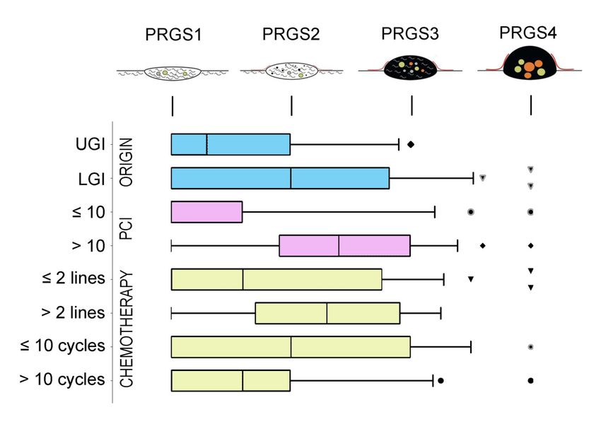
2 Toussaint et al.: Histological peritoneal response to systemic chemotherapy
2015 (start of PIPAC program in our department) to March 2019. The Assessment of histological response
systematic use of PRGS for evaluating response of PM to intraperitoneal
chemotherapy was started in our institution immediately after it became
All peritoneal biopsies were assessed by a board-certified pathologist
available in June 2016, and was used prospectively since then on a
specialized in peritoneal cancer specimens. Tumor regression was
routine basis. In addition, the PRGS was retrospectively assessed for the
evaluated using the Peritoneal Regression Grading Score (PRGS).
patients treated between January 2015 and June 2016. Only patients with
PRGS discriminates four categories based on the presence of residual
GI primary were considered for this analysis (n=47). Excluded were
tumor cells and the extent of regression features, as described pre-
patients without previous chemotherapy, without histological sampling
viously [4]. The PRGS was calculated as the mean of at least four
during surgery, missing PRGS assessment, or patients who refused to
biopsies from each abdominal quadrant, if technically possible [3, 4].
participate in the study (n=7). Only primary procedures were consid-
Also, the minimal (= the best regression) and the maximal (= the
ered, and patients with previous intraperitoneal chemotherapy of any
lesser regression) were documented.
kind were excluded. In the case of PIPAC, only biopsies from the first
procedure were selected for analysis. The Institutional Review Board of
the CHUV University Hospital approved the study (CER-VD 2019-00747).
Predefined subgroup analyses
Data management
Predefined stratifications were primary tumor types, number of lines,
and cycles of previous chemotherapy and treatment modality (PIPAC
Pertinent demographics, oncological and pathological data were vs. HIPEC). Patients were grouped into two entities: (1) upper gastro-
retrieved from a prospectively maintained institutional database and
intestinal tumors (UGI) group including patients with gastric cancer
entered in an a priori defined anonymized database containing the
and (2) lower gastrointestinal tumors (LGI) including patients with PM
following variables: Demographics: age, gender, primary tumor from rectal, colic, and small bowel origin.
origin, body mass index (kg/m2), ASA physical status classification
score, serum tumor markers (CA 19-9 (kU/l), CEA (µg/l), CA-125 (kU/l))
and KRAS/HER2 amplification; Chemotherapy regimen, number of
Statistics and analysis
lines and cycles, PCI (Peritoneal Cancer Index) and PRGS scores
(obtained during the first PIPAC or HIPEC).
PRGS was presented as mean ± SD for patients having four biopsies at
least, and in addition, the highest and lowest grading was reported
Surgical approach [4]. Continuous variables were presented as mean with standard
deviation (SD) or median with range or interquartile range (IQR) for
PIPAC was performed by use of a two-trocar technique in a strictly stan- skewed data. Categorical variables were reported as frequencies (%)
dardized way [5, 7]. Cytoreductive surgery and HIPEC were performed and compared with the chi-square test. Depending on the normality
through a midline laparotomy. In both approaches, laparoscopic or open, of distribution, Student’s t-test and Mann–Whitney U test or
first step of the procedure was a systematic and complete exploration of the Wilcoxon signed ranked test were used for float comparisons.
abdominal cavity with documentation of the Peritoneal cancer index (PCI). Statistical correlations were tested by use of Pearson’s rank corre-
Biopsies were collected in suspect areas representing, whenever possible, lation. A level of 0.05 was considered statistically significant.
at least four different areas of the abdomen. These were excisional biopsies Statistical analyses were performed, and figures were produced with
for the open HIPEC cases and small samples taken with biopsy forceps SPSS v20 software (Chicago, IL, USA), GraphPad Prism 7 (GraphPad
during the laparoscopic PIPAC cases [4, 5]. Of note, biopsies were taken Software, Inc., La Jolla, CA, USA), Python, NumPy, Pandas, and
before surgical resection or delivery of intra-peritoneal treatment. Seaborne (Anaconda, Berlin, Germany).
Table : Patients baseline demographics.
All patients (n=) LGI (n=) UGI (n=) p-Value
Median age (IQR) (–) (–) (–) .
Gender, male (%) (%) (%) .
ASA score .
(%) (%) (%)
(%) (%) (%)
Median PCI (IQR) (–) (–) (–) .
PIPAC, % (%) (%) (%) .
CRS + HIPEC, % (%) (%)
Prior chemotherapy received
Median lines (range) (–) (–) (–) .
Median cycles (range) (–) (–) (–) .
Median (IQR or range) or number (%) as appropriate. Statistical significance (p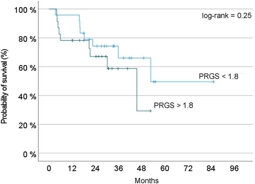
Toussaint et al.: Histological peritoneal response to systemic chemotherapy 3
Table : Regimen used for the last line of prior chemotherapy. Number of biopsies and variability
Number of Number of cycles
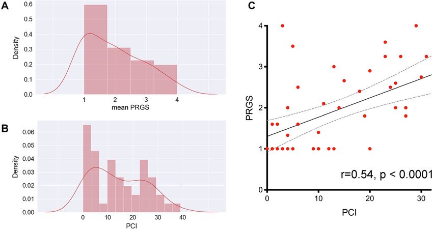
Overall, a median of four biopsies (range: 3–8) was
patients (median)
taken with a total of 196 analysed specimens. Dis-
FOLFOX rcepant PRGS values for the different tumor biopsies in
FOLFOX + bevacizumab
the same patient were documented for 24 out of 47 pa-
FOLFOX + cetuximab
FOLFIRI tients (51%).
FOLFIRI + bevacizumab
FOLFIRI + cetuximab
Other regimens/unknown –
Total – Macroscopic (PCI) and microscopic (PRGS)
FOLFOX leucovorin + fluorouracil, FOLFIRI
assessments
leucovorin + fluorouracil + irinotecan. Other: FOLFIRINOX
(leucovorin + fluorouracil + irinotecan + oxaliplatine), Panitumab, Median PRGS was 1.8 (IQR 1.0-2.63) for the entire cohort.
Eporubicine, Capécitabine, Docetaxel, Premetrexed, Carboplatine Median PCI was 12 (IQR 4-24) PCI and mean PRGS
correlated strongly to each other (p4 Toussaint et al.: Histological peritoneal response to systemic chemotherapy
Tumor response to chemotherapy, objective
histological regression
Twenty-four peritoneal biopsies (12%) showed no histological
regression (PRGS4), while PRGS 3, two or 1 (complete
regression) was diagnosed in 37 (19%), 39 (20%), and 69
(49%) specimens, respectively. The overall grade of regres-
sion did not corelate with the number of chemotherapy lines,
or cycles received previously. However, in a subgroup of pa-
tients treated with 10 and more chemotherapy cycles before
surgery, a complete histological regression (PRGS 1) was
documented more frequently than in patients treated with
fewer cycles (p=0.04). Interestingly, histological response to
Oxaliplatin correlated well with the increasing number of
cycles p=0.02 ϱ = −0.381. Figure 2 summarizes the sensitivity Figure 3: Probability of overall survival depending on PRGS.
Two groups of patients are compared: Blue curve = patients with a
analysis performed by tumor origin, the extent of peritoneal
PRGS inferior to the median value of the cohort; red curve: Patients
disease, and previous chemotherapy (number of lines, num- with a PRGS superior to this value, suggesting a poorer prognosis. In
ber of cycles received). this small cohort of patients, the difference observed did not reach
statistical significance (log-rank test, p=0.25).
PRGS and overall survival
our cohort, we pooled the patients with an unfavorable mean
We hypothesized that a favorable PRGS would correlate with
PRGS (Quartiles three and 4) vs those with high or complete
better overall survival. Considering the relatively small size of
histological regression (Quartiles one and 2). Figure 3 shows
the probability of overall survival, estimated with the Kaplan-
Meyer method. This curve suggests a predictive value of
PRGS for overall survival. However, this difference did not
reach statistical significance (log-rank, p=0.25).
Discussion
The Peritoneal Regression Grading Score (PRGS) was initially
developed to quantify PM’s histological response to palliative
intraperitoneal chemotherapy [8]. This study demonstrated
that PRGS can also be used for measuring the objective
response of PM to systemic, intravenous chemotherapy.
UGI vs LGI: p=0.1138, PCI ≤10 vs PCI >10: p=0.0001, Lines ≤2 vs Lines >2:
p=0.1414, Cycles ≤10 vs Cycles >10: p=0.1399 Several tumor regression systems (TRGs) have been
proposed to quantify the tumor response to neoadjuvant
Figure 2: Sensitivity analysis of histological regression (PRGS) of chemotherapy, in particular in oesogastric [9], ovarian [10]
peritoneal cancer after systemic chemotherapy. and rectal [11, 12] cancer. A tumor regression grading sys-
The horizontal box plots illustrate the PRGS stratified by tumor
tem was also established for colorectal liver metastases
origin, PCI, previous chemotherapy lines, and cycles.
PRGS1 = complete regression with the absence of tumor cells; [13]. The PRGS was the first score developed specifically for
PRGS2 = major regression features with only a few residual tumor PM, taking into account specific features such as their
cells; PRGS3 = minor regression with a predominance of residual frequent mucinous nature [3]. A generic, unique score for
tumor cells and few regressive features; PRGS4 = response. PRGS: assessing histological tumor response to chemotherapy in
median, 10, and 90 percentile with outlier’s data. LGI: Lower
PM makes sense because of the clinical impact of histo-
gastrointestinal tract; UGI: Upper gastrointestinal tract. The various
logical response to therapy and because the organ of
symbols (outlier’s) represented are automatically generated by the
program (GraphPad Prism 7). They are different in order to avoid metastasis (peritoneum) is the same [3]. The PRGS has been
confusing the lines. the object of a multi-institutional validation study [4] and isToussaint et al.: Histological peritoneal response to systemic chemotherapy 5
now diffusing into clinical practice [8, 14–20]. The PRGS is profiles of PM with the hope of identifying patterns with
increasingly used as secondary [21–24], or even as primary clinical significance. For example, it might be possible to
outcome criteria [25] in clinical studies on PM. identify patients with chemoresistant tumors who might not
In this study, the assessment of tumor activity using the benefit from cytoreductive surgery [30] or adapt the chemo-
PRGS correlated well with the macroscopic tumor spread, therapy regimen based on objective histological tumor
measured as the PCI. This correlation suggests an associa- response. The utility of next-generation sequencing to detect
tion between advanced disease extent and poor histological cancer-related mutations in peritoneal biopsies and perito-
regression. PRGS does not appear to be affected with the neal fluid after systemic chemotherapy and PIPAC treatment
number of previous cycles and lines or PM origin. In view of has been recently demonstrated [18]. Dynamic changes of
our small group of patients, it is hazardous to conclude that tumor gene expression during PIPAC in women with PM have
these three variables have no effect on the PRGS. shown prognostic significance [31]. Thus, complementing
To our knowledge, this has not been shown before and PRGS with molecular information could pave the way for
could be an indirect validation of PRGS. The PCI is widely individualized therapy of PM patients.
accepted in the oncological community because it has a We hypothesized that a favorable mean PRGS would
prognostic value [26] and because it can be used to determine correlate with better overall survival. The rationale for such
the surgical resectability of PM [27]. However, the PCI measures hypothesis is strong since tumor with no malignant cells
only the number and the size of tumor nodes throughout the detected in the histology (PRGS1) are indeed expected to be
peritoneal cavity without giving information on the number of less aggressive than highly vital tumors (PRGS 4). In a
viable tumor cells within these tumor deposits. The PRGS de- mouse model of PM from colorectal cancer, PRGS was a
livers additional information on the vitality of the tumor nodes. good measure of histological regression and was correlated
This information might be used, for example, to refine the with the efficacy of chemotherapy [32]. In clinical setting, a
indication to cytoreductive surgery by excluding patients with combined progression index based on PRGS and peritoneal
vital, highly aggressive tumors after neaodjuvant treatment. CPI+ was an independent predictor of worse prognosis for
This study showed that PRGS values were discrepant overall survival (HR = 5.24]), and progression-free survival
in 51% patients, documenting different grades of tumor (HR = 4.41) [33]. As expected, in this small cohort, the pa-
activity at different intraabdominal localizations. Thus, tients survival with a low (= favorable) PRGS was superior
the PRGS demonstrated different morphology of PM to the patients with a high PRGS. Since our study was not
simultaneously at various sites as a sign of tumor het- powered and had only exploratory value, this encouraging
erogeneity. As a consequence, multiple tumor biopsies finding should be interpreted with caution.
are needed to obtain reliable information on the activity of In conclusion, PRGS appears to be promising to assess
the peritoneal disease. There is currently no evidence treatment response in PM. The histological PRGS correlates
whether to consider the highest (worst) or the mean PRGS. with the macroscopical PCI, suggesting an association
Selecting the highest PRGS would follow the Union for between advanced disease extent and poor histological
International Cancer Control (UICC) recommendation for regression. The PRGS highlights the phenotypic heteroge-
tumor grading (G1 to G3) [28]. However, selecting the neity of PM within an individual patient. The PRGS can
highest PRGS would imply the loss of up to 75% of the assess the tumor response not only to intraperitoneal but
information, a highly debatable option in data science. also to systemic chemotherapy. The baseline needs to be
Whereas most groups are using the mean PRGS as a considered when evaluating intraperitoneal and/or sys-
measure, the present recommendations suggest reporting temic treatment’s potential incremental benefit. The PRGS
both the highest and the mean PRGS [3, 4]. might deliver important prognostic or even predictive in-
From the clinical perspective, the different PRGS values formation. However, the links between histological
in individual patients documented variable degrees of regression, molecular patterns, chemoresistance, and PM
response to systemic chemotherapy. Such variability is likely prognosis remain largely unclear and should be investi-
to be explained by the emergence of multidrug resistance by gated in proper clinicopathological studies.
clonal selection under therapy. However, knowledge about
PM’s clinical behavior or molecular patterns is scarce
compared to parenchamytous metastasis. Recently, the Research funding: None declared.
heterogeneity of PM was highlighted in patients with colo- Author contributions: All authors have accepted
rectal cancer: in these patients, recurrent peritoneal metas- responsibility for the entire content of this manuscript
tasis after radical treatment represented a more aggressive and approved its submission.
subset [29]. The next step is now to generate molecular Competing interests: Authors state no conflict of interest.6 Toussaint et al.: Histological peritoneal response to systemic chemotherapy
Informed consent: Informed consent was obtained from all 13. Rubbia-Brandt L, Giostra E, Brezault C, Roth AD, Andres A,
patients included in the study. Additional informed Audard V, et al. Importance of histological tumor response
assessment in predicting the outcome in patients with colorectal
consent was obtained from all patients for whom
liver metastases treated with neo-adjuvant chemotherapy
identifying information is included in this article. followed by liver surgery. Ann Oncol 2007;18:299–304.
Ethical approval: The Institutional Review Board of the 14. Sgarbura O, Villeneuve L, Alyami M, Bakrin N, Torrent JJ, Eveno C,
CHUV University Hospital approved the study (CER-VD et al. Current practice of pressurized intraperitoneal aerosol
2019-00747). chemotherapy (PIPAC): still standardized or on the verge of
diversification? Eur J Surg Oncol 2021;47:149–56.
15. Tabchouri N, Buggisch J, Demtroder CR, Thiery J, Rezniczek G,
Tempfer CB, et al. Pressurized intraperitoneal aerosol
References chemotherapy for colorectal peritoneal metastases. Ann Surg
Oncol 2021. https://doi.org/10.1245/s10434-020-09508-0.
1. Schule S, Mothes H, Settmacher U, Zanow J. Surgical treatment of 16. Taibi A, Teixeira Farinha H, Durand Fontanier S, Sayedalamin Z,
peritoneal metastases of colorectal cancer. Chirurg 2018;89: Hubner M, Sgarbura O. Pressurized intraperitoneal aerosol
663–8. chemotherapy enhanced by electrostatic precipitation (ePIPAC)
2. Sargent DJ, Rubinstein L, Schwartz L, Dancey JE, Gatsonis C, Dodd for patients with peritoneal metastases. Ann Surg Oncol 2021;28:
LE, et al. Validation of novel imaging methodologies for use as 3852–60.
cancer clinical trial end-points. Eur J Canc 2009;45:290–9. 17. Di Giorgio A, Schena CA, El Halabieh MA, Abatini C, Vita E,
3. Solass W, Sempoux C, Detlefsen S, Carr NJ, Bibeau F. Peritoneal Strippoli A, et al. Systemic chemotherapy and pressurized
sampling and histological assessment of therapeutic response in intraperitoneal aerosol chemotherapy (PIPAC): a bidirectional
peritoneal metastasis: proposal of the Peritoneal Regression approach for gastric cancer peritoneal metastasis. Surg Oncol
Grading Score (PRGS). Pleura Peritoneum 2016;1:99–107. 2020;34:270–5.
4. Solass W, Sempoux C, Carr NJ, Bibeau F, Neureiter D, Jager T, et al. 18. Nielsen M, Graversen M, Ellebaek SB, Kristensen TK, Fristrup C,
Reproducibility of the peritoneal regression grading score for Pfeiffer P, et al. Next-generation sequencing and histological
assessment of response to therapy in peritoneal metastasis. response assessment in peritoneal metastasis from pancreatic
Histopathology 2019;74:1014–24. cancer treated with PIPAC. J Clin Pathol 2021;74:19–24.
5. Hubner M, Grass F, Teixeira-Farinha H, Pache B, Mathevet P, 19. Ceribelli C, Debs T, Chevallier A, Piche MA, Bereder JM. Initial
Demartines N. Pressurized IntraPeritoneal aerosol chemotherapy experience of pressurized intraperitoneal aerosol
- practical aspects. Eur J Surg Oncol 2017;43:1102–9. chemotherapy (PIPAC) in a French hyperthermic
6. Kurtz F, Struller F, Horvath P, Solass W, Bosmuller H, intraperitoneal chemotherapy (HIPEC) expert center. Surg
Konigsrainer A, et al. Feasibility, safety, and efficacy of Endosc 2020;34:2803–6.
pressurized intraperitoneal aerosol chemotherapy (PIPAC) for 20. Ellebaek SB, Graversen M, Detlefsen S, Lundell L, Fristrup CW,
peritoneal metastasis: a registry study. Gastroenterol Res Pract Pfeiffer P, et al. Pressurized intraperitoneal aerosol
2018;2018:2743985. chemotherapy (PIPAC) of peritoneal metastasis from gastric
7. Rouche A, Hubner M, Grass F, Pache B, Demartines N, Blanc C. cancer: a descriptive cohort study. Clin Exp Metastasis 2020;37:
Anaesthesia in a toxic environment: pressurised intraperitoneal 325–32.
aerosol chemotherapy: a retrospective analysis. Turk J 21. NCT04000906 Available from: www.clinicaltrials.gov.
Anaesthesiol Reanim 2020;48:273–9. 22. NCT03172416 Available from: www.clinicaltrials.gov.
8. Ellebaek SB, Graversen M, Detlefsen S, Lundell L, Fristrup CW, 23. NCT04329494 Available from: www.clinicaltrials.gov.
Pfeiffer P, et al. Pressurized IntraPeritoneal aerosol 24. NCT03246321 Available from: www.clinicaltrials.gov.
chemotherapy (PIPAC)-directed treatment of peritoneal 25. NCT03287375 Available from: www.clinicaltrials.gov.
metastasis in end-stage colo-rectal cancer patients. Pleura 26. Avesani G, Arshad M, Lu H, Fotopoulou C, Cannone F, Melotti R,
Peritoneum 2020;5:20200109. et al. Radiological assessment of peritoneal cancer index on
9. Mandard AM, Dalibard F, Mandard JC, Marnay J, Henry-Amar M, preoperative CT in ovarian cancer is related to surgical outcome
Petiot JF, et al. Pathologic assessment of tumor regression after and survival. Radiol Med 2020;125:770–6.
preoperative chemoradiotherapy of esophageal carcinoma. 27. Shida D, Kobayashi H, Kameyama M, Hase K, Maeda K, Suto T,
Clinicopathologic correlations. Cancer 1994;73:2680–6. et al. Factors affecting R0 resection of colorectal cancer with
10. Bohm S, Faruqi A, Said I, Lockley M, Brockbank E, Jeyarajah A, synchronous peritoneal metastases: a multicenter prospective
et al. Chemotherapy response score: development and observational study by the Japanese society for cancer of the
validation of a system to quantify histopathologic response to colon and rectum. Int J Clin Oncol 2020;25:330–7.
neoadjuvant chemotherapy in tubo-ovarian high-grade serous 28. Brierley. The TNM classification of malignant tumours, 8th ed.
carcinoma. J Clin Oncol 2015;33:2457–63. Wiley Blackwell ed; 2017.
11. Rodel C, Martus P, Papadoupolos T, Fuzesi L, Klimpfinger M, 29. Breuer E, Hebeisen M, Schneider MA, Roth L, Pauli C,
Fietkau R, et al. Prognostic significance of tumor regression after Frischer-Ordu K, et al. Site of recurrence and survival after surgery
preoperative chemoradiotherapy for rectal cancer. J Clin Oncol for colorectal peritoneal metastasis. J Natl Cancer Inst 2021.
2005;23:8688–96. 30. Schneider MA, Eden J, Pache B, Laminger F, Lopez-Lopez V,
12. Dworak O, Keilholz L, Hoffmann A. Pathological features of rectal Steffen T, et al. Mutations of RAS/RAF proto-oncogenes impair
cancer after preoperative radiochemotherapy. Int J Colorectal Dis survival after cytoreductive surgery and HIPEC for peritoneal
1997;12:19–23. metastasis of colorectal origin. Ann Surg 2018;268:845–53.Toussaint et al.: Histological peritoneal response to systemic chemotherapy 7
31. Rezniczek GA, Jungst F, Jutte H, Tannapfel A, Hilal Z, Hefler LA, 33. Benzerdjeb N, Durieux E, Tantot J, Isaac S, Fontaine J, Harou O,
et al. Dynamic changes of tumor gene expression during repeated et al. Prognostic impact of combined progression index
pressurized intraperitoneal aerosol chemotherapy (PIPAC) in based on peritoneal grading regression score and peritoneal
women with peritoneal cancer. BMC Cancer 2016;16:654. cytology in peritoneal metastasis. Histopathology 2020;77:
32. Taibi A, Lo Dico R, Kaci R, Naneix AL, Malgras B, Mathonnet M, 548–59.
et al. Evaluation of a new histological grading system for
assessing the response to chemotherapy of peritoneal
metastases from colorectal cancer: a mouse model study. Eur Supplementary Material: The online version of this article offers
J Surg Oncol 2020;46:160–5. supplementary material (https://doi.org/10.1515/pp-2021-0118).You can also read