Correlation of ventricular septal defect height and outcomes after complete atrioventricular septal defect repair
←
→
Page content transcription
If your browser does not render page correctly, please read the page content below
Interactive CardioVascular and Thoracic Surgery (2021) 1–7 ORIGINAL ARTICLE
doi:10.1093/icvts/ivab263
Cite this article as: Fong LS, Youssef D, Ayer J, Nicholson IA, Winlaw DS, Orr Y. Correlation of ventricular septal defect height and outcomes after complete atrioven-
tricular septal defect repair. Interact CardioVasc Thorac Surg 2021; doi:10.1093/icvts/ivab263.
CONGENITAL
Correlation of ventricular septal defect height and outcomes after
complete atrioventricular septal defect repair
a,b, b
Laura S. Fong *, David Youssef , Julian Ayer a,b, Ian A. Nicholsonb, David S. Winlaw a,b
and
Downloaded from https://academic.oup.com/icvts/advance-article/doi/10.1093/icvts/ivab263/6387477 by guest on 30 November 2021
Yishay Orra,b
a
The University of Sydney Children’s Hospital at Westmead Clinical School, Sydney, NSW, Australia
b
Heart Centre for Children, Children’s Hospital at Westmead, Sydney, NSW, Australia
* Corresponding author. Heart Centre for Children, Children’s Hospital at Westmead, Hainsworth Street and Hawkesbury Road, Sydney, NSW 2145 Australia.
Tel. +61-2-98455455; e-mail: laura.fong@health.nsw.gov.au (L.S. Fong).
Received 15 March 2021; received in revised form 14 August 2021; accepted 9 September 2021
Abstract
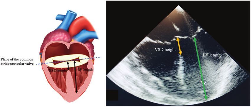
OBJECTIVES: There are limited data available on the height of the ventricular component of the septal deficiency (VSD) in patients under-
going complete atrioventricular septal defect (CAVSD) repair. VSD height may influence optimal choice of repair strategy with potential
consequences for long-term outcomes. We aimed to measure VSD height using 2-dimensional echocardiography and review its associa-
tion with postoperative outcomes.
METHODS: We retrospectively reviewed the preoperative echocardiograms of 45 consecutive patients who underwent CAVSD repair be-
tween May 2010 and December 2015 at a single centre. VSD height and left ventricular length on the four-chamber view were measured.
Demographic details and early and late outcomes including reoperation and long-term survival were studied.
C The Author(s) 2021. Published by Oxford University Press on behalf of the European Association for Cardio-Thoracic Surgery.
V
This is an Open Access article distributed under the terms of the Creative Commons Attribution-NonCommercial License (https://creativecommons.org/licenses/by-
nc/4.0/), which permits non-commercial re-use, distribution, and reproduction in any medium, provided the original work is properly cited. For commercial re-use,
please contact journals.permissions@oup.com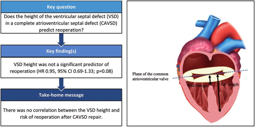
2                                         L.S. Fong et al. / Interactive CardioVascular and Thoracic Surgery

RESULTS: Twenty patients underwent modified single-patch repair and 25 patients underwent double-patch repair of CAVSD. VSD height
in the modified single-patch group ranged from 4.2 to 11.7 mm and in the double-patch group ranged from 5.1 to 14.9 mm. Nine patients
had a deep ‘scoop’ with a VSD height of >10 mm, (7 double patch, 2 modified single patch). VSD height did not correlate with a specific
Rastelli classification. There was no significant difference in the VSD height (P = 0.51) or the VSD height-to-left ventricular length ratio
(P = 0.43) between the 2 repair groups. There was no 30-day mortality. Eight patients required reoperation; however, VSD height was not a
significant predictor of reoperation (hazard ratio 0.95, 95% confidence interval 0.69–1.33; P = 0.08).
CONCLUSIONS: There was no correlation between VSD height and risk of reoperation after CAVSD repair. A deep ventricular scoop is un-
common in CAVSD patients.
Keywords: Complete atrioventricular septal defect • Complete atrioventricular canal • Atrioventricular septal defect

                                                                                                                                                     Downloaded from https://academic.oup.com/icvts/advance-article/doi/10.1093/icvts/ivab263/6387477 by guest on 30 November 2021
                                                                              outflow tract (LVOT) volumes in patients undergoing the DP and
    ABBREVIATIONS
                                                                              MSP technique [13, 14]. However, there are currently limited
                                                                              data available in the literature regarding the VSD height or
    2d2     Dimensional
                                                                              ‘scoop’ depth [5, 15] and its correlation with the chosen surgical
    CAVSD   Complete atrioventricular septal defect
                                                                              technique and clinical outcomes. These anatomical data are im-
    DP      Double patch
    HR      Hazard ratio                                                      portant as the depth of the septal ‘scoop’ may influence the likeli-
    LAVV    Left atrioventricular valve                                       hood of LVOTO or LAVV regurgitation following different repair
    LV      Left ventricular                                                  strategies [5]. This study aimed to determine the prevalence of a
    LVOT    Left ventricular outflow tract                                    deep ventricular ‘scoop’ in patients undergoing CAVSD repair
    LVOTO   Left ventricular outflow tract obstruction                        and review its association with postoperative outcomes and re-
    MSP     Modified single patch                                             pair technique (DP vs MSP).
    VSD     Ventricular septal defect
                                                                              PATIENTS AND METHODS
INTRODUCTION                                                                  Patient population
Recent studies [1] have demonstrated no significant difference in             A total of 47 consecutive patients who had undergone biventric-
long-term outcomes based on the surgical repair technique fol-                ular surgical repair of CAVSD between May 2010 and December
lowing repair of complete atrioventricular septal defect (CAVSD).             2015 were identified from institutional databases located at The
The 2 most commonly employed repair strategies are the modi-                  Children’s Hospital at Westmead, Sydney, Australia. Preoperative
fied single-patch (MSP) and the double-patch (DP) techniques.                 echocardiograms were available for analysis in 45 patients who
The DP technique has been the most extensively utilized tech-                 were included in the study. Patients with an associated diagnosis
nique and involves placement of separate atrial and ventricular               of tetralogy of Fallot and other conotruncal defects were ex-
septal patches. In contrast, the MSP technique, which was devel-              cluded. The study was approved by the Sydney Children’s
oped by Wilcox et al. [2] and Nunn [3, 4] independently in the                Hospital Network Human Research Ethics Committee (HREC/16/
1990s, has rapidly grown in popularity due to its relative techni-            SCHN/216) and the need for individual patient consent was
cal ease, as there is no requirement to size a ventricular septal             waived.
patch (as with the DP technique) or to divide bridging leaflets (as
in the classical single-patch technique). However, a large ventric-           Two-dimensional echocardiographic assessment
ular component to the septal deficiency, especially with a deep
ventricular septal defect (VSD), continues to be a concern for                Two separate operators independently measured the size of the
some surgeons when considering whether to perform the MSP                     ventricular component of the septal deficiency (VSD height) and
technique for CAVSD repair [5]. There has long been concern                   the left ventricular (LV) length on the four-chamber view at end-
both for the propensity to develop left ventricular outflow tract             diastole (Fig. 1). The VSD height was defined as the distance from
obstruction (LVOTO) and for the potential impact on left atrio-               the deepest point of the ventricular component at the level of
ventricular valve (LAVV) function with the MSP technique [6–10]               the atrioventricular junction to the intersection between the
due to the approximation of the valvar leaflets to the ventricular            atrioventricular valve components. Since the size of the hearts
septal crest. Although previous studies [1] demonstrate compara-              varied within this study, the measurements were standardized by
ble outcomes with both MSP and DP techniques, it is impossible                measuring the LV length, as a marker of LV size. LV length was
to control for surgeon preference in choosing repair technique                defined as the distance from the LV apex to the intersection of
based on subtleties of CAVSD anatomy such as VSD height and                   the atrioventricular valve components at the atrioventricular
depth of septal scoop.                                                        junction. The ratio of the VSD height to LV length was then calcu-
   Echocardiography is an essential part of the preoperative as-              lated for comparison. Since the LV apex may not be included
sessment of CAVSD patients prior to repair [11] and its evolution             in the four-chamber view, an assessment of foreshortening
over time has revolutionized the diagnosis and management of                  [where the 2-dimensional (2D) ultrasound plane does not cut
patients with CAVSD [12]. Previous echocardiographic studies                  through the true apex] was also made due to the risk of underes-
have demonstrated no significant difference in the LAVV annulus               timating the LV dimension on this view. The mean values of these
size, tenting height, size of vena contracta and left ventricular             2 measurements were then used as final discrete values.
L.S. Fong et al. / Interactive CardioVascular and Thoracic Surgery                                            3

                                                                                                                                                                 CONGENITAL
                                                                                                                                                                 Downloaded from https://academic.oup.com/icvts/advance-article/doi/10.1093/icvts/ivab263/6387477 by guest on 30 November 2021
Figure 1: Demonstration of measurements of the VSD height and LV length measured using 2-dimensional echocardiography on four-chamber view. LV: left ventricu-
lar; VSD: ventricular septal defect.

Surgical technique                                                                Ventricular septal defect height
The procedural details for repair of CAVSD have been reported                     The VSD height in the MSP group ranged from 4.2 to 11.7 mm
previously [16]. Briefly, all CAVSD repair operations were per-                   with a mean height of 7.3 ± 1.9 mm, and in the DP group, the
formed via a median sternotomy with aortobicaval cannulation                      VSD height ranged from 5.1 to 14.9 mm with a mean height of
and moderate hypothermia with either the DP or modified                           8.3 ± 2.4 (Table 2). Figure 2 demonstrates the broad range of VSD
single-patch (MSP) technique depending on the surgeon prefer-                     heights at the time of CAVSD repair, with 9 patients (7 DP, 2
ence. Patient follow-up data were obtained through hospital,                      MSP) having a deep ‘scoop’, defined as a VSD height >10 mm.
clinic, and cardiologist medical records. Overall median follow-                  There was no significant difference in mean VSD height (P = 0.51),
up duration was 4.2 (interquartile range 2.8–5.8) years.                          or deep ‘scoop’ (P = 0.13) between the 2 repair groups (P = 0.51,
                                                                                  Table 2).
Statistical analyses
                                                                                  Ratio of ventricular septal defect height to left
Continuous variables were described as median and range and
categorical variables were compared using the Pearson’s v2 or
                                                                                  ventricular length
Fisher’s exact test. Differences between continuous variables were
                                                                                  The ratio of the VSD height to LV length varied widely in both
analysed using the Mann–Whitney U-test for 2 samples. Risk fac-
                                                                                  groups, ranging from 14% to 47% (Fig. 3A). There was no signifi-
tors for time to first reoperation were modelled using Cox regres-
sion modelling with the associated hazard ratio (HR) estimated.                   cant difference in the VSD height-to-LV length ratio between the
P-value
4 L.S. Fong et al. / Interactive CardioVascular and Thoracic Surgery
Table 1: Patient and operative characteristics of complete atrioventricular septal defect repair
Characteristics MSP (n = 20) DP (n = 25) P-value
Age at AVSD repair (months), median (IQR) 4.2 (2.3–5.7) 3.5 (2.5–4.3) 0.52
Weight (kg), median (IQR) 4.5 (3.8–5.5) 4.5 (3.6–5.2) 0.59
Female gender, n (%) 11 (55) 11 (44) 0.46
Down syndrome, n (%) 9 (45) 10 (40) 0.74
Associated cardiovascular anomalies, n (%)
Coarctation of aorta 1 (5) 2 (8) 0.69
Downloaded from https://academic.oup.com/icvts/advance-article/doi/10.1093/icvts/ivab263/6387477 by guest on 30 November 2021
DOLAVV 0 3 (12) 0.11
Single papillary muscle 0 3 (12) 0.11
LVOTO 0 0
Preoperative ECHO LAVVR >_ 3 3 (15) 2 (8) 0.46
Rastelli class, n (%) 0.49
A 8 (40) 12 (48)
B 1 (5) 0
C 11 (55) 13 (52)
Prior PAB, n (%) 4 (20) 2 (8) 0.24
Management of zone of apposition, n (%) 0.20
Complete closure 20 (100) 23 (92)
Left open 0 2 (8)
Cardiopulmonary bypass time (min), mean ± SD 126 ± 45 167 ± 57 0.001
Aortic cross-clamp time (min), mean ± SD 88 ± 38 132 ± 50 0.0001
Postoperative ECHO LAVVR grade, n (%) 0.83
None 0 1 (4)
Trivial 3 (15) 3 (12)
Mild 13 (65) 16 (64)
Moderate 4 (20) 5 (20)
Severe 0 0
30-Day PPM insertion, n (%) 1 (5) 0 0.26
AVSD: atrioventricular septal defect; DOLAVV: double orifice left atrioventricular valve; DP: double patch; IQR: interquartile range; LAVVR >_ 3: moderate or greater
left atrioventricular valve regurgitation; LVOTO: left ventricular outflow tract obstruction; MSP: modified single patch; PAB: pulmonary artery banding; PPM: per-
manent pacemaker; SD: standard deviation.
Table 2: Echocardiographic features before complete atrioventricular septal defect repair
Characteristics MSP (n = 20) DP (n = 25) P-value
Mean VSD height, mean ± SD 7.3 ± 1.9 8.3 ± 2.4 0.51
Mean LV length, mean ± SD 27.1 ± 4.4 28.6 ± 7.4 0.69
Mean VSD height to LV length ratio, mean ± SD 0.28 ± 0.08 0.30 ± 0.08 0.43
Deep scoop (VSD height >10 mm), n (%) 2 (10) 7 (28) 0.13
DP: double patch; LV: left ventricular; MSP: modified single patch; VSD: ventricular septal defect.
(Table 3). One patient who had undergone DP repair required utilized, reoperation for LAVV dysfunction and LVOTO remain
late reoperation for LVOTO. important long-term concerns. With evolution of the MSP tech-
Two patients with a deep VSD required reoperation. Both nique, more focus has been placed on the extent and nature of
patients had undergone previous DP repair and required reoper- the VSD with regards to repair technique and the risk of LAVV
ation for LVOTO and severe LAVV regurgitation, respectively. dysfunction and development of LVOTO post-CAVSD repair.
Utilizing Cox regression, VSD height was not a significant predic- Although there is no clear evidence that VSD height impacts out-
tor of reoperation (HR 0.95, 95% confidence interval 0.69–1.33; comes following CAVSD repair, it is often considered as one of
P = 0.77). There was no 30-day mortality. Two patients died dur- the criteria guiding choice of repair technique. Backer et al. [10]
ing long-term follow-up due to cardiac failure and respiratory considered a VSD height of >12 mm to be an indication for DP
failure, respectively. repair. Importantly, our study suggests that a deep ventricular
scoop is uncommon in CAVSD patients, with the VSD beingL.S. Fong et al. / Interactive CardioVascular and Thoracic Surgery 5
CONGENITAL
Downloaded from https://academic.oup.com/icvts/advance-article/doi/10.1093/icvts/ivab263/6387477 by guest on 30 November 2021
Figure 2: (A) Graphical representation of VSD height and LV length in complete atrioventricular septal defect patients stratified by repair technique. (B) VSD height
stratified by need for reoperation and complete atrioventricular septal defect repair type. LV: left ventricular; VSD: ventricular septal defect. Double patch (red dia-
mond) and modified single patch (blue circle).
Figure 3: (A) Graphical representation of VSD height-to-LV length ratio stratified by CAVSD repair technique. (B) VSD height in CAVSD patients stratified by Rastelli
class and repair technique; CAVSD: complete atrioventricular septal defect; LV: left ventricular; VSD: ventricular septal defect. Double patch (red diamond) and modi-
fied single patch (blue circle).6 L.S. Fong et al. / Interactive CardioVascular and Thoracic Surgery
Limitations
Table 3: Reoperation after complete atrioventricular septal
defect repair This study was limited by its small sample size, retrospective na-
ture, single-centre nature and the inability to control for surgeon
Characteristics MSP (n = 20) DP (n = 25) P-value preference or decision-making in choice of repair technique. All
measurements were performed using available images stored
30-Day reoperation, n (%) 4 (20) 3 (12) 0.46
LAVV repair 1 1
from the patient’s preoperative transthoracic echocardiogram
LAVV replacement 2 0 and were therefore limited to the imaging data available. The 2D
Closure of residual VSD 1 0 definition of the transthoracic echocardiogram and any fore-
LAVV repair and closure 0 2 shortening may have resulted in underestimation of the LV
Downloaded from https://academic.oup.com/icvts/advance-article/doi/10.1093/icvts/ivab263/6387477 by guest on 30 November 2021
of residual VSD
length dimensions.
DP: double patch; LAVV: left atrioventricular valve; MSP: modified single
patch; VSD: ventricular septal defect.
CONCLUSIONS
ventricular septal crest, which may alter LAVV function or result There was no correlation observed between the VSD height and
in further elongation of the LVOT when the VSD is deep. risk of reoperation after CAVSD repair and mean VSD height was
Although numbers are small and the overall differences were not similar between repair techniques. Whilst a deep ventricular scoop
significant, there appears to be a preference amongst surgeons in is uncommon in CAVSD patients, there appears to be surgeon
our unit towards repair with the DP technique when there is a preference for the DP technique in these patients, and further
deep VSD scoop defined by a VSD height >10 mm. Seven of studies on this particular subgroup are needed. Measurement of
these patients were in the DP group compared with only 2 in the the VSD height should be an essential part of the standard preop-
MSP group; however, it is impossible to know whether the ap- erative assessment of CAVSD patients in the current era.
proach to these patients altered the overall outcome in this
study. A larger study is required to address the question of
whether the type of repair technique alters outcome in patients Funding
with a VSD height >10 mm.
Anatomical studies focusing on the morphological aspects of This study was supported by the National Heart Foundation of
CAVSD illustrate the close relationship between the VSD height Australia [Health professional scholarship 101616 to L.S.F.]
and LVOT diameter [15, 17]. A morphometric analysis by Adachi
et al. [5] found 16 of 43 atrioventricular septal defect heart speci-
mens had anterosuperior extension of the scoop, associated with Conflict of interest: none declared.
a skewed shape of the scoop and significantly narrower LVOT.
They suggested that not only the VSD height was important, but Author contributions
also the presence of anterosuperior extension could affect valvar
competence after MSP repair. However, assessing for the pres- Laura S. Fong: Conceptualization; Data curation; Formal analysis; Writing—
ence of anterosuperior extension can be quite challenging with original draft. David Youssef: Data curation; Writing—review & editing. Julian
Ayer: Supervision; Writing—review & editing. Ian A. Nicholson: Supervision.
conventional 2D echocardiography techniques, and subsequent David S. Winlaw: Supervision; Writing—review & editing. Yishay Orr: Formal
studies [1, 9] have failed to show a difference in reintervention analysis; Methodology; Supervision; Writing—review & editing.
for LVOTO between MSP and DP repair. With regard to LAVV
function, there is evidence that the LAVV annulus has more sys-
tolic contraction with the MSP technique compared to DP repair
Reviewer information
with no other identifiable differences in mechanics of LAVV func- Interactive CardioVascular and Thoracic Surgery thanks Amir-Reza
tion following repair with the 2 techniques [14]. Despite the small Hosseinpour, Cheul Lee, Christoph Schmitz and the other, anonymous
cohort size, this finding suggests that LAVV function is not nega- reviewer(s) for their contribution to the peer review process of this article.
tively impacted by use of the MSP technique as suggested by
equivalent rates of LAVV reintervention in our study and previous
studies [1]. REFERENCES
2D echocardiography has been the cornerstone of CAVSD di-
[1] Fong LS, Betts K, Bell D, Konstantinov IE, Nicholson IA, Winlaw DS et al.
agnosis and preoperative assessment [11], being both non- Complete atrioventricular septal defect repair in Australia: results over
invasive and widely accessible. However, recent advances [12] in 25 years. J Thorac Cardiovasc Surg 2019;30:30.
3-dimensional echocardiography may provide the opportunity [2] Wilcox BR, Jones DE, Frantz EG, Brink LW, Henry GW, Mill MR et al.
to better understand the anatomical defects of CAVSD due to Anatomically sound, simplified approach to repair of “complete” atrio-
ventricular septal defect. Ann Thorac Surg 1997;64:487–93; discussion
better depth perception and resolution allowing better sensitivity
493–4.
in determining complex leaflet and LVOT abnormalities [18], as [3] Nunn GR. Atrioventricular canal: modified single patch technique.
well as mechanisms for failed repair [19]. The issue with correlat- Semin Thorac Cardiovasc Surg Pediatr Card Surg Annu 2007;10:28–31.
ing preoperative CAVSD anatomy and long-term outcomes is the [4] Nicholson IA, Nunn GR, Sholler GF, Hawker RE, Cooper SG, Lau KC.
length of time it takes to accrue useful long-term follow-up data. Simplified single patch technique for the repair of atrioventricular septal
defect. J Thorac Cardiovasc Surg 1999;118:642–6.
Future studies correlating 3-dimensional echocardiography as- [5] Adachi I, Ho SY, McCarthy KP, Uemura H. Ventricular scoop in atrioven-
sessment and failed repair during long-term follow-up would be tricular septal defect: relevance to simplified single-patch method. Ann
beneficial. Thorac Surg 2009;87:198–203.L.S. Fong et al. / Interactive CardioVascular and Thoracic Surgery 7
[6] Backer CL, Stewart RD, Bailliard F, Kelle AM, Webb CL, Mavroudis C. [13] Al Senaidi KS, Ross DB, Rebeyka IM, Harder J, Kakadekar AP, Garros D
Complete atrioventricular canal: comparison of modified single-patch et al. Comparison of two surgical techniques for complete atrioventricu-
technique with two-patch technique. Ann Thorac Surg 2007;84:2038–46; lar septal defect repair using two- and three-dimensional echocardiog-
CONGENITAL
discussion 2038–46. raphy. Pediatr Cardiol 2014;35:393–8.
[7] Pan G, Song L, Zhou X, Zhao J. Complete atrioventricular septal defect: [14] Ugaki S, Khoo NS, Ross DB, Rebeyka IM, Adatia I. Modified single-patch
comparison of modified single-patch technique with two-patch tech- compared with two-patch repair of complete atrioventricular septal de-
nique in infants. J Card Surg 2014;29:251–5. fect. Ann Thorac Surg 2014;97:666–71.
[8] Yildirim O, Avsar M, Ozyuksel A, Akdemir M, Zeybek C, Demiroluk S [15] Penkoske PA, Neches WH, Anderson RH, Zuberbuhler JR. Further obser-
et al. Modified single versus double-patch technique for the repair vations on the morphology of atrioventricular septal defects.[Erratum
of complete atrioventricular septal defect. J Card Surg 2015;30: appears in J Thorac Cardiovasc Surg 1988;95(1):146]. J Thorac
595–600. Cardiovasc Surg 1985;90:611–22.
[9] Li D, Fan Q, Iwase T, Hirata Y, An Q. Modified single-patch technique [16] Fong LS, Betts K, Kannekanti R, Ayer J, Winlaw DS, Orr Y. Modified-single
Downloaded from https://academic.oup.com/icvts/advance-article/doi/10.1093/icvts/ivab263/6387477 by guest on 30 November 2021
versus two-patch technique for the repair of complete atrioventricular patch vs double patch repair of complete atrioventricular septal defects.
septal defect: a meta-analysis. Pediatr Cardiol 2017;38:1456–64. Semin Thorac Cardiovasc Surg 2019;12:12.
[10] Backer CL, Eltayeb O, Monge MC, Wurlitzer KC, Hack MA, Boles LH et al. [17] Ebels T, Ho SY, Anderson RH, Meijboom EJ, Eijgelaar A. The surgical
Modified single patch: are we still worried about subaortic stenosis? Ann anatomy of the left ventricular outflow tract in atrioventricular septal de-
Thorac Surg 2015;99:1671–5; discussion 1675–6. fect. Ann Thorac Surg 1986;41:483–8.
[11] Santoro G, Marino B, Di Carlo D, Formigari R, Santoro G, Marcelletti C et [18] Kutty S, Smallhorn JF. Evaluation of atrioventricular septal defects by
al. Patient selection for repair of complete atrioventricular canal guided three-dimensional echocardiography: benefits of navigating the third di-
by echocardiography. Eur J Cardiothorac Surg 1996;10:439–42. mension. J Am Soc Echocardiogr 2012;25:932–44.
[12] Colen TS, Jeffrey F. Three-dimensional echocardiography for the assess- [19] Takahashi K, Mackie AS, Thompson R, Al-Naami G, Inage A, Rebeyka IM et
ment of atrioventricular valves in congenital heart disease: past, present al. Quantitative real-time three-dimensional echocardiography provides
and future. Semin Thorac Cardiovasc Surg Pediatr Card Surg Annu 2015; new insight into the mechanisms of mitral valve regurgitation post-repair of
18:62–71. atrioventricular septal defect. J Am Soc Echocardiogr 2012;25:1231–44.You can also read