Multi energy spectral photon counting computed tomography (MARS) for detection of arthroplasty implant failure - Nature
←
→
Page content transcription
If your browser does not render page correctly, please read the page content below
www.nature.com/scientificreports
OPEN Multi‑energy spectral
photon‑counting computed
tomography (MARS) for detection
of arthroplasty implant failure
Lawrence Chun Man Lau1,8, Wayne Yuk Wai Lee1,2,8, Anthony P. H. Butler3,4,5,6,
Alex I. Chernoglazov6, Kwong Yin Chung1, Kevin Ki Wai Ho1, James Griffith7,
Philip H. Butler4,5,6* & Patrick Shu Hang Yung1*
To determine whether state-of-the-art multi-energy spectral photon-counting computed tomography
(MARS) can detect knee arthroplasty implant failure not detected by standard pre-operative imaging
techniques. A total knee arthroplasty (TKA) removed from a patient was reviewed. The extracted
prosthesis [NexGen Legacy Posterior Stabilized (LPS) TKA] was analyzed as were pre-operative
imaging examination and compared with a MARS-CT examination obtained of the extracted TKA
prosthesis. Radiographs, fluoroscopy, ultrasound and MRI preoperatively did not reveal the cause
of the implant failure. MARS CT images of the extracted prosthesis clearly showed the presence of
posteromedial polyethylene and tibial tray wear which is compatible with the clinical appearance of
the extracted TKA. MARS can identify polyethylene insert and metallic tibial tray wear as a cause of
TKA failure, that could not be identified with on standard pre-operative imaging. Although clinical
MARS CT system is still under development, this case does illustrate its potential clinical usefulness.
This is the first study to document how MARS CT imaging can detect orthopedic implant failure not
detected by standard current imaging techniques. This system has a potential clinical application in
orthopedic patients.
Osteoarthritis is the single most common cause of disability in elderly adults and the knee is the most common
involved site1. Symptomatic knee osteoarthritis is associated with pain, stiffness, and loss of mobility and can be
effectively managed by total knee arthroplasty (TKA) when conservative management fails. In an aging society,
TKA is in high demand with a projected 3.48 million TKA procedures performed annually in the United States
alone by 2 0302. Despite its frequent use, almost 20% of patients report suboptimal results after TKA with symp-
toms like persistent residual pain, stiffness and functional limitation3. Establishing the cause of symptoms and
pain in patients with TKA is cardinal to proper management as revision surgery with an uncertain diagnosis
has a poor outcome4.
Imaging is important in the diagnostic evaluation of TKA failure, mainly using modalities like radiography,
fluoroscopy, ultrasound (US), computed tomography (CT), and magnetic resonance imaging (MRI). However,
each of these modalities has its own limitations5. Radiography and fluoroscopy cannot delineate radiolucent
structures like the surrounding soft-tissue structures or polyethylene-made insert or baseplate. US can assess
effusions, and soft-tissue structures about the knee but cannot evaluate the prosthesis itself or the surrounding
bony structure. CT is limited by beam-hardening artefact around the metallic femoral and tibial components,
1
Department of Orthopaedics and Traumatology, Faculty of Medicine, The Prince of Wales Hospital, The Chinese
University of Hong Kong, Shatin, Hong Kong SAR, China. 2Li Ka Shing Institute of Health Sciences, The Prince of
Wales Hospital, The Chinese University of Hong Kong, Hong Kong, Hong Kong SAR, China. 3University of Otago,
2 Riccarton Ave, Christchurch 8140, New Zealand. 4School of Physical and Chemical Sciences, University of
Canterbury, Private Bag 4800, Christchurch 8140, New Zealand. 5The European Organization for Nuclear Research
(CERN), Geneva, Switzerland. 6MARS Bioimaging Ltd, 29a Clyde Rd, Christchurch, New Zealand. 7Department
of Radiology, Faculty of Medicine, The Prince of Wales Hospital, The Chinese University of Hong Kong, Shatin,
Hong Kong SAR, China. 8These authors contributed equally: Lawrence C. M. Lau and Wayne Y. W. Lee. *email:
phil.butler@marsbioimaging.com; patrickyung@cuhk.edu.hk
Scientific Reports | (2021) 11:1554 | https://doi.org/10.1038/s41598-020-80463-2 1
Vol.:(0123456789)www.nature.com/scientificreports/
limiting visualization of the polyethylene insert as well as the immediate surrounding bone and soft tissue. Simi-
larly, MRI is subject to metallic susceptibility obscuring the prosthesis and surrounding s tructures5.
The recently developed multi-energy spectral photon-counting computed tomography (MARS), by captur-
ing the characteristic attenuation of different materials at multiple energy r anges6,7 with a conventional single
X-ray source but equipped with multiple photon counting and energy resolving detectors, allows high efficiency
detection within the human diagnostic energy range (30–120 keV). As each material has a measurable energy-
dependent X-ray attenuation, spectroscopic imaging allows for multiple materials to be differentiated and quan-
tified from each other simultaneously8–11. From this multi-energy data, a ‘color’ image of the object of interest
can be generated, with high resolution structural information and also some compositional information12–14.
Thereby material-specific images can be produced in MARS with minimal beam-hardening artefact often seen
in standard CT imaging15,16.
The aim of this study was to demonstrate if MARS could detect knee arthroplasty implant failure that were
not detected by current imaging techniques.
Materials and methods
Ethical statement. This study complied with the Declaration of Helsinki after obtaining approval from the
Institutional Review Board of the local institution’s Research Ethical Committee [The Joint Chinese University
of Hong Kong—New Territories East Cluster Clinical Research Ethics Committee (The Joint CUHK-NTEC
CREC)] (CREC 2018.544). Experiments involving human participants (including the use of tissue samples) are
performed with informed consent obtained.
Prosthesis identification. A TKA prosthesis was identified from our extracted prosthesis bank with
accompanying clinical records. The inclusion criteria for TKA prostheses were as follows: (1) TKA retrieved
from symptomatic patient who had undergone revision surgery; (2) imaging and laboratory investigation had
failed to identify the cause of TKA failure before revision surgery; and (3) the cause of failure could be ascer-
tained based on morphologic assessment after extraction.
An extracted NexGen Legacy Posterior Stabilized (LPS) TKA (Zimmer Biomet, Warsaw, Indiana, USA) ful-
filled these inclusion criteria and was used for analysis and imaging. This TKA consists of a femoral component,
articular insert and tibial baseplate component. The femoral component was made of Zimaloy Cobalt-Chromium-
Molybdenum Alloy. The articular insert was made of Ultra-High Molecular-Weight Polyethylene (UHMWPE)
and the tibial baseplate was made of Tivanium Ti-6Al-4V Alloy. A small amount of cement and bone remained
attached to the base of the femoral and tibial components after extraction.
Clinical details and pre‑revision imaging. The TKA belonged to an elderly female who had intermittent
knee pain with clicking for 1 year. The TKA had been implanted twelve years earlier and she had remained well
until her current knee symptoms started. There was no history of trauma, or symptoms of infection. Physical
examination showed no signs of infection and full knee movement except for a distinct mid-range clunk. Sero-
logical inflammatory markers, white cell count and differential count were all normal.
Imaging of the TKA comprising radiographs (weight-bearing anteroposterior view, weight-bearing lateral
view in extension, 30° flexion and 45° flexion, skyline view, lower-limb scanogram), fluoroscopy, ultrasound,
and standard MRI on an 1.5 T imaging system was performed prior to revision surgery.
Revision surgery. Revision TKA surgery was performed. During the operation, posteromedial polyethyl-
ene insert wear, posteromedial metallic tibial tray wear and severe metallosis were found. The whole prosthesis
was removed, and extensive debridement was done. A constrained condylar knee arthroplasty was inserted.
Intra-operative tissue culture was negative for infection.
Imaging of extracted TKA prosthesis. The TKA protheses were imaged using a preclinical MARS scan-
ner (MARS bioimaging Ltd., Christchurch, New Zealand)7. The scanner comprises a microfocus poly‐ener-
getic X‐ray source and five Medipix 3RX energy‐resolving photon‐counting detectors within a continuous
rotating gantry. A 2‐mm–thick cadmium zinc telluride sensor (14.1 × 14.1 m m2) bump bonded at 110 μm to a
17,18
Medipix3RX readout chip was u sed . MARS imaging of TKA specimens and calibration phantoms was per-
formed using 5 charge‐summing mode energy thresholds of 7, 45, 55, 65, and 75 keV at 120 kVp (10). Imaging
data were reconstructed simultaneously into non‐overlapping energy bins across the measured spectrum, such
as 7–45.1, 45.1–54.9, 54.9–65.1, and 65.1–74.9 and 74.9 keV, at a spatial resolution of 0.09 × 0.09 × 0.09 mm.
Images of the TKA prostheses were calibrated using mass attenuation coefficients estimated from the calibration
phantom data to distinguish bone, cement, metallic femoral component and tibial baseplate and polyethylene
insert and furnish material decomposition maps (see Supplementary File 1). The images obtained from MARS
were then contrasted and compared with the results of previous imaging modalities obtained during clinical
investigation.
Results
Pre‑revision imaging of TKA prothesis. Radiographs of the knee were unremarkable (Fig. 1). Fluoro-
scopic screening noted a palpable clunk occurring during flexion–extension motion though but no discernible
change in femorotibial relationship when this clunk occurred. No anteroposterior or varus-valgus instability was
shown on fluoroscopy. Ultrasound revealed no evidence of iliotibial band syndrome or patellar clunk syndrome,
Scientific Reports | (2021) 11:1554 | https://doi.org/10.1038/s41598-020-80463-2 2
Vol:.(1234567890)www.nature.com/scientificreports/
Figure 1. Weightbearing radiographs in (a) Anteroposterior extension (b) lateral extension, (c) lateral 30°
flexion and (d) lateral 45° flexion weightbearing radiographs of knee. No definite polyethene thinning is evident.
Figure 2. T1-weighted (a) sagittal and (b) coronal MRI images showing severe metallic artefact.
with the clunk seeming to arise deeply from the tibiofemoral articulation rather than from the superficial struc-
tures. MRI revealed severe metallic artefact though was otherwise normal (Fig. 2).
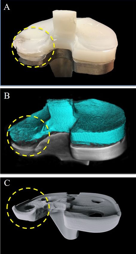
Post‑revision imaging of TKA prosthesis with MARS. The MARS-acquired images clearly demon-
strated clearly moderate-severity polyethylene insert wear, and posteromedial metallic tibial tray wear, compat-
ible with the actual appearance of the prosthesis (Fig. 3) (Supplementary Video 1).
Discussion
In the case presented, we showed how MARS-acquired CT images could clearly demonstrate polyethylene
insert wear and metallic tibial tray wear clearly, not demonstrable by other imaging techniques highlighting the
potential value of MARS imaging to aid clinical diagnosis.
Scientific Reports | (2021) 11:1554 | https://doi.org/10.1038/s41598-020-80463-2 3
Vol.:(0123456789)www.nature.com/scientificreports/
Figure 3. (a) Clinical photos, (b) MARS-CT images and (c) MARS-CT images with polyethelene digitally
removed. The wear at the postomedial aspect of the polyethylene impant and tibial metallic tray is clearly visible
(circle).
Although TKA has long-term success in most cases, some cases do develop complications and fail over
time. Common causes of TKA failure include aseptic loosening, instability, infection, polyethylene wear with
or without osteolysis, metallosis and extensor mechanism f ailure19. If severe, these causes of failure are easy to
Scientific Reports | (2021) 11:1554 | https://doi.org/10.1038/s41598-020-80463-2 4
Vol:.(1234567890)www.nature.com/scientificreports/
identify on radiography by, for example, a fracture line in periprosthetic fracture, or periprosthetic radiolucency
in aseptic loosening. However, in less severe cases, radiological changes are usually minimal or absent, as in
the case presented where pre-operative imaging failed to identify the cause of pain and clicking. Multi-energy
MARS CT was able to accurately reveal polyethylene wear without any significant metallic or other artefact
characteristic of standard M RI20.
This has potential clinical implications. As the polyethylene insert wear was not identified early on and
not exchanged, it continued to wear. Eventually, after the complete wear of part of the polyethylene insert, the
underneath metallic tibial tray was exposed and underwent further wear due to abrasion by the metallic condyle
of the femoral component. This produced metallosis that required extensive debridement. As the tibial tray and
the femoral component were damaged, they had to be removed and replaced with a new implant which was of
a constrained design to compensate the significant bone loss and instability created during the primary TKA
removal. If the presented patient had been able to undergo MARS CT and identify polyethylene insert wear
before its complete wear, the patient could have just undergone surgery that exchange the polyethylene insert,
which is a much smaller scale surgery and preserve more bone and soft tissue of the patient.
This case illustrates the potential use of MARS to identify polyethylene wear early potentially enabling less
extensive surgery to be performed, rather than major revision surgery with significant bone and soft tissue loss.
This constitutes a significant difference to the patient care and medical cost. Based on our finding, MARS could
play a unique role to gauge the remaining thickness of polyethylene insert before tibial tray is exposed. This
would allow surgeons to determine the urgency of revision surgery and advise the patient the timing of revision.
There are limitations to this presentation. No arthrography or CT arthrography study was performed pre-
operatively. These studies may have demonstrated the polyethylene wear21, though arthrography is, by definition,
a minimally invasive procedure not to be undertaken lightly especially in the prosthetic knee where the risk of
introducing infection is high. Although standard CT or MRI is currently not used to evaluate polyethylene wear
due to a rtifact22, dual energy CT or MR imaging with metallic artifact reduction may have allowed a more in-
depth assessment of the polyethylene implant though these techniques have not been evaluated in this regard.
We supplement our study by imaging the extracted TKA prothesis with high-resolution peripheral quantitative
computed tomography (HRpQCT) (XtremeCT II; Scanco Medical AG), which is another sophisticated form of
CT and it could not demonstrate the polyethylene wear as shown by the MARS CT (see Supplementary File 2).
In conclusion, this is the first study to show how MARS CT imaging can detect orthopedic implant failure
that is not detected by standard current imaging modalities. If confirmed in a clinical study, the accurate dem-
onstration of polyethylene wear could have considerable implications of patient care, potentially allowing wear
to be evaluated at an earlier stage, monitored accordingly with, if necessary, polyethylene insert replacement
before metallic tray wear and metallosis occurs. Further cadaveric investigation is warranted to optimize imag-
ing parameters and protocols, post-processing and analysis, which will lay the foundations for clinical study
with clinical MARS.
Data availability
No datasets were generated or analysed during the current study.
Received: 15 August 2020; Accepted: 21 December 2020
References
1. Turkiewicz, A. et al. Current and future impact of osteoarthritis on health care: A population-based study with projections to year
2032. Osteoarthr. Cartil. 22, 1826–1832. https://doi.org/10.1016/j.joca.2014.07.015 (2014).
2. Kurtz, S., Ong, K., Lau, E., Mowat, F. & Halpern, M. Projections of primary and revision hip and knee arthroplasty in the United
States from 2005 to 2030. J. Bone Jt. Surg. Am. 89, 780–785. https://doi.org/10.2106/JBJS.F.00222 (2007).
3. Bourne, R. B., Chesworth, B. M., Davis, A. M., Mahomed, N. N. & Charron, K. D. Patient satisfaction after total knee arthroplasty:
Who is satisfied and who is not?. Clin. Orthop. Relat. Res. 468, 57–63. https://doi.org/10.1007/s11999-009-1119-9 (2010).
4. Mont, M. A., Serna, F. K., Krackow, K. A. & Hungerford, D. S. Exploration of radiographically normal total knee replacements for
unexplained pain. Clin. Orthop. Relat. Res. https://doi.org/10.1097/00003086-199610000-00030 (1996).
5. Flierl, M. A., Sobh, A. H., Culp, B. M., Baker, E. A. & Sporer, S. M. Evaluation of the painful total knee arthroplasty. J. Am. Acad.
Orthop. Surg. 27, 743–751. https://doi.org/10.5435/JAAOS-D-18-00083 (2019).
6. Taguchi, K. & Iwanczyk, J. S. Vision 20/20: Single photon counting X-ray detectors in medical imaging. Med. Phys. 40, 100901.
https://doi.org/10.1118/1.4820371 (2013).
7. Anderson, N. G. & Butler, A. P. Clinical applications of spectral molecular imaging: Potential and challenges. Contrast Media Mol.
Imaging 9, 3–12. https://doi.org/10.1002/cmmi.1550 (2014).
8. Rajendran, K. et al. Quantitative imaging of excised osteoarthritic cartilage using spectral CT. Eur. Radiol. 27, 384–392 (2017).
9. Zainon, R. et al. Spectral CT of carotid atherosclerotic plaque: Comparison with histology. Eur. Radiol. 22, 2581–2588 (2012).
10. Ronaldson, J. P. et al. Toward quantifying the composition of soft tissues by spectral CT with Medipix3. Med. Phys. 39, 6847–6857
(2012).
11. Stamp, L. K. et al. Clinical utility of multi-energy spectral photon-counting computed tomography in crystal arthritis. Arthritis
Rheumatol. 71, 1158–1162 (2019).
12. Butler, P. H. et al. In Developments in X-Ray Tomography XII. 111130C (International Society for Optics and Photonics).
13. Bateman, C. et al. MARS-MD: Rejection based image domain material decomposition. J. Instrum. 13, P05020 (2018).
14. Mandalika, V. B. H. A. C., Alexander, I., Billinghurst, M., Bartneck, C., Hurrell, M. A., De Ruiter, N., Butler, A. P. H., Butler, P. H.
A hybrid 2D/3D user interface for radiological diagnosis. J. Digit. Imaging 56–73 (2018).
15. Amma, M. R. et al. In Developments in X-Ray Tomography XII. 111131D (International Society for Optics and Photonics).
16. Rajendran, K. et al. Reducing beam hardening effects and metal artefacts in spectral CT using Medipix3RX. J. Instrum. 9, P03015
(2014).
17. Ballabriga, R. et al. The Medipix3RX: A high resolution, zero dead-time pixel detector readout chip allowing spectroscopic imag-
ing. J. Instrum. 8, C02016 (2013).
18. Ballabriga, R. et al. Review of hybrid pixel detector readout ASICs for spectroscopic X-ray imaging. J. Instrum. 11, P01007 (2016).
Scientific Reports | (2021) 11:1554 | https://doi.org/10.1038/s41598-020-80463-2 5
Vol.:(0123456789)www.nature.com/scientificreports/
19. Mulcahy, H. & Chew, F. S. Current concepts in knee replacement: Complications. AJR Am. J. Roentgenol. 202, W76-86. https://
doi.org/10.2214/AJR.13.11308(2014).
20. Hargreaves, B. A. et al. Metal-induced artifacts in MRI. AJR Am. J. Roentgenol. 197, 547–555. https://doi.org/10.2214/AJR.11.7364
(2011).
21. Hsu, Y., Lin, C. H., Shu, G. H. F., Hsieh, T. J. & Chen, C. K. Fracture of the polyethylene tibial post in the posterior-stabilized total
knee prosthesis: Arthrographic and CT arthrographic diagnosis. Skelet. Radiol. 48, 1145–1148. https://doi.org/10.1007/s0025
6-019-03173-5 (2019).
22. Expert Panel on Musculoskeletal, I. et al. ACR appropriateness criteria((R)) imaging after total knee arthroplasty. J. Am. Coll.
Radiol. 14, S421–S448, https://doi.org/10.1016/j.jacr.2017.08.036 (2017).
Acknowledgements
This work was supported by CUHK MARS Scanner Project donated by Li Ka Shing (Canada) Foundation
(Project number: 7106251) and the Matching Grant Scheme launched by the HKSAR Government (Project
number: 8509245).
Author contributions
L.C.M.L. and W.Y.W.L. initiated and conducted the study and wrote the main manuscript text. L.C.M.L.,
W.Y.W.L., A.P.H.B., A.I.C. and P.H.B. produced the MARS images and wrote the imaging methodology. K.Y.C.,
K.K.W.H., J.G., P.S.H.Y. critically reviewed the manuscript and provided expert opinions. All authors reviewed
the manuscript.
Competing interests
Anthony P. H. Butler, Philip H. Butler own the MARS Bioimaging Ltd that produce the MARS scanner used
in this study. Alex I. Chernoglazov is employed by the MARS Bioimaging Ltd that produce the MARS scanner
used in this study. Lawrence C.M. Lau, Wayne Y.W. Lee, Kwong-Yin Chung, Kevin K.W. Ho, James Griffith and
Patrick S.H. Yung have no conflict of interest.
Additional information
Supplementary Information The online version contains supplementary material available at https://doi.
org/10.1038/s41598-020-80463-2.
Correspondence and requests for materials should be addressed to P.H.B. or P.S.Y.
Reprints and permissions information is available at www.nature.com/reprints.
Publisher’s note Springer Nature remains neutral with regard to jurisdictional claims in published maps and
institutional affiliations.
Open Access This article is licensed under a Creative Commons Attribution 4.0 International
License, which permits use, sharing, adaptation, distribution and reproduction in any medium or
format, as long as you give appropriate credit to the original author(s) and the source, provide a link to the
Creative Commons licence, and indicate if changes were made. The images or other third party material in this
article are included in the article’s Creative Commons licence, unless indicated otherwise in a credit line to the
material. If material is not included in the article’s Creative Commons licence and your intended use is not
permitted by statutory regulation or exceeds the permitted use, you will need to obtain permission directly from
the copyright holder. To view a copy of this licence, visit http://creativecommons.org/licenses/by/4.0/.
© The Author(s) 2021
Scientific Reports | (2021) 11:1554 | https://doi.org/10.1038/s41598-020-80463-2 6
Vol:.(1234567890)You can also read