Joint line plate fixation for tibial plateau fractures caused by hyperextension varus
←
→
Page content transcription
If your browser does not render page correctly, please read the page content below
EXPERIMENTAL AND THERAPEUTIC MEDICINE 21: 621, 2021

                            Joint line plate fixation for tibial plateau
                           fractures caused by hyperextension varus
          YU‑CHENG HUANG, JING JIAO, WEN‑JUN CHENG, FEI XIAO, WEI ZUO and JUN‑WEN WANG

                   Department of Orthopaedics, Wuhan Fourth Hospital, Puai Hospital, Tongji Medical College,
                      Huazhong University of Science and Technology, Wuhan, Hubei 430032, P.R. China

                                           Received May 7, 2020; Accepted October 6, 2020

                                                       DOI: 10.3892/etm.2021.10053

Abstract. The present study evaluated the outcomes of internal           with 70% of the knee joint; the bone is hard and most of the
fixation with a joint line plate in the treatment of tibial plateau      fractures involved in the medial platform are large condyle
fractures caused by hyperextension of the varus. The study               cleavages or collapses (1). Tibial plateau fractures are closely
included 25 cases (13 males and 12 females; age, 19‑71 years)            related to the injury mechanism, and for its classification,
of tibial plateau fracture caused by hyperextension of the varus,        Schatzker typing, Moore typing, Arbeitsgemeinschaftfür
which were treated at Puai Hospital, Tongji Medical College              Osteosynthesefragen/Orthopaedic Trauma Association
(Wuhan, China) between January 2015 and June 2017. Fractures             (AO/OTA) typing, three‑column typing and fixation theory
were treated with internal fixations of the inner cortex with a          are most widely accepted (2‑5). The correct understanding of
self‑clipped joint line plate made of steel. After the surgery,          the injury mechanism may effectively guide intra‑operative
patients were examined immediately and at 3, 6 and 12 months.            reduction, enabling the accurate choice of type, location and
Healing was evaluated by X‑ray examination. All cases were               direction of internal fixators, which is crucial for fracture
cured during follow‑up. After surgery, one patient developed             prognosis (4,6,7). At present, 69% of Schatzker IV fractures
partial necrosis of the skin margin of the incision and recovered        are posterior coronal fractures (8), for which the mechanism
after a dressing change. Furthermore, one patient with a                 of injury is mostly caused by flexion internal force. Thus,
concomitant peroneal nerve injury and hypoesthesia recovered             research on typing and subtypes of Schatzker IV type frac‑
after treatment with neurotrophic drugs. No screw loosening,             tures is intensive (9,10) and there are numerous treatment
fractures or failure of the internal fixations occurred. According       methods (11‑13). However, clinical reports on the typing and
to the X‑ray results, there were significant differences in the tibial   treatment of extended tibial plateau fractures are rare (14,15).
plateau angle (TPA) and medial posterior slope angle (m‑PSA)             Extended tibial plateau fractures cannot be fully explained
between the pre‑operative stage and 12 months post‑operatively           by Schatzker typing, Moore typing or AO/OTA typing (16);
(P0.05). In conclusion,            characteristics of which are as follows: Loss of posterior slope
internal fixation with a joint line plate is an appropriate treatment    angle (PSA), a posterior cortical tension fracture, compres‑
for tibial plateau fractures involving the anteromedial margin           sion of the anterior bone and varus deformity. Fractures of
with good clinical efficacy.                                             the tibial plateau are caused by extension or hyperextension,
                                                                         combined with a force on the varus. The medial column is
Introduction                                                             the side subjected to pressure, which means that it bears the
                                                                         impact of the internal condyle of the femur. When subjected to
Since the lower limb is not aligned with the anatomical                  strong forces, comminuted fracture blocks of the medial and
axis of the tibia, the inner side of the tibial plateau is loaded        medial anterior border often occur and the articular surface
                                                                         may be compressed leading to its collapse; therefore, this
                                                                         type of fracture may involve the posterior column and even
                                                                         a three‑column comminuted fracture. The articular surface
                                                                         features a backward inclination and may even turn to a forward
Correspondence to: Dr Jun‑Wen Wang, Department of Orthopaedics,
                                                                         tilt, and the medial meniscus is squeezed and may be damaged
Wuhan Fourth Hospital, Puai Hospital, Tongji Medical College,
Huazhong University of Science and Technology, 473 Hanzheng,
                                                                         or torn. The lateral and posterolateral structure is the tension
Wuhan, Hubei 430032, P.R. China                                          side, frequently complicating lateral collateral ligament or
E‑mail: jingui4776663@163.com                                            posterolateral complex injury and posterior cruciate ligament
                                                                         injury (17‑20).
Key words: tibial fracture, internal fracture fixation, bone plates,          As a special type of fracture, tibial plateau fractures
hyperextension, varus                                                    involving the anteromedial margin caused by a hyperextension
                                                                         varus injury in a clinical setting have rarely been reported in
                                                                         the literature (16). This type of fracture is usually caused by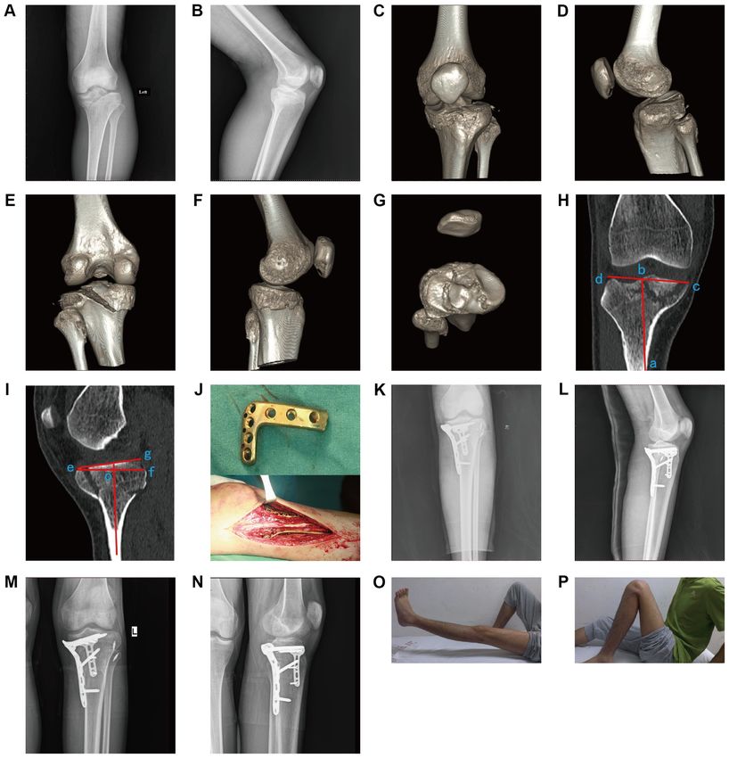
2                               HUANG et al: TREATMENT METHOD FOR TIBIAL PLATEAU FRACTURE

serious, violent injury and is characteristic of endangering the          The surgical incision types were as follows: Simple
medial column, posterior column or even the three columns of          anteromedial incision (n=4 cases); simple posterior medial
the tibial plateau, which complicate the injury of the medial         incision (n=7); anteromedial and backside inverted ‘L’ incision
and lateral meniscus, posterolateral complex and posterior            (n=2); anterolateral and posterior medial incision (n=10);
cruciate ligament (14,21). The key to surgical treatment of           anterolateral and backside inverted ‘L’ incision (n=1); and
tibial plateau fractures involving the anteromedial margin            anterolateral and anteromedial, as well as backside inverted ‘L’
caused by a hyperextension varus injury is the accurate               incision (n=1). All 25 patients received a bone graft during the
reduction of the posterior cortex and the effective support           operation, including autologous iliac bone grafts (n=6 cases),
and fixation of the anterior compression bone, particularly the       allograft bone grafts (n=17) and artificial bone grafts, using
reduction and strong fixation of the anteromedial comminuted          hydroxyapatite material (n=2).
bone, which is a difficulty for the treatment of this type of
fracture. If the treatment is improper, it inevitably results in      Surgical methods. Prior to the operation, proper posture
loss of reduction, thus affecting the stability of fixation and       selection, surgical approach and reset order were carefully
increasing the incidence of complications. From January 2015          planned and chosen. The supine position was used for
to June 2017, 27 patients with tibial plateau fractures caused        patients who required an anteromedial approach, posterior
by hyperextension varus were treated with a self‑clipped joint        medial approach or a conventional internal and external
line plate at Puai Hospital, Tongji Medical College (Wuhan,           combined approach. The prone position was taken first after
China). Among the 27 patients, 2 were lost to follow‑up, and          the posterior side inverted ‘L’ incision was performed. After
25 had complete patient data and recovered well throughout            the internal column and the posterior column were fixed, the
the follow‑up. In the present study, a retrospective analysis of      position was changed to a supine position. The anterome‑
the 25 patients is provided.                                          dial and/or anterolateral incision was used to reset and fix
                                                                      the anteromedial fracture block and the lateral column. For
Materials and methods                                                 patients with complex, three‑column fractures, the floating
                                                                      position may be used to observe the intra‑operative reduction.
Subjects. The inclusion criteria were as follows: i) Adult            After exposure, the internal and external condyle fractures
patients with fresh, closed fractures; ii) patients diagnosed         were opened and the meniscus was exposed when the knee
as having a tibial plateau fracture involving the anteromedial        joint was turned outward. In reference to pre‑operative routine
margin caused by a hyperextension varus injury by X‑ray films         MRI examination, if there was a meniscus injury, absorbable
and CT; iii) patients with normal knee joint motion prior to          sutures were first used to repair the injury. There were 2 cases
injury; and iv) patients with no severe vascular or nerve inju‑       in which the medial meniscus margins had a vertical radial
ries. Patients with open fracture, pathological fracture, severe      rupture and 4 cases in which the lateral meniscus margins had
vascular or nerve injury or complicating osteofascial compart‑        a vertical longitudinal rupture; all meniscus ruptures were
ment syndrome were excluded from the study.                           repaired with absorbable sutures. During meniscus repair,
    A total of 25 patients were included in the present               it is possible to evaluate the height and shape of the tibial
study, comprising 13 males and 12 females, aged between               plateau articular surface, which is conducive to the anatomical
19 and 71 years with a mean age of 45.2 years. All patients had       reduction of tibial plateau fractures.
closed fractures, although 2 patients had fractures with other             The collapsed articular surface was restored using tools of
complicated parts. The average time from injury to internal           the periosteum, the top bar was placed through the fractured
fixation was 10.7 days (range, 5 to 17 days). The causes of injury    window and a bone allograft, autogenous bone or artificial
were as follows: Motor vehicle collision (n=6 cases), high‑fall       bone graft was implanted at the collapse site. When the
injury (fall from over standing height, n=5), fall from an electric   articular surface was satisfactorily restored, the Kirschner
bicycle (n=9), fall from a bicycle (n=3) and fall injury (fall from   wire was temporarily fixed to the subchondral bone. After the
standing height or less, n=2). The patients' classification based     fixation was confirmed by the C‑arm X‑ray machine, proper
on the three‑column theory was as follows (22): Simple medial         internal fixation materials were selected. The rebuilt titanium
column fracture (n=4 cases), fracture of the medial and lateral       plate, 1/3 tube plate, rebuilt titanium locking plate or 3.5 mm
columns (n=5), fracture of the medial and posterior columns           proximal tibia plate (PTP) titanium plate were clipped in
(n=9) and three‑column fractures (n=7). The classification based      accordance with the prebending of the medial tibia to form
on AO/OTA was as follows: B2.3 (n=4 cases), B3.2 (n=9), C2.3          the joint line plate, which was then fixed to the anteromedial
(n=8) and C3.3 (n=4). The basic characteristics of the patients       margin of the tibia plateau. According to the fracture condi‑
are presented in Table SI.                                            tions, combined with other internal fixation materials, such
    Patients with simple medial column fractures were treated         as the proximal tibial T‑type locking titanium plate, the distal
with a plaster cast prior to the operation and the other patients     radius T‑type titanium plate, the PTP titanium plate and the
were treated with calcaneal traction. All patients received           reconstruction titanium plate, final fixation was completed. For
routine pre‑operative swelling treatment and prevention of            unstable bone fragments involving articular surface fixation,
deep venous thrombosis of the lower extremities. Pre‑operative        small hollow lag screws or absorbable sutures may be used
three‑dimensional CT and MRI examinations were performed              for the fix. After the bone structure was fixed, the stability of
to assess the extent of fracture displacement and comminution,        the knee joint ligament was examined. In 2 cases in which an
and to assess ligament and meniscus injury. The blood                 avulsion fracture at the insertion point of the posterior cruciate
circulation and sensory‑motor function of the lower limbs was         occurred, the ligaments were fixed with hollow screws after
closely monitored prior to the operation.                             reduction. In 4 cases in which the lateral collateral ligament
EXPERIMENTAL AND THERAPEUTIC MEDICINE 21: 621, 2021 3
was avulsed from the fibular head, exposing the fibula capit‑ (range, 15‑250 ml). The mean Rasmussen score immediately
ulum through the lateral incision, wire anchors were placed after the operation was 15.6 points (range, 12‑18 points).
and the avulsion was repaired by suture. No medial collateral Fracture healing was achieved in all 25 patients. The average
ligament, anterior cruciate ligament or posterior cruciate liga‑ time of healing and full weight‑bearing was 12.1 weeks
ment rupture were identified in any of the patients. The wound (range, 8‑14 weeks) and 15.8 weeks (range, 13‑20 weeks),
drainage tube was routinely retained after the operation. respectively. The HSS scores of all the patients at 12 months
post‑operatively averaged 87.6 (range, 68‑96), yielding an
Post‑operative treatment and follow‑up. All patients were excellent to good rate of 91.5%. The average range of motion
treated with a cotton pad to bandage the wound to reduce of the affected knee ranged from 2.3˚ to 125.1˚ 12 months after
bleeding after the operation. At the same time, the affected the operation. The data of the status of fracture healing after
limb was raised to reduce wound and limb swelling. the operation are presented in Table SI.
Low‑molecular‑weight heparin was administered 12 h after the
operation to prevent deep vein thrombosis of the lower limb. TPA and posterior slope angle of the tibial plateau prior to
After 48 h, the wound drainage tube was removed and knee operation and after the operation. The tibial plateau TPA and
joint flexion and extension function exercises were performed m‑PSA immediately as well as 12 months after the operation
under the protection of braces. were significantly different from those prior to the operation
All patients underwent examination immediately after the (P0.05; Fig. 1A and B). No significant difference was present
recorded. Any complications after the operation and during in the pre‑operative l‑PSA between the stage immediately
follow‑up were recorded. Due to the medial tibial plateau after the operation and 12 months after the operation (P>0.05;
bearing most of the load of the knee joint, if the fracture block Fig. 1C). Partial incision necrosis occurred post‑operatively in
of the anteromedial platform was seriously crushed, the time 1 case, which uneventfully healed after wound management.
of partial weight load was postponed accordingly. It may Typically, the fractures of a 19‑year‑old male with hyperexten‑
be suggested that the patient should be supported for up to sion varus of tibial plateau fractures (Schatzker V type) caused
3 months after the operation. The time‑point of full‑weight by a motor vehicle collision (Fig. 2) and a 33‑year‑old female
load‑bearing was finally determined by X‑ray according to the with hyperextension varus of tibial plateau fractures (Schatzker
degree of fracture grinding and the healing conditions. V type) caused by a high‑fall injury (Fig. 3) both were healed,
The tibial plateau angle (TPA) and PSA were measured by and there was no loss of reduction at 12 months post‑surgery.
X‑ray. The TPA and PSA of the tibial plateau prior to operation, In addition, there were 2 cases complicated with pre‑operative
immediately after operation and 12 months after the operation common peroneal nerve injury reported with a dorsal sense of
were compared. Knee joint function of the affected limb was numbness, which healed after 3 months of neurotrophic drug
measured 12 months after the operation and was evaluated administration. No screw loosening, plate breakage or fixation
according to the knee joint Hospital for Special Surgery (HSS) failure was detected during follow‑up.
standard score (23).
Discussion
Statistical analysis. The statistical software SPSS 19.0
(IBM, Corp.) was used for statistical analyses. First, the Internal fixation materials commonly used for clinical medial
Shapiro‑Wilk test was used to determine whether the measure‑ platform repairs, such as 3.5 or 4.5 mm locking titanium plates
ment data had a normal distribution. The tibial plateau TPA, or reconstruction titanium plates cannot be fixed to the antero‑
the medial PSA (m‑PSA) and the lateral PSA (l‑PSA) had medial margin and cannot form a strong support and fixation
normal distribution data, and the variance was homogeneous; for the broken fracture block of the anteromedial cortex due
therefore, values were expressed as the mean ± standard devia‑ to the limited coverage or support area (24‑27). In particular,
tion. Comparisons of the tibial plateau TPA and PSA prior to cases in which the anteromedial cortical bone fracture extends
the operation, immediately after the operation and 12 months to the anterolateral side, resulting in the overall comminuted
after the operation were analyzed by repeated‑measures collapse of the tibial plateau, are lacking effective fixation,
ANOVA with further pairwise comparisons by the least‑signif‑ which inevitably results in inadequate elevation of the medial
icant difference t‑test. P4 HUANG et al: TREATMENT METHOD FOR TIBIAL PLATEAU FRACTURE
Figure 1. (A) TPA, (B) medial PSA (m‑PSA) and (C) lateral PSA (l‑PSA) of all patients prior to surgery, immediately after surgery and at 12 months after the
operation. The TPA and m‑PSA immediately after surgery were significantly different from those prior to the operation. There was no significant difference in
the tibial plateau TPA or the m‑PSA at 12 months after the operation vs. immediately after the operation. No significant difference in the l‑PSA was observed
among the stages prior to surgery, immediately after the operation and 12 months after surgery. *P0.05). TPA, tibial plateau angle;
PSA, posterior slope angle.
applied a 1/3 tubular steel plate preflexion and transverse cartilage, which may provide a powerful mechanical support
fixation to the posterior lateral joint in comminuted fractures for the formation of the comminuted fracture blocks of the
of the posterolateral joint margin of the tibial plateau and anteromedial margin (31).
called it ‘hoop plating’. Lv et al (30) used a 2.4‑mm radial Giordano et al (29) proposed that the ‘hoop plate’ may
distal locking plate for the treatment of a posterior lateral also be used for the joint edge, which may appear the same
collapse fracture of the tibial plateau. The plate was placed as the concept of the joint line plate of the present study, but
closely to the articular surface, which achieved a satisfactory actually it is not. The hoop plate is used to treat posterior
clinical effect. However, the 2.4 or 2.7 mm radial distal plate condyle fractures of the tibial plateau, of which the 1/3 tube
system has low strength and small coverage, is mostly used for plate is pre‑bent to the posterior side of the tibial plateau to
the internal fixation of upper limb fractures and is not reported strengthen the effect of a ‘buttress plate’, meaning that ‘hoop
for the treatment of comminuted fractures of the medial tibial plates’ only have a fixed function as the collar. The joint line
medial cortex. plate is also placed on the joint edge in the present study of
In the present study, the concept of the circumference the anteromedial tibial plateau joint and may also be used for
line steel plate was proposed: The shape of the internal fixa‑ other parts of the fracture. At the same time, the idea of the
tion material is an arc, semi‑ring or even a ring fixed to the joint line plate in fixing bone fragments of the joint edge is
articular cartilage along the joint edge to fix the broken frac‑ emphasized, similar to the ‘hoop plate’ or locking plate, which
ture block. The idea is to fix the comminuted fracture blocks exerts the angle stabilizing effect of locking screws to form
to the main fracture block or other stable fracture blocks of a ‘bamboo raft fixation’ effect, having a supporting function
the joint surface to form the whole condyle. Through its own on the metaphyseal. At present, there is no such specialized
metaphyseal support or other metaphyseal plate bridging, type of joint line plate used for the anteromedial margin of
the condyle was fixed to the diaphysis. At the same time, the the tibial plateau. In the present study, a self‑clipped joint line
plate was fixed to the anteromedial margin and the screw was plate was made based on the size and position of the fracture
located below the articular surface, particularly with the angle blocks, including the use of reconstruction titanium plates
stability of the locking plate, forming ‘bamboo raft fixation’ (n=6 cases), 1/3 tube type steel plate fixation (n=5), reconstruc‑
technology and the cross fixation technique for the articular tion locking plate fixation (n=10) and a 3.5‑mm system PTPEXPERIMENTAL AND THERAPEUTIC MEDICINE 21: 621, 2021 5
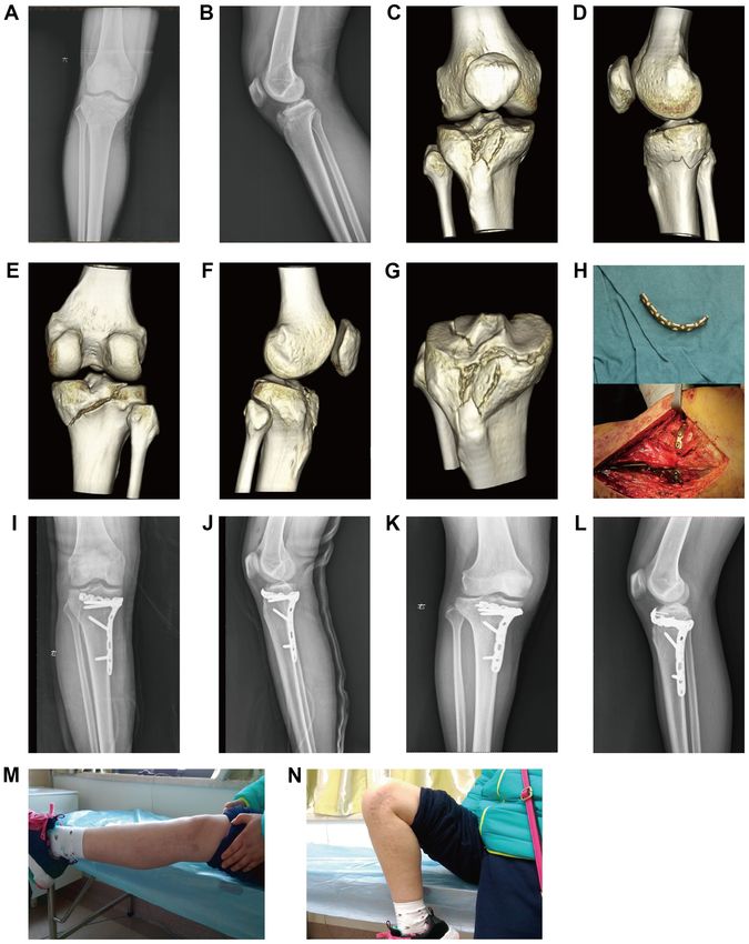
Figure 2. Representative case of a 19‑year‑old male with hyperextension varus of tibial plateau fractures (Schatzker V type) caused by an electric bicycle
collision. Pre‑operative radiographs in frontal (A) and side (B) position. Pre‑operative three‑dimensional CT displaying medial and lateral column fractures.
(C) Anterior view. (D) Lateral view from left side. (E) Posterior view. (F) Lateral view from right side. (G) Transverse view. (H) ∠abc represents the tibial
plateau angle and (I) ∠gef represents the posterior slope angle by CT. (J) Image of a 2.7‑mm T‑shaped Locking plate that was horizontally placed and its
location during the operation. (K and L) Radiography was performed immediately after the operation. (M and N) Radiographs at 12 months post‑surgery
indicated that the fractures were healed and there was no loss of reduction. (O and P) General functional position of the patient 12 months after the operation.
titanium plate fixation (n=4). Among the 25 patients included and PSA, and may be compared with that of healthy limbs.
in the follow‑up, 20 cases achieved anatomical reduction and It is crucial that the bone graft in the bone defect area is
the tibial plateau TPA and PSA recovered satisfactorily after sufficient. If there are certain defects, it is suggested that an
the operation. At one year after the operation, the tibial plateau autogenous iliac bone graft may be used, with the advantages
TPA, m‑PSA and l‑PSA exhibited no significant changes from of having powerful support, effects of good bone formation,
the angles immediately after the surgery and the reduction bone induction and conduction, and fastening of the fracture
was not lost. It was proven that with the fixation of the medial during healing (32). During the operation, the medial collateral
supporting plate, the self‑clipped joint plate was able to ligament, the ‘goose foot’ and other medial stable structures
meet the requirements of the early functional exercise of the should be protected to avoid post‑operative instability of the
affected limb and the recovery of the knee joint function was knee joint (33). When prebending the joint line plate, the plate
satisfactory after the operation. should be closely adhered to the bone to reduce irritation to
During the operation, the height of the anteromedial the skin and soft tissue and the influence of the flexion activity
platform fracture should be fully restored, along with the TPA of the knee joint after the operation. During the operation, if it6 HUANG et al: TREATMENT METHOD FOR TIBIAL PLATEAU FRACTURE Figure 3. Representative case of a 33‑year‑old female with hyperextension varus of tibial plateau fractures (Schatzker V type) caused by a fall injury. Pre‑operative radiographs in (A) frontal and (B) side position. Pre‑operative three‑dimensional CT revealing three‑column fractures. (C) Anterior view. (D) Lateral view from left side. (E) Posterior view. (F) Lateral view from right side. (G) Transverse view. (H) Image of a 3.5‑mm rebuilt titanium plate that was horizontally placed and its location during the operation. Radiographs immediately post‑surgery in (I) frontal and (J) side position. Radiographs at 12 months post‑surgery in (K) frontal and (L) side position indicated that the fractures were healed and there was no loss of reduction. (M and N) General functional position of the patient 12 months after the operation. is necessary to reduce the dissection of the soft tissue around this type of fracture and particularly the lateral collateral the incision, the use of an electric knife and clamping the edge ligament may be torn or the fibular head may have an avulsion of the skin should be avoided in order to reduce complica‑ fracture. Internal and external stress tests should be performed tions such as incision fat liquefaction, skin necrosis or wound after the internal fixation of the intraoperative fracture infection. There may be a posterolateral complex injury in reduction, the stability of the lateral collateral ligament should
EXPERIMENTAL AND THERAPEUTIC MEDICINE 21: 621, 2021                                                      7

be checked and one‑stage surgical repair should be performed                4. Zhang BB, Sun H, Zhan Y, He QF, Zhu Y, Wang YK and Luo CF:
                                                                               Reliability and repeatability of tibial plateau fracture assessment
if necessary (34).                                                             with an injury mechanism‑based concept. Bone Joint Res 8:
    Deficits of the present study are that the number of cases                 357‑366, 2019.
with the application of the joint line plate was limited and                5. Ramponi DR and McSwigan T: Tibial plateau fractures. Adv
                                                                               Emerg Nurs J 40: 155‑161, 2018.
the follow‑up time was relatively short. Further studies with               6. Graham P: Tibial plateau fracture. Orthop Nurs 36: 303‑305, 2017.
more cases and further follow‑up observations are required to               7. Lu C, Ye G, Liu W, Wu H, Wu G and Chen J: Tibial plateau fracture
acquire more accurate clinical treatment results. In addition,                 related to unicompartmental knee arthroplasty: Two case reports
                                                                               and literature review. Medicine (Baltimore) 98: e17338, 2019.
the self‑clipped joint line plate has no supporting functions               8. Chang SM, Zhang YQ, Yao MW, Du SC, Li Q and Guo Z:
on the epiphysis, which is necessary to cooperate with other                   Schatzker type IV medial tibial plateau fractures: A
internal fixation materials to complete the final fixation.                    computed tomography‑based morphological subclassification.
                                                                               Orthopedics 37: e699‑e706, 2014.
Therefore, according to the idea of joint line plating, it is               9. Chang SM, Hu SJ, Zhang YQ, Yao MW, Ma Z, Wang X, Dargel J
esteemed to develop an anatomic type of joint line plating to                  and Eysel P: A surgical protocol for bicondylar four‑quadrant
meet clinical requirements.                                                    tibial plateau fractures. Int Orthop 38: 2559‑2564, 2014.
                                                                           10. Yan B, Yin W, Zhang X, Liu D, Gui K, Sun J, Chen Y and Ni M:
                                                                               Effectiveness analysis of surgical treatment of Schatzker type
Acknowledgements                                                               tibial plateau fractures. Zhongguo Xiu Fu Chong Jian Wai Ke Za
                                                                               Zhi 31: 1305‑1310, 2017 (In Chinese).
                                                                           11. Cherney S and Gardner MJ: Bicondylar tibial plateau fractures:
Not applicable.                                                                Assessing and treating the medial fragment. J Knee Surg 27:
                                                                               39‑45, 2014.
Funding                                                                    12. He X, Ye P, Hu Y, Huang L, Zhang F, Liu G, Ruan Y and Luo C:
                                                                               A posterior inverted L‑shaped approach for the treatment of
                                                                               posterior bicondylar tibial plateau fractures. Arch Orthop
This study was supported by grants from the Health and                         Trauma Surg 133: 23‑28, 2013.
Family Planning Commission of Wuhan City for Clinical                      13. Lu KH, Lu EW, Lin CW, Yang JS and Yang SF: New insights
                                                                               into molecular and cellular mechanisms of zoledronate in human
Medicine Research Project (grant no. WX16D27).                                 osteosarcoma. Pharmacol Ther 214: 107611, 2020.
                                                                           14. Kołodziejczyk K, Kuliński K, Fedorowicz K, Langner M,
Availability of data and materials                                             Czubak J and Pomianowski S: Difficulties in treating complex
                                                                               knee injuries with fracture of posterior tibial plateau. Ortop
                                                                               Traumatol Rehabil 20: 293‑300, 2018.
The datasets used and/or analyzed during the current study                 15. Yang X, Xu F, Yin Z and Wang Q: Clinical observation of
are available from the corresponding author on reasonable request.             3.5 mm T support plate fixation for simple posterolateral tibial
                                                                               plateau fracture by posterolateral inverted L‑shaped approach.
                                                                               Zhongguo Xiu Fu Chong Jian Wai Ke Za Zhi 31: 815‑819, 2017
Authors' contributions                                                         (In Chinese).
                                                                           16. Firoozabadi R, Schneidkraut J, Beingessner D, Dunbar R and
                                                                               Barei D: Hyperextension varus bicondylar tibial plateau fracture
YCH performed the experiments, analyzed the data and wrote                     pattern: Diagnosis and treatment strategies. J Orthop Trauma 30:
the paper; JJ contributed to the conception of the study; WJC,                 e152‑e157, 2016.
FX and WZ participated in the execution of the experiment;                 17. Cohen AP, King D and Gibbon AJ: Impingement fracture of the
                                                                               anteromedial tibial margin: A radiographic sign of combined
JWW contributed to analysis and manuscript preparation. All                    posterolateral complex and posterior cruciate ligament disrup‑
authors read and approved the final manuscript.                                tion. Skeletal Radiol 30: 114‑116, 2001.
                                                                           18. Bennett DL, George MJ, El‑Khoury GY, Stanley MD and
                                                                               Sundaram M: Anterior rim tibial plateau fractures and postero‑
Ethics approval and consent to participate                                     lateral corner knee injury. Emerg Radiol 10: 76‑83, 2003.
                                                                           19. Chiba T, Sugita T, Onuma M, Kawamata T and Umehara J:
The study was approved by the ethics committee of the Wuhan                    Injuries to the posterolateral aspect of the knee accompanied
                                                                               by compression fracture of the anterior part of the medial tibial
Fourth Hospital, Puai Hospital, Tongji Medical College,                        plateau. Arthroscopy 17: 642‑647, 2001.
Huazhong University of Science and Technology (Wuhan,                      20. Yoo JH, Kim EH, Yim SJ and Lee BI: A case of compression
China). All patients provided written informed consent.                        fracture of medial tibial plateau and medial femoral condyle
                                                                               combined with posterior cruciate ligament and posterolateral
                                                                               corner injury. Knee 16: 83‑86, 2009 .
Patient consent for publication                                            21. Hong F, Wang N and Chen GJ: Posterior medial approach
                                                                               inverted L‑shaped incision combined with reconstruction plate
                                                                               for posterior condylar fracture of tibial plateau. Zhongguo Gu
All patients agreed to publish their images and scans.                         Shang 29: 1027‑1032, 2016 (In Chinese).
                                                                           22. Luo CF, Sun H, Zhang B and Zeng BF: Three‑column fixation for
Competing interests                                                            complex tibial plateau fractures. J Orthop Trauma 24: 683, 2010.
                                                                           23. Insall JN, Ranawat CS, Aglietti P and Shine J: A comparison of
                                                                               four models of total knee‑replacement prostheses. J Bone Joint
The authors declare that they have no competing interests.                     Surg Am 58: 754‑65, 1976.
                                                                           24. Dreyfuss D, Allon R, Izacson N and Hutt D: A Comparison
                                                                               of locking plates and intramedullary pinning for fixation of
References                                                                     metacarpal shaft fractures. Hand (NY) 14: 27‑33, 2019.
                                                                           25. Wu CM, Chen YA, Liao HT, Chen CH, Pan CH and Chen CT:
                                                                               Surgical treatment of isolated zygomatic fracture: Outcome
 1. Duan KD and Huang JR: Progress in diagnosis and treatment                  comparison between titanium plate and bioabsorbable plate.
    of posterior condylar fracture of tibial plateau. Zhongguo Gu              Asian J Surg 41: 370‑376, 2018.
    Shang 32: 1173‑1176, 2019 (In Chinese).                                26. Darrow BG, Weigel JP, Greenacre CB, Xie X, Liaw PK and
 2. Lowe DT, Milone MT, Gonzalez LJ and Egol KA: Repair of tibial              Biskup JJ: Ex vivo biomechanical comparison of titanium locking
    plateau fracture (Schatzker II). JBJS Essent Surg Tech 9: e25, 2019.       plate, stainless steel nonlocking plate, and Tie‑in external fixator
 3. Kfuri M and Schatzker J: Revisiting the Schatzker classification           applied by a dorsal approach on ostectomized humeri of pigeons
    of tibial plateau fractures. Injury 49: 2252‑2263, 2018.                   (Columba livia). J Avian Med Surg 33: 29‑37, 2019.
8 HUANG et al: TREATMENT METHOD FOR TIBIAL PLATEAU FRACTURE
27. Schliemann B, Seifert R, Theisen C, Gehweiler D, Wähnert D, 32. Naito K, Sugiyama Y, Obata H, Mogami A, Obayashi O and
Schulze M, Raschke MJ and Weimann A: PEEK versus tita‑ Kaneko K: Screw fixation and autogenous bone graft for an irre‑
nium locking plates for proximal humerus fracture fixation: ducible distal ulna fracture associated with distal radius fracture.
A comparative biomechanical study in two‑ and three‑part J Hand Surg Asian Pac Vol 22: 236‑239, 2017.
fractures. Arch Orthop Trauma Surg 137: 63‑71, 2017. 33. Shi Z, Zhang X, Zhang K, Jiang R, Yang W and C. L: Short‑term
28. Maeda H, Noguchi M, Suehiro M, Mihara E and Chou Z: effect of non‑absorbable sutures repairing iatrogenic medial
Analysis of surgical treatment results in patients with tibial collateral ligament injury during total knee arthroplasty. Chin
plateau fractures. Orthoped Traumatol 42: 805‑809, 2010. J Bone Joint Surg 12: 126‑130, 2019.
29. Giordano J, Schatzker M and Kfuri M: The ‘Hoop’ plate for 34. Mihalko WM, Saleh KJ, Krackow KA and Whiteside LA:
posterior bicondylar shear tibial plateau fractures: Description of Soft‑tissue balancing during total knee arthroplasty in the varus
a new surgical technique. J Knee Surg 30: 509‑513, 2017. knee. J Am Acad Orthop Surg 17: 766‑774, 2009.
30. Lv TR, Q C and X. L: Treatment of posterolateral depression
fractures of tibial plateau with improved postemlateral approach This work is licensed under a Creative Commons
and 2.4 mm distal radiuslocking plate. Chin J Orthop Trauma 18: Attribution-NonCommercial-NoDerivatives 4.0
851‑856, 2016. International (CC BY-NC-ND 4.0) License.
31. G F, ZJ P and H. L: Dual locking plate fixation for Type C3
tibial plateau fractures involving the posterior column. Chin J
Orthop 34: 695‑702, 2014.You can also read