Histomorphometric analysis of liver biopsies of treated patients with Gaucher disease type
←
→
Page content transcription
If your browser does not render page correctly, please read the page content below
Original Article
Histomorphometric analysis of liver biopsies of treated patients with
Gaucher disease type 1
Rodrigo Tzovenos Starosta1,2 , Marina Siebert3,4 , Filippo Pinto e Vairo5,6 ,
Bruno Lafaiete de Lima Costa7, Christiano Tomaso Ponzoni7,
Ida Vanessa Doederlein Schwartz1,8,9 , Carlos Thadeu Schmidt Cerski4,7
How to cite: Starosta RT, Siebert M, Vairo FP, et al. Histomorphometric analysis of liver biopsies of treated patients with
gaucher disease type 1. Autops Case Rep [Internet]. 2021;11:e2021306. https://doi.org/10.4322/acr.2021.306
ABSTRACT
Gaucher disease (GD) is an autosomal recessive lysosomal disorder caused by a disturbance in the metabolism of
glucocerebroside in the macrophages. Most of its manifestations – hepatosplenomegaly, anemia, thrombocytopenia,
and bone pain – are amenable to a macrophage-target therapy such as enzyme replacement. However, there is
increasing evidence that abnormalities of the liver persist despite the specific GD treatment. In this work, we adapted
histomorphometry techniques to the study of hepatocytes in GD using liver tissue of treated patients, developing the
first morphometrical method for canalicular quantification in immunohistochemistry-stained liver biopsies, and exploring
histomorphometric characteristics of GD. This is the first histomorphometric technique developed for canalicular analysis
on histological liver biopsy samples.
Keywords
Gaucher Disease; Image Cytometry; Hepatocytes; Bile Canaliculi; Biopsy, Large-Core Needle
INTRODUCTION
Gaucher disease (GD) is one of the most common dysregulated macrophagic activation2, 3 and invasion of
lysosomal disorders with an estimated prevalence of tissues by Gaucher cells.4, 5 Macrophage‑targeted therapy
1:60,000 in the general population and 1:800 in the through infusions of recombinant glucocerebrosidase
Ashkenazi Jewish population.1 The pathophysiology (GCase) which is uptaken via the mannose receptor
of GD is classically defined as a disturbance in the pathway 6 has been successful in alleviating the key
processing of sphingolipids inside the macrophages,1, clinical findings in GD – anemia, thrombocytopenia,
2
the main manifestations of GD being a function of hepatosplenomegaly – and improving quality of life
1
Universidade Federal do Rio Grande do Sul, Graduate Program in Genetics and Molecular Biology, Porto Alegre, RS, Brasil
2
Washington University, Department of Pediatrics, Saint Louis, MO, USA
3
Hospital de Clínicas de Porto Alegre, Laboratorial Research Unit, Experimental Research Center, Porto Alegre, RS, Brasil
4
Universidade Federal do Rio Grande do Sul, Graduate Program in Science in Gastroenterology and Hepatology, Porto Alegre, RS, Brasil
5
Mayo Clinic, Center for Individualized Medicine, Rochester, MN, USA
6
Mayo Clinic, Department of Clinical Genomics, Rochester, MN, USA
7
Hospital de Clínicas de Porto Alegre, Department of Surgical Pathology, Porto Alegre, RS, Brasil
8
Universidade Federal do Rio Grande do Sul, Department of Genetics, Porto Alegre, RS, Brasil
9
Hospital de Clínicas de Porto Alegre, Medical Genetics Service, Porto Alegre, RS, Brasil
Copyright: © 2021 The Authors. This is an Open Access article distributed under the terms of the Creative
Commons Attribution License, which permits unrestricted use, distribution, and reproduction in any medium,
provided the original work is properly cited.
Histomorphometric analysis of liver biopsies of treated patients with Gaucher disease type 1
in these patients.7 However, there are still features of (SSP) of HCPA. The archive of the SSP was also searched
GD that are not fully explained by the involvement of for liver biopsy samples with a diagnosis of “healthy
macrophages. It is known that patients with GD, even liver tissue” or “steatohepatitis grade 1” (“clinical liver
after long-term treatment, tend to have a higher liver disease”) by an expert in Liver Pathology (CTSC).
stiffness (which is a surrogate measurement for fibrosis) Samples were processed and stained with
than controls.8-11 Moreover, the biliary phenotype of immunohistochemistry (IHC) for CD10 (to highlight
GD has been subject of increasing focus: patients with the bile canaliculi) according to the SSP protocol.
GD have an increased incidence of gallstones,12-15 and Briefly, samples were cut into 3 µm-thick sections and
an abnormal bile composition with increased level of deparaffinized. Antigen recovery was performed with
glucocerebroside (glucosylceramide; GlcCer) and other
CC1 buffer at pH 9.0 and 95ºC for 2 minutes followed
sphingolipids such as glucosylsphingosine (GlcSph,
by peroxidase blocking with OptiView Peroxidase
alias lyso-GL1).12, 16 Biliary excretion of GlcCer is being
Inhibitor (Ventana Medical Systems). The primary
suggested as a protective factor against hepatocellular
rabbit anti-human anti-CD10 monoclonal antibody
storage of this substance;16, 17 however, this export of
(clone SP67; Roche Diagnostics, Tucson, Arizona,
lysosomal GlcCer into the bile canaliculi may be one of
USA) was incubated for 28 minutes at 36ºC. After
the possible mechanisms of injury to the biliary system
primary antibody incubation, the reaction was detected
in GD through interference with the composition of
with the OptiView DAB IHC Detection Kit (Ventana
bile and the function of transporters in the hepatocytic
Medical Systems) and slides were counterstained with
apical membrane. This interaction between GlcCer and
hematoxylin and bluing reagent (Li2CO3 + Na2CO3).
bile transporters has been demonstrated in studies of
the same transporters in cancer multidrug resistance.18, 19 Imaging
Histomorphometry, or histological morphometry,
Stained and mounted slides were
is the quantification of morphology at the tissue level.
microphotographed with the CellSens software
It is a well-established technique with applications in
(Olympus Corporation) at a magnification of 1000x.
the research of many tissues, including the liver, where
Each slide had 6 random high-power fields captured
it has been used for the quantification of characteristics
and saved as tagged image file format (.tiff) images.
such as fibrosis, immunohistochemical markers, and
steatosis.20, 21, 22 It has also been used as a grading and Histomorphometry
prognostic marker in a variety of tumors.23-25
Each stained image was converted from the
Histomorphometry is still an underutilized, incipient
native RGB color format to 8-bit using the ImageJ
technique in canalicular pathology26. Although some
software.29 The pixel-to-µm conversion was calculated
studies have used morphometric parameters such as
from the scale generated by CellSens. For canalicular
canalicular length and total canalicular area in a research
histomorphometry, IHC-stained RGB images were
context, making use of tools such as immunofluorescence
treated with the IHC Toolbox plugin (https://imagej.nih.
and transmission electron microscopy 27, 28, there are no
gov/ij/plugins/ihc-toolbox) to isolate IHC-positive areas
studies on the use of histomorphometry in clinical liver
(i.e., hepatic canaliculi), as shown in Figure 1.
samples. In this study, we adapted histomorphometry
to the study the canalicular parameters in liver biopsies Manual thresholding was used to select all IHC-
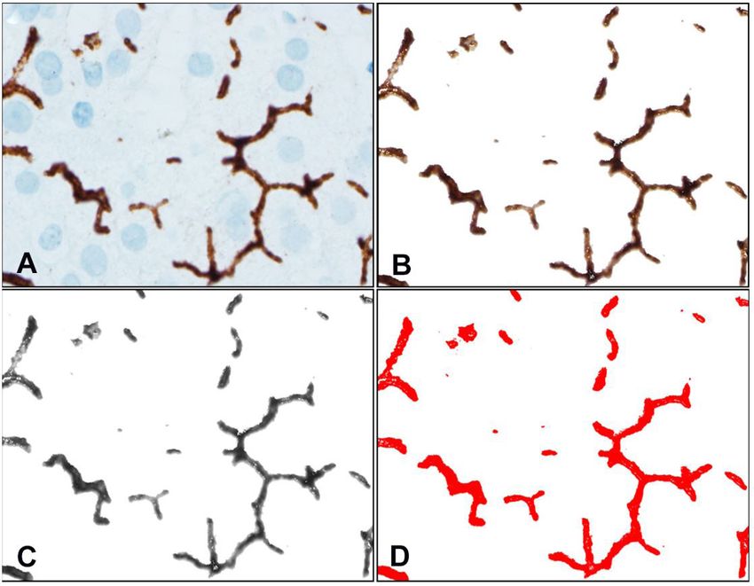
of patients with GD. positive regions of interest (ROIs). ROIs withStarosta RT, Siebert M, Vairo FP, et al. Figure 1. A – RGB photomicrograph, 1000x, showing IHC staining anti-CD10 highlighting the canaliculi in brown; B – extraction of canaliculi from background using the IHC Toolbox plugin on ImageJ; C – Transformation of extracted canaliculi from RGB to 8-bit; D – Thresholding of 8-bit image selecting all IHC-positive areas, with no overlap with the background. Figure 2. A – schematic depiction of a less-branching canaliculus, for clarification of the perimeter-to-Feret ratio. The solid red line is the canaliculus perimeter (P1). The solid green line represents the projection of the canaliculus on the Y axis of the image field, which is the Feret Y (Y1) measure. The solid blue line represents the projection of the canaliculus on the X axis of the image field, which is the Feret X measure (X1). The Feret diameter of a canaliculus (F1) is the arithmetic mean of X1 and Y1; B – schematic depiction of a more-branching canaliculus. The solid red line is the canaliculus perimeter (P2). The projection of this canaliculus in the Y (Y2) and X (X2) axes are depicted as the solid green and blue lines, respectively, and the Feret diameter of this canaliculus (F2) is the arithmetic mean of X2 and Y2. As shown, the increase in branching affects more the perimeter than the Feret diameter of a canaliculus: P1 < P2, F1 ≈ F2. In this way, P1/F1 < P2/F2. These images are theoretical simplified schemes for clarification and are per se not representative of either group. Autops Case Rep (São Paulo). 2021;11:e2021306 3-7
Histomorphometric analysis of liver biopsies of treated patients with Gaucher disease type 1

     MGV was corrected to absorbance (corrected MGV,                       Ethical Approval
“cMGV”) with the formula cMGV = 255 – MGV. Due
                                                                                This study was approved by the HCPA Research
to the background correction obtained with the IHC
                                                                           Ethics Committee under the number #18- 0654.
Toolbox plugin, canalicular cMGV was not normalized.
Because of the pattern similarity between the hepatic                      Research consent was waived by the HCPA Research
canaliculi and the bone trabeculae, we used the                            Ethics Committee because of the retrospective nature
Map_BoneMicrostructure plugin (https://imagej.nih.                         of the analyses.
gov/ij/plugins/microstructure) to obtain mean canaliculi
thickness in µm for whole images.                                          RESULTS
Statistical Analysis
                                                                           Patients
      Because of the small sample sizes, all variables
were considered parametric for statistical analysis. For                         Liver biopsy samples of five type 1 GD patients
descriptive statistics, variables are described as mean                    were retrieved. Patient characteristics are displayed in
± SD. The Student’s T-test was used for comparisons                        Table 1. Two patients (pts 3 and 4) had clinical liver
between group means. A subgroup analysis was                               disease. Liver biopsy samples of seven controls were
conducted post-hoc to the group comparisons. For this                      retrieved (healthy = 4; steatohepatitis = 3).
analysis, subjects were divided into those with clinical
                                                                           Canalicular Histomorphometry
liver disease (steatohepatitis or cirrhosis) and those
without. Analytic statistics were not performed in the                          Results of histomorphometrical analysis are
subgroup analysis due to the small sample sizes.                           displayed in Table 2. No significant statistical difference

Table 1. Characteristics of GD patients
                                                           Age at
                                                             Treatment at biopsy
 Patient       Sex          GBA Genotype                                                    Biopsy diagnosis
                                                          biopsy (y)(months)
     1         F    p.Glu388Lys/p.Ser405Asn                  48
                                                             Miglustat (11)          Macrovesicular steatosis
     2         F    p.Asn409Ser/p.Leu483Arg       63         Miglustat (22)          Hemosiderosis, presence of
                                                                                     Gaucher cells
     3        M p.Asn409Ser/RecNciI               56         Imiglucerase  30 IU/Kg/ Steatohepatitis with mild activity
                                                             biweekly (69)
     4        M p.Asn409Ser/RecNciI               61         Imiglucerase 30 IU/Kg/ Cirrhosis, hemosiderosis, presence
                                                             biweekly (72)           of Gaucher cells
     5        M p.Asn409Ser/RecNciI               42         Taliglucerase alfa 30   Macrovesicular steatosis
                                                             IU/Kg/biweekly (216)
All patients are diagnosed with GD type 1. Y = years-old; F = female; M = male; RecNciI = recombinant allele with the
GBA1 pseudogene, includes the p.Leu483Arg, p.Ala495Pro, and p.Val499Val variants.

Table 2. Results of histomorphometrical analysis.
                                          Gaucher disease                                       Control group

      Parameter                                                No clinical                                                        p-value
                                            Clinical liver                          Total        Clinical liver
                           Total (n=5)                            liver                                           Healthy (n=4)
                                            disease (n=2)                           (n=7)        disease (n=3)
                                                              disease (n=3)
Area (µm2)                 8.21 ± 4.76       7.71 ± 3.33       8.52 ± 6.27       8.23 ± 6.30      2.96 ± 1.05     12.19 ± 5.48     0.639
cMGV                     114.29 ± 31.67     97.96 ± 4.59     125.18 ± 39.39 113.62 ± 47.67       65.48 ± 11.30 149.72 ± 20.14      0.876
Perimeter (µm)            11.42 ± 2.17      12.33 ± 3.81      10.81 ± 0.92      12.77 ± 6.36      7.98 ± 1.93     16.36 ± 6.19     0.870
Feret diameter (µm)        3.52 ± 0.73       3.83 ± 1.09       3.31 ± 0.56       3.46 ± 1.95      1.86 ± 0.47      4.65 ± 1.73     0.085
Perimeter-to-Feret         2.89 ± 0.20       2.83 ± 0.03       2.93 ± 0.27       2.94 ± 0.05      2.95 ± 0.05      2.93 ± 0.05     0.060
ratio
Solidity                   0.75 ± 0.03       0.76 ± 0.01       0.74 ± 0.04       0.73 ± 0.04      0.68 ± 0.005     0.76 ± 0.007    0.343
Thickness (µm)             8.69 ± 1.34       9.24 ± 1.31       8.32 ± 1.50       8.46 ± 1.48      9.49 ± 0.94      7.10 ± 0.60     0.793
The p-values are a result of the comparison between Gaucher disease (total) and control group (total). No analyses were performed
for the subgroups.

4-7                                                                                            Autops Case Rep (São Paulo). 2021;11:e2021306
Starosta RT, Siebert M, Vairo FP, et al.
was found between the groups for any of the variables. and demonstrated. Although significant differences
The closest parameter to statistical significance was the were not found, this study paves the way for further
Perimeter-to-Feret ratio (p=0.06). investigation of canalicular pathology in GD and in
other diseases.
DISCUSSION
CONCLUSION
Histomorphometry is a digital image analysis
approach that relies on the identification and analysis No significant differences were found between
of morphological elements in a histological section.30 GD and control samples. An almost-significant p-value
It is a technique used for diagnosis of neoplasms was found for perimeter-to-Feret ratio, indicating
and to aid in tailoring cancer treatment.31 We used that further exploring this new parameter in larger
histomorphometry to explore canalicular parameters
samples might yield valuable results. This is the first
in a sample of patients with GD type 1.
histomorphometric technique developed for canalicular
Although no statistically significant differences analysis on routine liver biopsy samples.
were found in this study, the borderline p-value
for the Perimeter-to-Feret ratio is promising, and, if Research Highlights
confirmed in further studies with bigger populations,
• The perimeter-to-Feret ratio is informative on
might indicate canalicular dysfunction as part of the
canalicular branching on liver biopsy samples.
pathogenesis of GD.
On subgroup analysis, patients with GD and • Canalicular parameters are amenable to
no clinical liver disease seemed to have a reduced quantification by histomorphometry on liver
canalicular area, cMGV, perimeter, and Feret diameter biopsy samples.
than healthy controls – however, because of statistical
analyses not being possible due to the small sample
ACKNOWLEDGEMENTS
size, conclusions from the subgroup analysis are limited.
In contrast with the common cholestatic pattern of This study was supported by the Coordenação
many diseases which consists of canalicular dilation,32
de Aperfeiçoamento de Pessoal de Nível Superior
reduced branching has not been studied as much due
(CAPES) and the Fundo de Apoio a Pesquisa e
to the lack of methods to perform a detailed analysis
of these structures. It is known that patients with GD Eventos do Hospital de Clínicas de Porto Alegre
have increased secretion of GlcCer in bile, leading to (FIPE-HCPA). We are grateful to the statistical
physiological changes such as upregulation of GBA2, counseling provided by Dr. Michael Wallendorf
coding for a bile acid 3-O-glucosidase that can also at Washington University.
metabolize GlcCer and GlcSph, 33 producing toxic
compounds such as sphingosine.34 It is possible that
this process leads to biliary injury, thus impacting on REFERENCES
the normal canalicular structure.
1. Stirnemann J, Belmatoug N, Camou F, et al. A review of
The main limitation of this study is the small
Gaucher Disease pathophysiology, clinical presentation
sample size. GD is a rare metabolic disorder, and with and treatments. Int J Mol Sci. 2017;18(2):441. http://
the current technology available in clinical practice dx.doi.org/10.3390/ijms18020441. PMid:28218669.
for follow-up of these patients – such as transient
2. Aflaki E, Moaven N, Borger DK, et al. Lysosomal storage
elastography and magnetic resonance imaging –
and impaired autophagy lead to inflammasome activation
liver biopsies are seldom performed because of the in Gaucher macrophages. Aging Cell. 2016;15(1):77-88.
invasiveness of the procedure and the increased http://dx.doi.org/10.1111/acel.12409. PMid:26486234.
bleeding risk in this group.
3. Boven LA, van Meurs M, Boot RG, et al. Gaucher cells
In summary, this is the first report of the application
demonstrate a distinct macrophage phenotype and
of histomorphometry in the study of liver canaliculi in resemble alternatively activated macrophages. Am J Clin
a metabolic disorder. A new parameter for canalicular Pathol. 2004;122(3):359-69. http://dx.doi.org/10.1309/
analysis, the perimeter-to-Feret ratio, was developed BG5VA8JRDQH1M7HN. PMid:15362365.
Autops Case Rep (São Paulo). 2021;11:e2021306 5-7Histomorphometric analysis of liver biopsies of treated patients with Gaucher disease type 1
4. Adar T, Ilan Y, Elstein D, Zimran A. Liver involvement in III. Mol Genet Metab Rep. 2020;22:100564. http://dx.doi.
Gaucher disease - review and clinical approach. Blood org/10.1016/j.ymgmr.2019.100564. PMid:32099816.
Cells Mol Dis. 2018;68:66-73. PMid:27842801.
16. Pentchev PG, Gal AE, Wong R, et al. Biliary excretion
5. James SP, Stromeyer FW, Chang C, Barranger JA. of glycolipid in induced or inherited glucosylceramide
LIver abnormalities in patients with Gaucher’s disease. lipidosis. Biochim Biophys Acta. 1981;665(3):615-8.
Gastroenterology. 1981;80(1):126-33. http://dx.doi. http://dx.doi.org/10.1016/0005-2760(81)90279-4.
org/10.1016/0016-5085(81)90202-X. PMid:7450398. PMid:7295755.
6. Tekoah Y, Tzaban S, Kizhner T, et al. Glycosylation 17. Crawford JM. Role of vesicle-mediated transport
and functionality of recombinant β-glucocerebrosidase pathways in hepatocellular bile secretion. Semin
from various production systems. Biosci Rep. Liver Dis. 1996;16(2):169-89. http://dx.doi.
2013;33(5):e00071. http://dx.doi.org/10.1042/ org/10.1055/s-2007-1007230. PMid:8781022.
BSR20130081. PMid:23980545.
18. Lee WK, Kolesnick RN. Sphingolipid abnormalities
7. Oliveira FL, Alegra T, Dornelles A, et al. Quality of in cancer multidrug resistance: chicken or egg? Cell
life of brazilian patients with Gaucher disease and Signal. 2017;38:134-45. http://dx.doi.org/10.1016/j.
fabry disease. JIMD Rep. 2012;7:31-7. http://dx.doi. cellsig.2017.06.017. PMid:28687494.
org/10.1007/8904_2012_136. PMid:23430492. 19. Gouazé-Andersson V, Yu JY, Kreitenberg AJ, Bielawska A,
8. Nascimbeni F, Cassinerio E, Dalla Salda A, et al. Giuliano AE, Cabot MC. Ceramide and glucosylceramide
Prevalence and predictors of liver fibrosis evaluated by upregulate expression of the multidrug resistance
vibration controlled transient elastography in type 1 gene MDR1 in cancer cells. Biochim Biophys Acta.
Gaucher disease. Mol Genet Metab. 2018;125(1-2):64- 2007;1771(12):1407-17. http://dx.doi.org/10.1016/j.
72. http://dx.doi.org/10.1016/j.ymgme.2018.08.004. bbalip.2007.09.005. PMid:18035065.
PMid:30115580. 20. Sandoval C, Vásquez B, Souza-Mello V, Adeli K,
Mandarim-de-Lacerda C, Sol M. Morphoquantitative
9. Serai SD, Naidu AP, Burrow TA, Prada CE, Xanthakos S,
effects of oral β-carotene supplementation on liver of
Towbin AJ. Correlating liver stiffness with disease severity
C57BL/6 mice exposed to ethanol consumption. Int J Clin
scoring system (DS3) values in Gaucher disease type 1
Exp Pathol. 2019;12(5):1713-22. PMid:31933989.
(GD1) patients. Mol Genet Metab. 2018;123(3):357-
63. http://dx.doi.org/10.1016/j.ymgme.2017.10.013. 21. Leite C, Starosta RT, Trindade EN, et al. Elastic fibers
PMid:29361370. density: a new parameter of improvement of NAFLD in
bariatric surgery patients. Obes Surg. 2020;30(10):3839-
10. Pedroso MLA, Didoné CN Fo, Radunz V, Barros JA. Liver
46. http://dx.doi.org/10.1007/s11695-020-04722-x.
damage in a patient with Gaucher’s disease type 1 and
PMid:32451920.
alpha-1 antitrypsin deficiency: a potential epigenetic
effect? J Gastrointestin Liver Dis. 2019;28(1):121-3. 22. Leite C, Starosta RT, Trindade EN, Trindade MRM, Álvares-
PMid:30851181. da-Silva MR, Cerski CTS. Corrected integrated density:
a novel method for liver elastic fibers quantification in
11. Lipiński P, Szymańska-Rożek P, Socha P, Tylki-Szymańska chronic hepatitis C. Surg Exp Pathol. 2020;3(1):4. http://
A. Controlled attenuation parameter and liver stiffness dx.doi.org/10.1186/s42047-020-0055-6.
measurements using transient elastography by FibroScan
in Gaucher disease. Mol Genet Metab. 2020;129(2):125- 23. Mendaçolli PJ, Brianezi G, Schmitt JV, Marques ME,
31. PMid:31704237. Miot HA. Nuclear morphometry and chromatin textural
characteristics of basal cell carcinoma. An Bras Dermatol.
12. Taddei TH, Dziura J, Chen S, et al. High incidence 2015;90(6):874-8.
of cholesterol gallstone disease in type 1 Gaucher
disease: characterizing the biliary phenotype of type 1 24. Yang W, Tian R, Xue T. Nuclear shape descriptors by
Gaucher disease. J Inherit Metab Dis. 2010;33(3):291- automated morphometry may distinguish aggressive
300. http://dx.doi.org/10.1007/s10545-010-9070-1. variants of squamous cell carcinoma from relatively
PMid:20354791. benign skin proliferative lesions: a pilot study. Tumour
Biol. 2015;36(8):6125-31. http://dx.doi.org/10.1007/
13. Zimmermann A, Popp RA, Al-Khzouz C, et al. Cholelithiasis s13277-015-3294-5. PMid:25753477.
in Patients with Gaucher Disease type 1: risk factors and
the role of ABCG5/ABCG8 gene variants. J Gastrointestin 25. Mello MR, Metze K, Adam RL, et al. Phenotypic subtypes
Liver Dis. 2016;25(4):447-55. of acute lymphoblastic leukemia associated with different
nuclear chromatin texture. Anal Quant Cytol Histol.
14. Rosenbaum H, Sidransky E. Cholelithiasis in patients with 2008;30(2):92-8. PMid:18561745.
Gaucher disease. Blood Cells Mol Dis. 2002;28(1):21-7.
26. Patel K. Noninvasive tools to assess liver disease. Curr
15. Starosta RT, Vairo FPE, Dornelles AD, et al. Liver Opin Gastroenterol. 2010;26(3):227-33. http://dx.doi.
involvement in patients with Gaucher disease types I and org/10.1097/MOG.0b013e3283383c68. PMid:20179592.
6-7 Autops Case Rep (São Paulo). 2021;11:e2021306Starosta RT, Siebert M, Vairo FP, et al.
27. Hall AR, Le H, Arnold C, et al. Aluminum exposure from 31. Barsoum I, Tawedrous E, Faragalla H, Yousef GM. Histo-
parenteral nutrition: early bile canaliculus changes of the genomics: digital pathology at the forefront of precision
hepatocyte. Nutrients. 2018;10(6):723. http://dx.doi. medicine. Diagnosis. 2019;6(3):203-12.
org/10.3390/nu10060723. PMid:29867048.
32. Li MK, Crawford JM. The pathology of cholestasis.
28. Takemura A, Izaki A, Sekine S, Ito K. Inhibition of bile Semin Liver Dis. 2004;24(1):21-42. http://dx.doi.
canalicular network formation in rat sandwich cultured org/10.1055/s-2004-823099. PMid:15085484.
hepatocytes by drugs associated with risk of severe liver
injury. Toxicol In Vitro. 2016;35:121-30. http://dx.doi. 33. Yildiz Y, Hoffmann P, Vom Dahl S, et al. Functional
org/10.1016/j.tiv.2016.05.016. PMid:27256767. and genetic characterization of the non-lysosomal
glucosylceramidase 2 as a modifier for Gaucher disease.
29. Schneider CA, Rasband WS, Eliceiri KW. NIH Image to ImageJ: Orphanet J Rare Dis. 2013;8(1):151. http://dx.doi.
25 years of image analysis. Nat Methods. 2012;9(7):671-5. org/10.1186/1750-1172-8-151. PMid:24070122.
http://dx.doi.org/10.1038/nmeth.2089. PMid:22930834.
34. Mistry PK, Liu J, Sun L, et al. Glucocerebrosidase 2
30. Madabhushi A, Lee G. Image analysis and machine learning gene deletion rescues type 1 Gaucher disease. Proc
in digital pathology: challenges and opportunities. Med Natl Acad Sci USA. 2014;111(13):4934-9. http://dx.doi.
Image Anal. 2016;33:170-5. org/10.1073/pnas.1400768111. PMid:24639522.
This study was carried out at the services of Surgical Pathology and of Medical Genetics of Hospital de Clínicas
de Porto Alegre, Universidade Federal do Rio Grande do Sul, Porto Alegre, RS, Brazil.
Authors’ contributions: Rodrigo Tzovenos Starosta, Ida Vanessa Doederlein Schwartz, and Cerski CTS designed
the study. Ida Vanessa Doederlein Schwartz and Carlos Thadeu Schmidt Cerski obtained ethical approval and
funding for the study. Rodrigo Tzovenos Starosta and Carlos Thadeu Schmidt Cerski selected the patients and
controls. Rodrigo Tzovenos Starosta, Christiano Tomaso Ponzoni, and Bruno Lafaiete de Lima Costa designed
and tested the histomorphometry protocol. Rodrigo Tzovenos Starosta and Marina Siebert performed the
histomorphometric analysis. Rodrigo Tzovenos Starosta, Marina Siebert, Filippo Pinto e Vairo, Ida Vanessa
Doederlein Schwartz, and Carlos Thadeu Schmidt Cerski interpreted the data. Rodrigo Tzovenos Starosta wrote
the first version of the manuscript. Marina Siebert, Filippo Pinto e Vairo, Christiano Tomaso Ponzoni, Bruno
Lafaiete de Lima Costa, Ida Vanessa Doederlein Schwartz, and Carlos Thadeu Schmidt Cerski provided essential
input into the manuscript and wrote the next versions.
Ethics Statement: Ethical approval was conceded by the Research Ethics Committee of the Hospital de Clínicas
de Porto Alegre (CEP-HCPA) under the number #18-0654. Because of the retrospective nature of the sample
analyses, informed research consent was waived by the HCPA Research Ethics Committee.
Conflict of interest: All authors declare no competing interests regarding this manuscript.
Financial support: Funding for this study was provided by the Coordenação de Aperfeiçoamento de Pessoal
de Nível Superior (CAPES) and the Fundo de Apoio a Pesquisa e Eventos do Hospital de Clínicas de Porto
Alegre (FIPE-HCPA).
Submitted on: January 12th, 2021
Approved on: June 15th, 2021
Correspondence
Rodrigo Tzovenos Starosta
Saint Louis Children’s Hospital
1 Children’s Place, 63108, Saint Louis, MO, USA,
Phone: +1 314 454 6093
r.starosta@wustl.edu
Autops Case Rep (São Paulo). 2021;11:e2021306 7-7You can also read