Neuronal intermediate filament inclusion disease may be incorrectly classified as a subtype of FTLD-FUS
←
→
Page content transcription
If your browser does not render page correctly, please read the page content below
Free Neuropathology 1:9 (2020)                                                                                                                                            Kevin F. Bieniek et al
doi: https://doi.org/10.17879/freeneuropathology-2020-2639                                                                                                                         page 1 of 10

                                                                                                                                                                    Original Paper

Neuronal intermediate filament inclusion disease may be incorrectly
classified as a subtype of FTLD-FUS

Kevin F. Bieniek1, Keith A. Josephs2, Wen-Lang Lin3, Dennis W. Dickson3
1
    Department of Pathology & Laboratory Medicine, University of Texas Health Science Center, San Antonio, TX,
    USA
2
    Department of Neurology (Behavioral Neurology & Movement Disorders) Mayo Clinic, Rochester, MN, USA
3
    Department of Neuroscience (Neuropathology), Mayo Clinic, Jacksonville, FL, USA

Corresponding author:
                                                 1st
Keith A Josephs, MD, MST, MSc · Mayo Clinic · 200 Street S.W. · Rochester, MN, 55902 · USA · Tel: +1-507-538-1038 · Fax: +1-507-
538-6012
josephs.keith@mayo.edu

Submitted: 02 February 2020                      ·     Accepted: 05 March 2020                    ·    Copyedited by: Aivi T. Nguyen                     ·        Published: 11 March 2020

Abstract
Background: The majority of cases of frontotemporal lobar degeneration (FTLD) are characterized by focal cor-
tical atrophy with an underlying tau or TDP-43 proteinopathy. A subset of FTLD cases, however, lack tau and
TDP-43 immunoreactivity, but have neuronal inclusions positive for ubiquitin, referred to as atypical FTLD
(aFTLD-U). Studies have demonstrated that ubiquitin-positive inclusions in aFTLD-U are immunoreactive for
fused in sarcoma (FUS). As such, the current nosology for this entity is FTLD-FUS, which is thought to include
not only aFTLD-U but also neuronal intermediate filament inclusion disease (NIFID) and basophilic inclusion
body disease.
Objective: To compare pathological features of cases of aFTLD-U and NIFID.
Methods: We reviewed the neuropathology of 15 patients (10 males and 5 females; average age at death 54
years (range 41-69 years)) with an antemortem clinical diagnosis of a frontotemporal dementia and pathologi-
cal diagnosis of aFTLD-U (n=8) or NIFID (n=7). Sections were processed for immunohistochemistry and immu-
noelectron microscopy with FUS, TDP-43, and α-internexin (αINX) antibodies.
Results: Eight cases had pathologic features consistent with FTLD-FUS, with severe striatal atrophy (7/8 cases),
as well as FUS-positive neuronal cytoplasmic and vermiform intranuclear inclusions, but no αINX immunoreac-
tivity. Five cases had features consistent with NIFID, with neuronal inclusions positive for both FUS and αINX.
Striatal atrophy was present in only two of the NIFID cases. Two cases had αINX-positive neuronal inclusions
consistent with NIFID, but both lacked striatal atrophy and FUS immunoreactivity. Surprisingly, one of these
two NIFID cases had lesions immunoreactive for TDP-43.
Discussion: While FUS pathology remains a prominent feature of aFTLD-U, there is pathologic heterogeneity,
including rare cases of NIFID with TDP-43- rather than FUS-positive inclusions.

Keywords: FUS, TDP-43, Atypical frontotemporal lobar degeneration, NIFID, Electron microscope

Copyright: © 2020 The author(s). This is an open access article distributed under the terms of the Creative Commons Attribution 4.0 International License (https://creativecommons.org/licenses/by/4.0/),
which permits unrestricted use, distribution, and reproduction in any medium, provided the original author and source are credited, a link to the Creative Commons license is provided, and any changes
are indicated. The Creative Commons Public Domain Dedication waiver (https://creativecommons.org/publicdomain/zero/1.0/) applies to the data made available in this article, unless otherwise stated.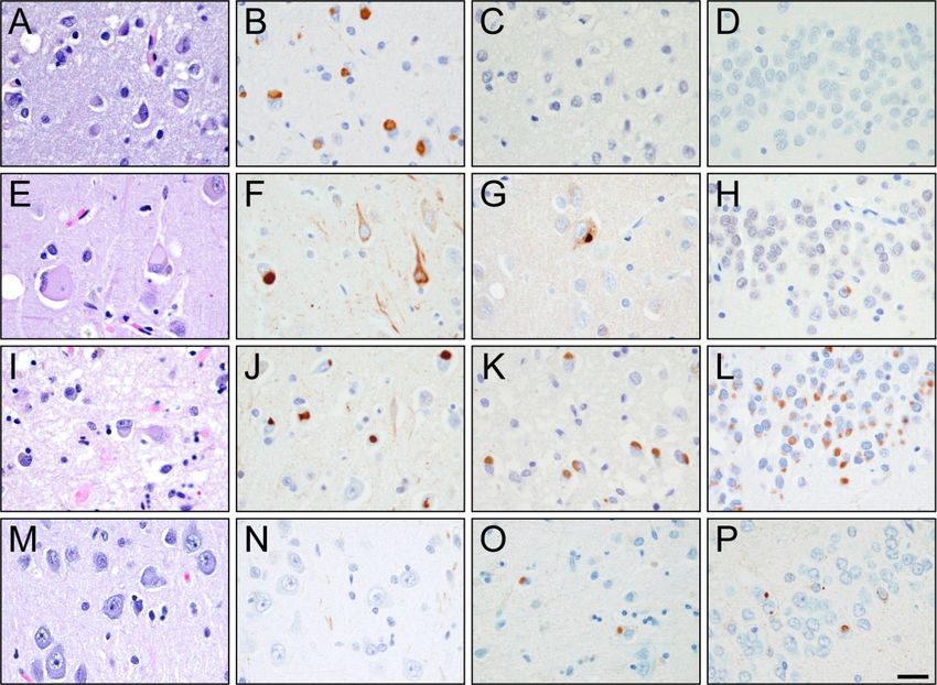
Free Neuropathology 1:9 (2020)                                                                        Kevin F. Bieniek et al
doi: https://doi.org/10.17879/freeneuropathology-2020-2639                                                     page 2 of 10

Introduction                                                   NIFID or aFTLD-U. A total of 15 cases were identified. All
                                                               15 cases were evaluated by a single expert neuropa-
                                                               thologist (DWD).
      The pathological term frontotemporal lobar de-
generation (FTLD) assumes the presence of focal frontal
and anterior temporal lobar atrophy(1). Histologically,        Clinical data
the majority of FTLDs are pathologically classified into
two broad categories of tau-positive FTLD (FTLD-tau)                 The medical records of all 15 cases were reviewed
and TDP-43-positive FTLD (FTLD-TDP) based on the pres-         by one clinician with expertise in neurodegenerative
ence of tau and TDP-43 immunoreactive inclusions,              diseases (KAJ) to abstract demographic and clinical in-
respectively(2, 3). A subset of FTLD cases however, lack       formation. Data abstracted included sex, age at onset,
tau and TDP-43 immunoreactivity, and instead have              prominent symptoms during the disease course, family
neuronal inclusions that are immunoreactive to ubiqui-         history of any neurodegenerative diseases and final
tin(4). Such cases have been referred to in the literature     clinical diagnosis prior to death.
as atypical FTLD with ubiquitin inclusion (aFTLD-U)(5). In
the past decade, immunohistochemical studies have              Pathological methods
revealed that the ubiquitinated protein in cases of
aFTLD-U is the fused in sarcoma (FUS) protein(6). Inter-             All 15 cases underwent histologic and ultrastruc-
estingly, there are two other relatively rare FTLD patho-      tural evaluation. Tissue sections were stained with he-
logical variants that also have neuronal inclusions that       matoxylin and eosin, Luxol fast blue-periodic acid Schiff
are immunoreactive to FUS(7, 8). These include the             (LFB-PAS) and Bielschowsky silver stains. Immunohisto-
entity neuronal intermediate filament inclusion disease        chemical staining was performed using standard meth-
(NIFID)(9), previously known as neurofilament inclusion        ods. The deparaffinized and rehydrated sections were
body disease(10), and basophilic inclusion body dis-           steamed in distilled water for 30 min and im-
ease(11) that have also been referred to as the general-       munostained in batches to assure consistency with a
ized variant of Pick’s disease(12). As a result, aFTLD-U,      DAKO Autostainer (DAKO, Carpinteria, CA) using 3,
NIFID and basophilic inclusion body disease are all cur-       3’diaminobenzidine as the chromogen. After im-
rently classified as subtypes of FTLD-FUS(3, 13).              munostaining, the sections were lightly counterstained
                                                               with hematoxylin. The following antibodies were used:
      In keeping with pathologically lumping these three       phosphorylated neurofilament (SMI-31, 1:20,000; Co-
entities as FTLD-FUS, is the fact that all three subtypes of   vance, Berkeley, CA); ubiquitin (mouse monoclonal Ubi-
FTLD-FUS, particularly aFTLD-U and NIFID, are strongly         1, 1:40,000; EnCor Biotechnology, Alachua, FL; rabbit
associated with a clinical presentation of the behavioral      polyclonal UBQ(20), 1:500 and rabbit polyclonal UH-
variant of frontotemporal dementia (bvFTD)(13-15).             19(21), 1:2,500); phospho-tau (CP13, 1:100; Peter Da-
Basophilic inclusion body disease is more strongly asso-       vies, Albert Einstein College of Medicine, Bronx, NY);
ciated with the juvenile form of amyotrophic lateral           alpha-synuclein (NACP(22), 1:3,000), alpha-internexin
sclerosis(16). In addition, aFTLD-U and NIFID have both        (1:100; EnCor Biotechnology, Alachua, FL); TDP-43
been found to be associated with striatal atrophy on           (MC2085, Dr. Petrucelli, 1:1500), pTDP-43 (S409/410,
MRI(17, 18). Hence, aFTLD-U and NIFID have a lot of            Cosmo Bio Co., 1:5000) and rabbit polyclonal anti-FUS
features in common. One study has directly compared            (1:500; HPA008784; Sigma, St. Louis. MO and Bethyl Lab;
aFTLD-U and NIFID(19). Hence, little is known about            A300-302A; Montgomery, TX). The Sigma antibody gave
which clinical and pathologically differences between          consistent and better staining and was used throughout
the two variants may further help to distinguish them,         the study. The presence or absence of motor neuron
and whether all cases of aFTLD-U and NIFID do indeed           disease was assessed and defined as previously de-
show FUS immunoreactivity. In this study, we set out to        scribed, including stains for activated microglia(23).
address these two unknowns in a cohort of 15 FTLD
cases that including aFTLD (n=8) and NIFID (n=7).
                                                               Electron microscopy

Materials and methods                                                Small pieces of formalin-fixed brains were im-
                                                               mersed in 2.5% glutaraldehyde-0.1 M cacodylate buffer
                                                               overnight at 4ºC. After washing in buffer, they were
Subject selection                                              post-fixed in aqueous 2% osmium tetroxide for 1 hr,
                                                               washed and fixed in 1% uranyl acetate-50% ethanol for
       The neuropathological databases at the Mayo Clin-       30 min, followed by dehydration in 70%, 80%, 95%,
ic, Jacksonville, Florida were queried to identify all cases   100% ethanols and propylene oxide. They were infiltrat-
of FTLD that had been given a pathological diagnosis of
Free Neuropathology 1:9 (2020)                                                                                   Kevin F. Bieniek et al
doi: https://doi.org/10.17879/freeneuropathology-2020-2639                                                                page 3 of 10

ed and embedded in Epon 812. Thin sections were                         10 min, cooled to room temperature for 15 min followed
stained with uranyl acetate and lead citrate and exam-                  by immunogold labeling. The Sigma anti-FUS was used at
ined in a Philips 208S electron microscope fitted with a                1:20 in PBS.
bottom-mount CCD camera (Orius 831, Gatan,
Pleasanton, CA).
                                                                        Results
Immunoelectron microscopy
                                                                              Demographics and clinical data for all 15 cases are
      Small pieces of formalin-fixed brains were dehy-
                                                                        shown in Table 1. There were 10 males and 5 females
drated in serial washes of 30%, 50%, 70%, 90% ethanol
                                                                        with median age at death of 54 years (range 41-69
for 10 min each, infiltrated and embedded in LR White
                                                       o                years). The median disease duration was 5 years (range
resin. They were polymerized in a vacuum oven at 50 C
                                                                        3-13 years). The most common final clinical diagnosis in
for 2 days. Thin sections were collected on Formvar-
                                                                        this series was behavioral variant of frontotemporal
coated nickel grids. Grids were floated with section-
                                              o                         dementia (bvFTD)(4, 24), rendered in 10 (67%) cases.
sides down on citrate buffer, pH 6.0, in a 100 C oven for

Table 1: Demographic and clinical features of all 15 cases

                           Age/
 Case             Sex                 Duration      Prominent symptoms/signs                                      Diagnosis
                           death
        ‡
 NIFID1            F         54            4        Behavioral & executive changes, mute                          bvFTD
 NIFID2*           M         51            5        Falls, pyramidal & extrapyramidal                             PLS
 NIFID3            F         50            4        Behavioral change, OCB, hyper-religious                       bvFTD
                                                    Apraxic dysgraphia, exacerbated startle, pyramidal and
 NIFID4            M         49            3                                                                      CBS
                                                    extrapyramidal signs
                                                    Falls, stiffness, difficult walking, speech apraxia, myo-
 NIFID5            M         67            3                                                                      MSA-P
                                                    clonic arm jerks, bradykinesia
                                                    Personality change, easily distractible, OCD-like behav-
 NIFID6            M         41            4                                                                      bvFTD
                                                    iors, disinhibited
                                                    Dynamic aphasia, later mute, difficulty walking, swal-
 NIFID7            F         61            2                                                                      PSP
                                                    lowing. Non-ambulatory
            †
 aFTLD-U1          M         68            5        Aphasia, apraxia, extrapyramidal                              CBS
            †
 aFTLD-U2          M         42            4        Personality change, hypersexual, hyperphagic                  bvFTD
 aFTLD-U3          F         42           10        Personality change, hypersexual, mute (could sing)            bvFTD
            †                                       Personality change, hallucinations, insomnia, hyper-
 aFTLD-U4          M         53           10                                                                      bvFTD
                                                    phagic, stereotypy
 aFTLD-U5          M         69            5        Behavioral change with lack of initiative, more placid        bvFTD
                                                    Personality change, mute, hypersexual, docile, hyper-
 aFTLD-U6          M         58           11                                                                      bvFTD
                                                    phagic, poor hygiene, hyper oral
 aFTLD-U7          F         55            5        Personality change, poor hygiene, socially withdrawn          bvFTD
                                                    Personality change, disorganized behavior, increased
 aFTLD-U8          M         45           13                                                                      bvFTD
                                                    appetite
ALS = amyotrophic lateral sclerosis; bvFTD = behavioral variant frontotemporal dementia; CBS = corticobasal syndrome
OCB = obsessive compulsive behavior; PSP = progressive supranuclear palsy, SND = striatonigral degeneration
† Previously published (Josephs et al. Acta Neuropathol 2008; 116: 159-167)
‡ Previously published (Josephs et al. Brain 2003; 126: 2291-2303
* Previously published (Josephs et al. Acta Neuropathol 2005; 109: 427-32)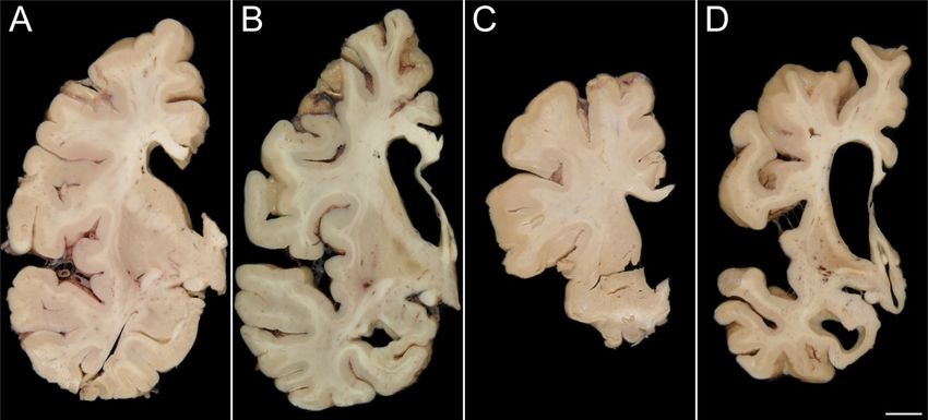
Free Neuropathology 1:9 (2020)                                                                                Kevin F. Bieniek et al
doi: https://doi.org/10.17879/freeneuropathology-2020-2639                                                             page 4 of 10

     For the other 5 cases, the final clinical diagnoses                Light microscopy
were corticobasal syndrome in two cases(25), and one
each diagnosed with progressive supranuclear palsy(26),                       Results of the light microscopic examination are
primary lateral sclerosis(27) and multiple system atro-                 shown in Table 2 and Figure 2. None of the 15 cases had
phy-Parkinsonian type (MSA-P)(28).                                      evidence of tau deposition, and none met criteria for
                                                                        Alzheimer’s or Lewy body disease. All 15 cases were
      The median age of onset of the NIFID cases was 52
                                                                        immune-reactive to ubiquitin. Thirteen of the 15 cases
years old (range: 41-61 years) while for aFTLD-U it was
                                                                        had neuronal inclusions that were immune-reactive to
54 years old (range: 42-69 years). Disease duration in
                                                                        FUS. Seven of the 15 cases had eosinophilic inclusions
NIFID was only 3.5 years (range: 2.0-5.0 years) and was
                                                                        that were seen on hematoxylin and eosin and were
much shorter than the median disease duration of the
                                                                        immunostained for α-internexin in keeping with their
aFTLD-U group which was 10 years (range: 4-13 years)
                                                                        pathological diagnosis of NIFID. None of the remaining
(P
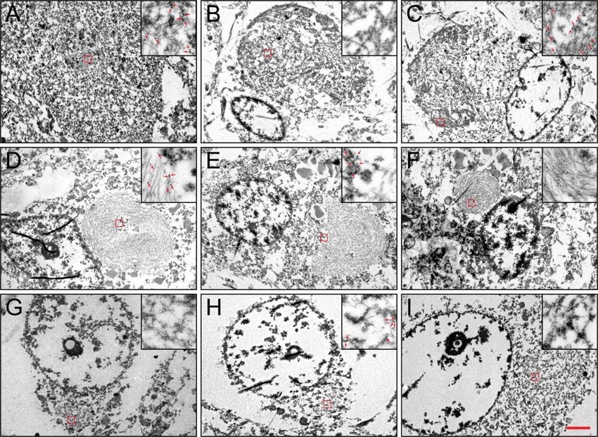
Free Neuropathology 1:9 (2020)                                                                                  Kevin F. Bieniek et al
doi: https://doi.org/10.17879/freeneuropathology-2020-2639                                                               page 5 of 10

Figure 1: Striatal atrophy on gross examination

Striatal atrophy was not seen in many NIFID cases (A; Case 5) and was only observed in two cases (B; Case 6). Conversely, only one
aFTLD-U case lacked marked striatal atrophy (C; Case 14) while the rest of the aFTLD-U cases demonstrated severe atrophy (D; Case 13)
[bar: 1 cm].

Figure 2: Alpha-internexin and FUS pathology

Hematoxylin and Eosin (A/E/I/M), α-internexin (B/F/J/N) and FUS (C/G/K/O) in the frontal cortex and FUS (D/H/L/P) in the hippocampus
of Cases 4 (A-D), 5 (E-H), 7 (I-L), and 15 (M-P) [bar:100 μm]
Free Neuropathology 1:9 (2020)                                                                                  Kevin F. Bieniek et al
doi: https://doi.org/10.17879/freeneuropathology-2020-2639                                                               page 6 of 10

Figure 3: FUS negative TDP-43 positive NIFID case

Neuronal eosinophilic (A/B) inclusions in the hippocampus (A/C/E/G) and frontal cortex (B/D/F/H) of Case 3 are negative for FUS (C/D)
but positive for TDP-43 (E/F/H), as well as αINX (G/H). α-internexin (brown) and TDP-43 (blue) colocalizes on double-labeling immuno-
histochemistry (H)[bar:100 μm]

Electron and immunoelectron microscopy                                 in loose arrangement and all were immuno-positive for
                                                                       FUS.
      Results of the electron microscopic examination
are shown in Figure 4. In the FUS-positive NIFID cases,
we found that FUS was localized to loose granulofila-                  Discussion
ments that were in close proximity to intermediate fila-
ment inclusions that contained tightly packed uncoated                       In this study we found pathological evidence for
filaments unlabeled by FUS antibody. The two types of                  aFTLD-U to be a homogeneous entity that is strongly
filaments did not mix. The compact intermediate fila-                  associated with a clinical diagnosis of bvFTD, and patho-
ment inclusions were similar to those we previously                    logically by FUS immunoreactivity and striatal atrophy.
reported(29). In the TDP-positive NIFID case (#3), the                 On the other hand, we found NIFID to be more hetero-
TDP-positive NCIs were composed of granulofilaments in                 geneous with more variable clinical presentations. Fur-
tightly packed bundles or loose orientations. Cytoplas-                thermore, NIFID does not always appear to be associat-
mic organelles, e.g. mitochondria, were occasionally                   ed with FUS immunoreactivity and is typically not asso-
encompassed by these inclusions.                                       ciated with striatal atrophy. Interestingly, we found
      These granulofilaments were not labeled with FUS.                evidence of overlap between a case that would meet
The characteristic compact intermediate filament inclu-                criteria for FTLD-TDP as well as NIFID.
sions were not as widespread as the TDP-positive inclu-                      In this study, aFTLD-U was a very homogeneous
sions in this case. Importantly, they were located in                  entity and the evidence supports aFTLD-U being classi-
separate neurons. In addition to their ultrastructural                 fied as FTLD-FUS. From a clinical standpoint aFTLD-U is
difference from the TDP-positive inclusions, these in-                 strongly associated with clinical features of bvFTD as
termediate filament inclusions were not labeled by TDP-                previously reported(14, 30) and hence should be consid-
43. Immunohistochemistry showed that the intermedi-                    ered in patients presenting with bvFTD especially in the
ate filament inclusions were immuno-negative for FUS.                  presence of striatal atrophy. Indeed striatal atrophy has
The TDP-negative, FUS-negative NIFID case (#4) had                     been reported in aFTLD-U on antemortem MRI imag-
compact intermediate filament inclusions similar to                    ing(17). NIFID, on the other hand, as currently defined
those described above. These compact intermediate                      does not appear to be as distinct an entity clinically and
filament inclusions were immuno-negative to FUS and                    pathologically as aFTLD-U. Supportive of this statement
TDP-43. Neuronal cytoplasmic inclusions in all the                     is the fact that of the seven NIFID cases in
aFTLD-U cases consisted of granulofilaments inclusions
Free Neuropathology 1:9 (2020) Kevin F. Bieniek et al
doi: https://doi.org/10.17879/freeneuropathology-2020-2639 page 7 of 10
Figure 4: Electron Microscopy
Variable α-internexin (A/D/G), FUS (B/E/H), and TDP-43 (C/F/I) immunoreactivity on electron microscopy in the neuronal inclusions of
Cases 3 (A-C), 2 (D-F), and 9 (G-I)[bar: 2 μm main; 0.3 μm inset]
this study, patients were given five different clinical To understand what may be happening with NIFID
diagnoses at the last evaluation prior to death. Secondly, it is worth further discussion. Neuronal intermediate
striatal atrophy, although present in two NIFID case was filament inclusion body disease is a type of FTLD with
absent in the rest. Interestingly, both NIFID cases with previously reported clinical presenting features of
striatal atrophy showed FUS immunoreactivity, one with bvFTD, corticobasal syndrome, and motor neuron dis-
a clinical diagnosis of bvFTD. It therefore appears that ease, especially the primary lateral sclerosis variant(10,
striatal atrophy is predictive of FUS, but FUS is not nec- 31, 32). Typically, patients with NIFID have a relatively
essarily predictive of striatal atrophy. Regardless, NIFID rapidly progressive course, becoming mute and unable
should also be considered in the differential diagnosis of to ambulate, dying around 3 ½ years after onset(10).
bvFTD with striatal atrophy and should suggest the pres- These features were observed in this cohort of seven
ence of FTLD-FUS. The one feature that may be helpful NIFID cases. From a pathological standpoint, in NIFID,
in predicting FTLD-FUS NIFID from FTLD-FUS aFTLD-U in neuronal cytoplasmic inclusions (NCIs) are easily visible
patients with bvFTD and striatal atrophy may be the on hematoxylin and eosin, show variable staining to
rapidity of progression, with faster progression being ubiquitin and silver stains but are strikingly immunoreac-
more suggestive of FTLD-FUS NIFID. tive to type IV intermediate filaments, including neuro-
filament and α-internexin(9, 10, 33, 34). These NCI,
however, are not morphologically homogeneous and itFree Neuropathology 1:9 (2020) Kevin F. Bieniek et al
doi: https://doi.org/10.17879/freeneuropathology-2020-2639 page 8 of 10
has been known from the original description of sions as we previously reported(4), and immunoelectron
NIFID(10) that NCI appeared to separate into two types: microscopy revealed FUS immunoreactivity. Of note is
rounded inclusions that are similar to Pick bodies, hence the fact that compact hyaline inclusions were absent in
called ‘Pick-body like (PBL) inclusions,’ and smaller more all eight aFTLD-U cases. This finding would also support
compact inclusions with a glass like appearance called our hypothesis that FUS immunoreactivity in NIFID and
‘compact hyaline inclusions’(7, 10, 19, 29). Could this NCI aFTLD-U is associated with the presence of the granu-
inclusion type difference be playing any role in the het- lofilamentous inclusions while α-internexin immunore-
erogeneity we observed in this study? The ultrastructual activity is associated with the presence of the compact
analysis in our seven NIFID cases shows that these two hyaline inclusions. It remains unclear however; why
different types of NCI also have different ultrastructual some granulofilamentous inclusions show exclusive FUS
appearances. The PBL inclusions were ultrastructually immunoreactivity (e.g. aFTLD-U) and others show exclu-
granulofilamentous while the compact hyaline inclusions sive TDP-43 immunoreactivity (e.g. FTLD-TDP). Only one
had a more tightly compact appearance. With immunoe- study to date has reported both FUS and TDP-43 immu-
lectron microscopy we found that the granulofilamen- noreactivity in the same NCIs(39).
tous inclusions were immunoreactive to FUS while the
compact hyaline inclusions were immunoreactive to Overall, the data from this study support the im-
intermediate filament. Interestingly all seven NIFID cases portance of FUS in the pathogenesis of aFTLD-U and the
had compact hyaline inclusions, and all seven showed α- classification of aFTLD-U as an FTLD-FUS. However, the
internexin immunoreactivity. However, unlike in previ- role of FUS in NIFID is less clear, with evidence support-
ous reports(35, 36), two of our seven NIFID cases did not ing NIFID being somewhat different from aFTLD-U with a
show FUS immunoreactivity and one of these two NIFID concern that NIFID, at least not all cases, may be incor-
cases showed predominantly, almost exclusively, com- rectly classified as a subtype of FTLD-FUS. Supporting
pact hyaline inclusions (Case #4). This NIFID case was this statement is the fact that another FUS negative case
immuno-negative to FUS, suggesting that the absence of with α-internexin positive inclusions has been de-
the granulofilamentous inclusions may be the explana- scribed(40). In this other case, however, a SOD1 muta-
tion for the absence of FUS immunoreactivity. The other tion was identified and some inclusions were immuno-
FUS negative case (Case #3) is also unique. In this case, reactive to SOD1. On a different note, the presence of
both granulofilamentous inclusions and compact hyaline intermediate filament inclusions and TDP-43 immunore-
inclusions were present. As expected, the presence of active inclusions in the same case makes one contem-
the compact hyaline inclusions was associated with α- plate the current sub-classification of FTLD into strict
internexin immunoreactivity. Surprisingly though, the categories. The relationship between FUS and interme-
granulofilamentous inclusions in this case (Case #3) diate filament is reminiscent of the relationship between
showed immunoreactivity to TDP-43 but not FUS. It is Alzheimer’s disease pathology and Lewy body disease.
therefore possible that this is a case of FTLD-TDP in Alzheimer’s disease pathology can occur in isolation, as
which compact hyaline inclusions happen to also be can Lewy body disease, but there are instances in which
present and hence accounts for the α-internexin immu- both Alzheimer’s disease and Lewy body disease co-
noreactivity observed. This would not be counterintui- occur. It would be naïve to argue that when two pathol-
tive since granulofilamentous morphology is the typical ogies co-occur, that one pathology is more important or
appearance of the NCIs in FTLD-TDP(37). On the other more relevant than the other.
hand, we cannot exclude the possibility that this is a In summary, aFTLD-U and NIFID share some but
case of NIFID with TDP-43 immunoreactivity given that not all features. Striatal atrophy, while appearing to be a
TDP-43 immunoreactivity has been described in many characteristic feature of aFTLD-U, does not appear to be
different diseases(38). a feature of NIFID although it can occur in some cases.
Compared to our NIFID cases, all eight aFTLD-U Further analyses are needed to better understand the
cases showed a homogeneous pattern of FUS immuno- relationship of aFTLD-U to NIFID and of NIFID to FUS.
reactivity similar to what has been previously report- Such analysis should include the assessment of other
ed(6, 8, 19, 36). In our eight aFTLD-U cases, ultrastruc- proteins that have been reported to be associated with
tural analysis demonstrated granulofilamentous inclu- aFTLD-U and NIFID including TAF15 and EWS(41), as well
as transportin1(42).Free Neuropathology 1:9 (2020) Kevin F. Bieniek et al
doi: https://doi.org/10.17879/freeneuropathology-2020-2639 page 9 of 10
References
1. Cairns NJ, Bigio EH, Mackenzie IR, Neumann M, Lee VM, Hatanpaa with a distinct clinical form of frontotemporal dementia but is not
KJ, et al. Neuropathologic diagnostic and nosologic criteria for related to mutations in the FUS gene. Acta Neuropathol.
frontotemporal lobar degeneration: consensus of the Consortium for 2011;122(1):99-110.
Frontotemporal Lobar Degeneration. Acta Neuropathol. 2007;114(1):5-
22. 16. Matsumoto S, Kusaka H, Murakami N, Hashizume Y, Okazaki H,
Hirano A. Basophilic inclusions in sporadic juvenile amyotrophic lateral
2. Mackenzie IR, Neumann M, Bigio EH, Cairns NJ, Alafuzoff I, Kril J, et sclerosis: an immunocytochemical and ultrastructural study. Acta
al. Nomenclature for neuropathologic subtypes of frontotemporal Neuropathol. 1992;83(6):579-83.
lobar degeneration: consensus recommendations. Acta Neuropathol.
2009;117(1):15-8. 17. Josephs KA, Whitwell JL, Parisi JE, Petersen RC, Boeve BF, Jack CR,
Jr., et al. Caudate atrophy on MRI is a characteristic feature of FTLD-
3. Mackenzie IR, Neumann M, Bigio EH, Cairns NJ, Alafuzoff I, Kril J, et FUS. Eur J Neurol. 2010;17(7):969-75.
al. Nomenclature and nosology for neuropathologic subtypes of
frontotemporal lobar degeneration: an update. Acta Neuropathol. 18. Seelaar H, Klijnsma KY, de Koning I, van der Lugt A, Chiu WZ,
2010;119(1):1-4. Azmani A, et al. Frequency of ubiquitin and FUS-positive, TDP-43-
negative frontotemporal lobar degeneration. J Neurol.
4. Josephs KA, Lin WL, Ahmed Z, Stroh DA, Graff-Radford NR, Dickson 2010;257(5):747-53.
DW. Frontotemporal lobar degeneration with ubiquitin-positive, but
TDP-43-negative inclusions. Acta Neuropathol. 2008;116(2):159-67. 19. Lashley T, Rohrer JD, Bandopadhyay R, Fry C, Ahmed Z, Isaacs AM,
et al. A comparative clinical, pathological, biochemical and genetic
5. Mackenzie IR, Foti D, Woulfe J, Hurwitz TA. Atypical frontotemporal study of fused in sarcoma proteinopathies. Brain. 2011;134(Pt 9):2548-
lobar degeneration with ubiquitin-positive, TDP-43-negative neuronal 64.
inclusions. Brain. 2008;131(Pt 5):1282-93.
20. Dickson DW, Wertkin A, Mattiace LA, Fier E, Kress Y, Davies P, et al.
6. Neumann M, Rademakers R, Roeber S, Baker M, Kretzschmar HA, Ubiquitin immunoelectron microscopy of dystrophic neurites in
Mackenzie IR. A new subtype of frontotemporal lobar degeneration cerebellar senile plaques of Alzheimer's disease. Acta Neuropathol.
with FUS pathology. Brain. 2009;132(Pt 11):2922-31. 1990;79(5):486-93.
7. Neumann M, Roeber S, Kretzschmar HA, Rademakers R, Baker M, 21. Lee S, Park YD, Yen SH, Ksiezak-Reding H, Goldman JE, Dickson DW.
Mackenzie IR. Abundant FUS-immunoreactive pathology in neuronal A study of infantile motor neuron disease with neurofilament and
intermediate filament inclusion disease. Acta Neuropathol. ubiquitin immunocytochemistry. Neuropediatrics. 1989;20(2):107-11.
2009;118(5):605-16.
22. Gwinn-Hardy K, Mehta ND, Farrer M, Maraganore D, Muenter M,
8. Munoz DG, Neumann M, Kusaka H, Yokota O, Ishihara K, Terada S, et Yen SH, et al. Distinctive neuropathology revealed by alpha-synuclein
al. FUS pathology in basophilic inclusion body disease. Acta antibodies in hereditary parkinsonism and dementia linked to
Neuropathol. 2009;118(5):617-27. chromosome 4p. Acta Neuropathol. 2000;99(6):663-72.
9. Cairns NJ, Zhukareva V, Uryu K, Zhang B, Bigio E, Mackenzie IR, et al. 23. Josephs KA, Parisi JE, Knopman DS, Boeve BF, Petersen RC, Dickson
alpha-internexin is present in the pathological inclusions of neuronal DW. Clinically undetected motor neuron disease in pathologically
intermediate filament inclusion disease. Am J Pathol. proven frontotemporal lobar degeneration with motor neuron disease.
2004;164(6):2153-61. Arch Neurol. 2006;63(4):506-12.
10. Josephs KA, Holton JL, Rossor MN, Braendgaard H, Ozawa T, Fox 24. Neary D, Snowden JS, Gustafson L, Passant U, Stuss D, Black S, et al.
NC, et al. Neurofilament inclusion body disease: a new proteinopathy? Frontotemporal lobar degeneration: a consensus on clinical diagnostic
Brain. 2003;126(Pt 10):2291-303. criteria. Neurology. 1998;51(6):1546-54.
11. Yokota O, Tsuchiya K, Terada S, Ishizu H, Uchikado H, Ikeda M, et al. 25. Boeve BF, Lang AE, Litvan I. Corticobasal degeneration and its
Basophilic inclusion body disease and neuronal intermediate filament relationship to progressive supranuclear palsy and frontotemporal
inclusion disease: a comparative clinicopathological study. Acta dementia. Ann Neurol. 2003;54 Suppl 5:S15-9.
Neuropathol. 2008;115(5):561-75.
26. Litvan I, Agid Y, Calne D, Campbell G, Dubois B, Duvoisin RC, et al.
12. Munoz-Garcia D, Ludwin SK. Classic and generalized variants of Clinical research criteria for the diagnosis of progressive supranuclear
Pick's disease: a clinicopathological, ultrastructural, and palsy (Steele-Richardson-Olszewski syndrome): report of the NINDS-
immunocytochemical comparative study. Ann Neurol. 1984;16(4):467- SPSP international workshop. Neurology. 1996;47(1):1-9.
80.
27. Zhai P, Pagan F, Statland J, Butman JA, Floeter MK. Primary lateral
13. Josephs KA, Hodges JR, Snowden JS, Mackenzie IR, Neumann M, sclerosis: A heterogeneous disorder composed of different subtypes?
Mann DM, et al. Neuropathological background of phenotypical Neurology. 2003;60(8):1258-65.
variability in frontotemporal dementia. Acta Neuropathol.
2011;122(2):137-53. 28. Papp MI, Kahn JE, Lantos PL. Glial cytoplasmic inclusions in the CNS
of patients with multiple system atrophy (striatonigral degeneration,
14. Urwin H, Josephs KA, Rohrer JD, Mackenzie IR, Neumann M, olivopontocerebellar atrophy and Shy-Drager syndrome). J Neurol Sci.
Authier A, et al. FUS pathology defines the majority of tau- and TDP- 1989;94(1-3):79-100.
43-negative frontotemporal lobar degeneration. Acta Neuropathol.
2010;120(1):33-41. 29. Uchikado H, Li A, Lin WL, Dickson DW. Heterogeneous inclusions in
neurofilament inclusion disease. Neuropathology. 2006;26(5):417-21.
15. Snowden JS, Hu Q, Rollinson S, Halliwell N, Robinson A, Davidson
YS, et al. The most common type of FTLD-FUS (aFTLD-U) is associatedFree Neuropathology 1:9 (2020) Kevin F. Bieniek et al
doi: https://doi.org/10.17879/freeneuropathology-2020-2639 page 10 of 10
30. Rohrer JD, Lashley T, Schott JM, Warren JE, Mead S, Isaacs AM, et 37. Lin WL, Dickson DW. Ultrastructural localization of TDP-43 in
al. Clinical and neuroanatomical signatures of tissue pathology in filamentous neuronal inclusions in various neurodegenerative
frontotemporal lobar degeneration. Brain. 2011;134(Pt 9):2565-81. diseases. Acta Neuropathol. 2008;116(2):205-13.
31. Bigio EH, Lipton AM, White CL, 3rd, Dickson DW, Hirano A. 38. Josephs KA, Mackenzie I, Frosch MP, Bigio EH, Neumann M, Arai T,
Frontotemporal and motor neurone degeneration with neurofilament et al. LATE to the PART-y. Brain. 2019;142(9):e47.
inclusion bodies: additional evidence for overlap between FTD and
ALS. Neuropathol Appl Neurobiol. 2003;29(3):239-53. 39. Deng HX, Zhai H, Bigio EH, Yan J, Fecto F, Ajroud K, et al. FUS-
immunoreactive inclusions are a common feature in sporadic and non-
32. Cairns NJ, Grossman M, Arnold SE, Burn DJ, Jaros E, Perry RH, et al. SOD1 familial amyotrophic lateral sclerosis. Ann Neurol.
Clinical and neuropathologic variation in neuronal intermediate 2010;67(6):739-48.
filament inclusion disease. Neurology. 2004;63(8):1376-84.
40. Nakamura M, Bieniek KF, Lin WL, Graff-Radford NR, Murray ME,
33. Josephs KA, Uchikado H, McComb RD, Bashir R, Wszolek Z, Castanedes-Casey M, et al. A truncating SOD1 mutation, p.Gly141X, is
Swanson J, et al. Extending the clinicopathological spectrum of associated with clinical and pathologic heterogeneity, including
neurofilament inclusion disease. Acta Neuropathol. 2005;109(4):427- frontotemporal lobar degeneration. Acta Neuropathol.
32. 2015;130(1):145-57.
34. Uchikado H, Shaw G, Wang DS, Dickson DW. Screening for 41. Neumann M, Bentmann E, Dormann D, Jawaid A, DeJesus-
neurofilament inclusion disease using alpha-internexin Hernandez M, Ansorge O, et al. FET proteins TAF15 and EWS are
immunohistochemistry. Neurology. 2005;64(9):1658-9. selective markers that distinguish FTLD with FUS pathology from
amyotrophic lateral sclerosis with FUS mutations. Brain. 2011;134(Pt
35. Armstrong RA, Gearing M, Bigio EH, Cruz-Sanchez FF, Duyckaerts C, 9):2595-609.
Mackenzie IR, et al. The spectrum and severity of FUS-immunoreactive
inclusions in the frontal and temporal lobes of ten cases of neuronal 42. Brelstaff J, Lashley T, Holton JL, Lees AJ, Rossor MN, Bandopadhyay
intermediate filament inclusion disease. Acta Neuropathol. R, et al. Transportin1: a marker of FTLD-FUS. Acta Neuropathol.
2011;121(2):219-28. 2011;122(5):591-600..
36. Mackenzie IR, Munoz DG, Kusaka H, Yokota O, Ishihara K, Roeber S,
et al. Distinct pathological subtypes of FTLD-FUS. Acta Neuropathol.
2011;121(2):207-18.You can also read