Burn Clinical Pathway - JOHNS HOPKINS ALL CHILDREN'S HOSPITAL - Johns Hopkins All Children's Hospital
←
→
Page content transcription
If your browser does not render page correctly, please read the page content below
JOHNS HOPKINS ALL CHILDREN’S HOSPITAL

            Burn
Clinical Pathway
Johns Hopkins All Children’s Hospital
                        Trauma-Burn Clinical Pathway

         Table of Contents
              1. Rationale
              2. Background
              3. Diagnosis
              4. Clinical Management
                    a. Table 1- Treatment Based on Burn Type
                    b. Table 2- Treatment and Wound Care based on Burn Depth
              5. Documentation Reminders
              6. Follow-Up Care
              7. Patient/Family Education
              8. Emergency Center Burn Clinical Pathway Algorithm
              9. High Risk Burns Clinical Pathway Algorithm
              10. References
              11. Outcome Measures
              12. Clinical Pathways Team Information
              13. Appendix 1: Lund-Browder
              14. Appendix 2: Discharge Instructions

                                                                                                Updated: June 2021
                                                 Owners: Chris Snyder, MD; Danielle Mercurio, MD; Katie Deemer, RN
This pathway is intended as a guide for physicians, physician assistants, nurse practitioners and other healthcare providers. It should be
adapted to the care of specific patient based on the patient’s individualized circumstances and the practitioner’s professional judgment.

                                                                                                                               1
Johns Hopkins All Children's Hospital
Burn Clinical Pathway
Rationale:
This clinical pathway was developed by a consensus group of JHACH physicians, advance
practice providers, and nurses to standardize the management of children with burn injuries and
achieve the best possible outcome. It addresses the following clinical questions or problems:
1. When to activate a trauma team activation for a burn patient?
2. How to diagnose and classify burns?
3. How to treat burns?
4. When to consider transfer to a Burn Center for definitive treatment?
Background
Burns are the 3rd leading cause of injury deaths for children aged 5-9 years and the 5th for ages
1-4 years in the United States. Roughly 25% of all burn injuries occur in children under the age
of 15 years. This practice guideline will outline the essential elements of evaluation and
management of burns, to include the criteria of transferring patients to a burn center.
Diagnosis
Primary Assessment should include consideration for mechanism of injury (MOI) and other
trauma and control of life threats. These may include, but are not limited to, inhalation injury,
hypovolemia, hypoglycemia, and hypothermia.
After primary assessment and control of any life-threatening injuries, it is imperative to
distinguish the depth of burn injury. Cutaneous burns are classified according to depth of tissue
injury: Superficial (1st degree), Superficial-Partial Thickness (2nd degree), Deep Partial
Thickness (2nd Degree), Full-thickness (3rd degree), Full-thickness-extension to deep tissues (4th
degree).
A thorough estimation of burn size is essential to determine initial management, fluid
resuscitation and consideration for transfer to a burn center. The extent of burns is expressed
as the total percentage of body surface areas (TBSA). There are several methods to calculate
TBSA.
Palm Method: Small or patchy burns can be approximated by using the surface area of the
patient’s palm. The palm of the patient’s hand, excluding the fingers, is approximately 0.5
percent of total body surface area and the entire palmar surface including fingers is 1 percent in
children and adults.
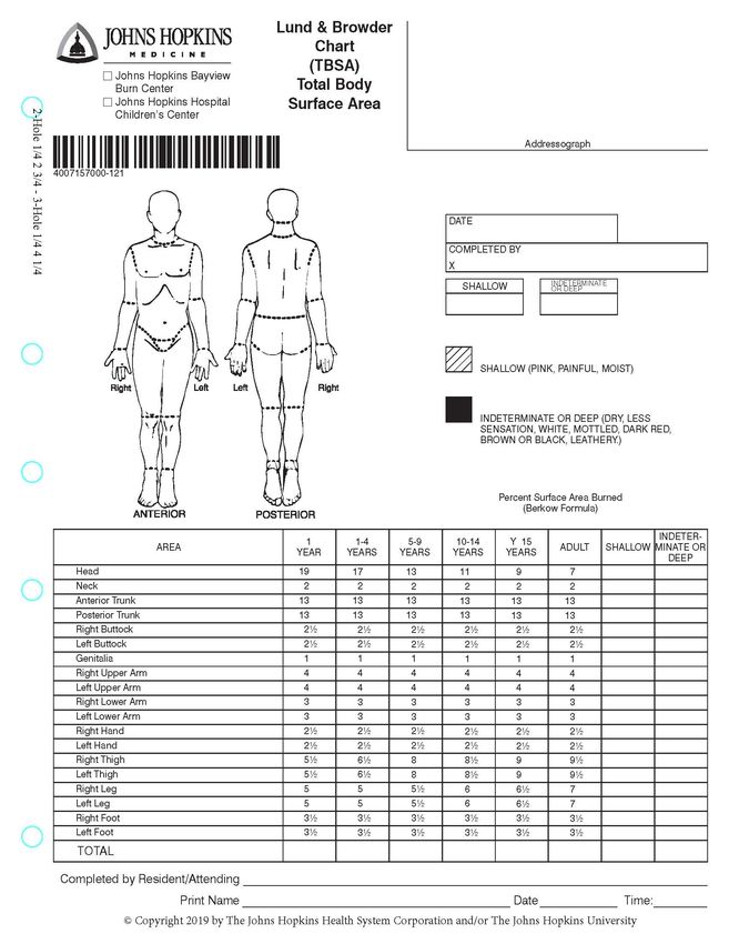
Lund-Browder: The most accurate method for estimating TBSA for both children and adults.
See Appendix.
2Burns are classified into four categories based on the burn source. This will affect the clinical
management of the burn.
a. Thermal
b. Chemical
c. Electrical
d. Radiation
Clinical Management
Radiographic Studies:
a. Consider CT imaging of the head if the patient has altered mental status or
associated head trauma from a fall. Do not let this delay therapeutic intervention for
Carbon Monoxide (CO) or Cyanide (CN) poisoning.
b. Consider plain chest films for smoke inhalation
c. Consider plain films if concern for concomitant trauma or foreign body/debris in
wounds.
Lab recommendations: CBC, BUN, electrolytes, lactate, and coagulation profile when TBSA
>20%.
Fluid Resuscitation: Formal fluid resuscitation using American Burn Association (ABA)
Consensus Formula should be initiated for patients with partial thickness or worse burns
covering 20% or more TBSA.
a. 2-4 ml/kg/% TBSA Burn (using LR) in the 1st 24 hours
1. Give ½ in the First 8 hours post burn
2. Give ½ in the Next 16 hours post burn
a. Adults: 2 ml/kg/% TBSA
b. Children: 3ml/kg/% TBSA
c. Electrical: 4ml/kg/% TBSA
b. Ringers lactated solution is fluid of choice for formal fluid resuscitation
c. Avoid crystalloid boluses
d. Place foley catheter in patients receiving formal fluid resuscitation and monitor urine
output for a goal of 1cc/kg/hr in patients < 30 kg and 0.5cc/kg/hr in patients >30 kg.
e. If the patient is less than < 30kg, add maintenance fluids that include dextrose (D5LR
or D5NS)
Pain Management
a. Small burn injuries: acetaminophen and nonsteroidal anti-inflammatory drugs.
b. Deeper burn: OTC analgesics in combination with opioids
1) Intranasal fentanyl 1.5 mcg/kg
2) Oxycodone 0.1 to 0.2 mcg/kg for patients 50 kg.
3) Referral to a burn center is needed when pain is too severe (or anticipated) to
perform dressing changes and wound care without parental narcotics or
conscious sedation.
Additional medications:
a. Tetanus booster warranted if not received in last 12 months (deep superficial partial
thickness)
b. Antibiotics: Antibiotic for prophylaxis use is not routinely recommended.
3Concerns for Non-accidental Burn Injuries:
a. Does the injury match the reported history?
b. Is the age of the burn consistent with the history?
1) Most caregivers would reach out for burn care immediately. If there is a delay to
care, is there a clear and adequate reason?
i. Does it appear as if a reasonable treatment plan has been implemented
prior to your involvement if there was a delay?
c. Is the pattern suggestive of an abusive injury?
1) Cigarette burns and lighter burns5
2) Submersion burns with sharp demarcations, sparing of skin folds/buttocks are
concerning for forced submersion
3) Clear patterns with curling iron, radiators/heaters, stoves, etc. concerning as
contact with such surfaces should be transient and elicit immediate removal to
neurologically normal child
American Burn Association Burn Center Referral Criteria: (May be telephone consultation to
determine if patient transfer is warranted)
a. Partial thickness burns >10% TBSA
b. Burns that involve the face, hands, feet, genitalia, perineum, or major joints.
c. Third degree burns
d. Electric burns including lightning injury
e. Chemical burns
f. Inhalation Injury
Florida Trauma Burn Centers:
1. Tampa General Hospital:
(813) 844-7000
https://www.tgh.org/services/burn
2. Shands Health Care:
(352) 265-8000
https://ufhealth.org/uf-health-shands-burn-center
3. Jackson Memorial Hospital & Ryder Trauma Center:
(305) 243-6256
https://jacksonhealth.org/burn-center/#gref
4Table 1: Recommendations of Treatment Based On Burn Type
General • Remove clothing that is hot, burned, or exposed to chemicals/smoke.
Considerations • Remove jewelry that may become constricting.
• Observe for smoke inhalation in patients who have suffered thermal burns.
• There is NO role for topical steroids or antibiotics in the initial treatment of
minor burns, as this may increase the risk of infection and impair healing.
• NO Silver sulfadiazine cream as it is cytotoxic and slows wound healing
• Wrap fingers and toes individually
• Maintain neutral to extended anatomical position with access to fingertips
and toes for circulation to avoid contractures.
Circumferential • At risk for Acute Compartment Syndrome
Burns • ACS may necessitate fasciotomy
• May require escharotomy if there is distal ischemia. Rarely required in the
immediate aftermath of a burn.
• Consider trauma team activation
• Frequent assessment of extremity perfusion
• High risk finding for non-accidental trauma. Please see JHACH Physical
Child Abuse Clinical Pathway
Chemical Burn • Don personal protective equipment: gloves, non-permeable gown (trauma
with Powder gowns) and mask with face shield.
• Remove all patient’s clothing and jewelry
• Brush of all visible powder off skin using gloved hands
• Await EC or Trauma Team recommendations
• Consult Poison Control for assistance identifying chemicals and treatment
recommendations. 1-800-222-1222
Suspected or • Consider EKG, cardiac enzymes, CK, and urinalysis to check for myoglobin
reported due to rhabdomyolysis.
electrical • Any patient in contact with a high voltage source should have continuous
burns cardiac monitoring.
• Await recommendations for wound care.
Carbon • For victims of house fires with carbon monoxide poisoning, oxygen saturation
Monoxide by pulse oximetry will often be normal or elevated and falsely reassuring
Poisoning relative to directly measure oxygen saturation.
• Treat the patient with high flow oxygen until carboxyhemoglobin levels drop
to less than 10%.
5Inhalation • The leading injury in the upper airway is thermal injury: results in erythema,
Injury ulcerations and edema
• Aggressive fluid administration to treat burn shock may promote early edema
formation
• Increased production to thick secretions can cause distal airway obstruction,
atelectasis, and impaired gas exchange
• Tracheobronchial injury is usually caused by chemicals in smoke: symptoms
include coughing and wheezing, soot containing airway secretions, increased
work of breathing resulting in hypoventilation, erythema, and increased
pulmonary shunting from lobar collapse or atelectasis
• May have normal chest radiograph
• Consider trauma team activation
• Remove all smoke covered clothing
• Frequent respiratory assessments
• May require intubation or tracheostomy. These could be difficult and the
critical airway team should be utilized (Call 7-4300 and request surgical
critical airway team)
Patterned • High risk finding for non-accidental trauma. Please JHACH Physical Child
Burns Abuse Clinical Pathway
Burn-like Diseases that mimic the appearance of burn-injuries and may vary in depth (e.g.
Diseases superficial, partial thickness, or full thickness)
Superficial
• Staphylococcal Scalded Skin Syndrome
• Toxic Epidermal Necrolysis
• Epidermolysis Bullosa
Partial to Full Thickness
• Stevens-Johnson Syndrome
• Pemphigus Vulgaris
• Necrotizing Fasciitis
6Table 2: Recommendations of Treatment and Wound Care Based On Burn Depth
Superficial • Appearance: Minor Damage, Erythematous (Pink to Reddened Skin), painful,
(1st Degree) Dry, without blisters.
• Course: Heals without scarring in 3-5 days.
• Treatment: Moisturize for comfort, topical antimicrobial not needed for intact
epidermis.
Partial • Appearance: Partial destruction of dermis. Bright pink to cherry red skin with
Thickness blisters. Skin moist, wet, very painful & edematous.
Burns • Course: Heals in 7-10 days with minimal
(2nd Degree)
• Treatment:
1. Do not aspirate or rupture intact blisters as increases risk of infection and
causes pain.
2. Cleanse with normal saline (baby shampoo PRN).
3. Apply Mepilex Ag to the wound. Mepilex Ag should overlap the wound by
at least 2 cm onto the surrounding skin. Apply the adherent side to the
wound. Remove the film and mold the dressing in place-do not stretch.
Hold the Mepilex Ag in place with dry gauze or wrap, secure with tubular.
No tape to skin. Mepilex is left in place for 5 days. Change gauze only as
needed.
Deep Partial • Appearance: Greater than 50% of dermis. Moderate eschar/ slough or
Thickness ruptured blisters. Less painful because nerves are damaged (not complete
(2nd Degree) anesthesia).
• Course: Heals in 2-3 weeks, severe scarring can occur, risk of skin scar
contracture. May require skin grafting
• Treatment:
1. Debride/remove eschar and necrotic skin, including ruptured blisters.
Extensive debriding is rarely necessary and may be deferred until initial f/u
visit.
2. Cleanse wound with normal saline plus baby shampoo
3. Apply Mepilex Ag to the wound. Mepilex Ag should overlap the wound by
at least 2 cm onto the surrounding skin. Apply the adherent side to the
wound. Remove the film and mold the dressing in place-do not stretch.
Hold the Mepilex Ag in place with dry gauze or wrap, secure with tubular.
No tape to skin. Mepilex is left in place for 5 days. Change gauze only as
needed.
Full • Appearance: Damage to all skin layers (3rd degree) and may involve fat,
Thickness muscle, bone (4th degree). Skin is severely charred, waxy white, pale,
Burns leathery. No bleeding, painless (complete anesthesia).
(3rd and 4th • Course: High risk for infection and fluid loss
degree)
• Treatment: Immediate referral to burn center and/or Plastic Surgery consult.
7Documentation Reminders
The following items should be documented within the medical record.
a. History of Burn Injury
1. When? Initial time of burn important if fluid resuscitation warranted
2. How? Fire, steam, chemical, electrical, hot material
3. Where? Enclosed space (inhalation injury)?
b. Burn TBSA, type, and depth in the EPIC Burn Narrator utilizing the Lund-Browder
method.
c. Medical History: comorbidities (IDDM, immunosuppression)
d. Medication History: steroid use, blood thinners, diabetic meds, etc.
e. Allergies
f. Last Tetanus: booster warranted if not received in last 12 months (deep superficial
partial thickness)
g. Wound Assessment
h. Treatment performed
i. Follow-up
j. Referrals
Follow-Up Care
Patients with superficial or partial thickness burns,Johns Hopkins All Children's Hospital
Emergency Center Burn Clinical Pathway
Emergency Center Burn Patient
Assess
• Burn Size using the Lund-Browder method
• Patient Age
• Patient Comorbidities
• Past Medical and Surgical History
Is the burn full-thickness or circumferential?
OR
Is there concern for smoke inhalation?
OR
Is the burn partial thickness and cover more than 10% TBSA?
YES NO
See High Risk Burns Is the burn partial to full-thickness?
Clinical Pathway
YES NO
• Provide adequate pain control • Refer to Table 1 & Table
• Verify Tetanus Status-Inoculate PRN 2 for recommendations of
• Wash patient with cool, soapy water (baby care
shampoo) & debride dead epidermis with • Provide patient/family
washcloth education for discharge
• Leave blisters intact instructions
• For partial/deep partial thickness burns: use
Mepilex Ag dressings
Does the burn meet these special
considerations?
• Burns involve face, hands, feet, genitalia,
perineum, or major joints
• Electric burns including lightning injury
• Chemical burns
• Pain to severe to accomplish dressing
changes without parenteral narcotics or
conscious sedation
• Burns require grafting
YES
Consultation to Burn Center NO
Discharge Home
1) Follow-Up with Wound Clinic
Does Patient Require 2) Patient/family education discharge
Admission for burn care? instructions
YES
Transfer to Burn CenterJohns Hopkins All Children's Hospital
High Risk Burns Clinical Pathway
Patient’s with High Risk Burns
• Partial to full-thickness burns >10% TBSA
• Partial to full-thickness circumferential burn
• Smoke Inhalation
Activate Trauma Alert
Establish need for IV Access
• Perform Primary and Secondary Trauma Survey
• Provide adequate pain control
• Verify Tetanus Status-Inoculate PRN
• Wash patient with cool, soapy water (baby shampoo) & debride dead epidermis with washcloth
• For partial/deep partial thickness burns: use Mepilex Ag dressings
• Wrap Patient in Dry Sheets
• Maintain Normothermia
Burn 10%- 20% TBSA Burn >20% TBSA
Give Maintenance IV Fluid Initiate Burn Center Transfer
& Oral Rehydration • Formal Fluid Resuscitation using ABA Consensus Formula
• Avoid Crystalloid Boluses
• Foley Catheter
o Record Urine Output Hourly
Telephone Consultation • Adjust Fluid Rate Up or Down Based on Urine Output
with Burn Center for Formulation of o Goal urine output of 1cc/kg/hr for pt’s 30 kg
Special Considerations and Additional Information:
Consider Intubation Prior to Transfer IF: American Burn Association Criteria for injuries
• Extensive Head, Neck, Facial Burns requiring referral to a Burn Center
• >40% TBSA Burn • Partial thickness burns >10% TBSA
• Stridor, Wheezing, Hoarse Voice, Dyspnea • Burns that involve face, hands, feet, genitalia, perineum,
• CoHbg> 30% and Obtunded or major joints
• If Persistent Lactic Acidosis, Consider Cyanide Poisoning • Third degree burns
and Cyano Kit Use • Electric burns including lightning injury
• Extension 7-4300 Surgical Critical Airway Team • Chemical Burns
• Inhalation Injury
Consider Escharotomy or Fasciotomy after Consult American Burn Association Consensus Formula
with Burn Surgeon: • 2-4 ml/kg/% TBSA Burn (using LR) in the 1st 24 hr
- Give ½ in the First 8 Hrs Post Burn
• Full Thickness Circumferential Full Thickness Burns - Give ½ in the Next 16 Hrs Post Burn
AND • Adults: 2 ml/kg/% TBSA
• Clinical Signs of Compartment Syndrome • Children: 3ml/kg/% TBSA
OR • Electrical: 4ml/kg/% TBSA
• Full Thickness Chest Burns with Restricted Respiratory
Excursion In Adults:
• Adjust Fluid Rate for Goal UO 0.5kg/hr
In Children: (References
1. American Burn Association. Burn Injury Fact Sheet. Copyright 2018.
https://ameriburn.org/wp-content/uploads/2017/12/nbaw-factsheet_121417-1.pdf
(Accessed on February 26, 2020)
2. American Burn Association White Paper. Surgical management of the burn wound and
use of skin substitutes. Copyright 2009. www.ameriburn.or (Accessed on January 04,
2010).
3. Edwards, C. (2020) Surface and Burn Trauma. In J. S. Blansfield, Trauma Nursing Core
Curse Provider Manual Eighth Edition (pp 209-230). Emergency Nurses Association.
4. Griffin, B.R.; Frear, C.C.; Bable, F.; Oakley, E.; Kimble, R.M. (2019). Cool Running
Water First Aid Decreases Skin Grafting Requirements in Pediatric Burns: A Cohort
Study of Two Thousand Four Hundred Ninety-Five Children. Annals of Emergency
Medicine, 75(1) 75-85. https://doi.org/10.1016/j.annemergmed.2019.06.28.
5. S Harel, B Burkey, AD Nanassy, M Marcolongo, E Phillips, C Campbell, M McColgan.
Flame time of a cigarette lighter to achieve temperature capable of inflicting a burn. Sept
2017, Burns, 43(6): 1227-1232.
6. Western Trauma Association. Preferred Triage and Initial Management of the Burned
Patient. Journal of Trauma and Acute Care Surgery. 87(5):1239-1243, November, 2019.
Outcome Measures:
• Emergency center length of stay
• Total number of burn patients seen annually
• Total number of burn patients transferred out annually
• Patient who meet trauma team activation criteria activated
11Clinical Pathway Team
Burns Clinical Pathway
Johns Hopkins All Children’s Hospital
Owner(s): (Christopher Snyder, MD, Trauma Medical Director, Danielle Mercurio, MD,
Emergency Center, Katie Deemer, RN, Trauma PI Coordinator)
Also Reviewed by:
Specialists: Kristine Rogers, APRN, Wound Clinic
Emergency Center: Lisa Odendal, MD
Others: Michael Harrington, MD, Tampa General Hospital Plastic Surgery
Clinical Pathway Management Team: Joseph Perno, MD; Courtney Titus, PA-C
Date Approved by JHACH Clinical Practice Council: October 20th, 2020
Date Available on Webpage: November 23rd, 2020
Last Revised: June 21,2021
Disclaimer
Clinical Pathways are intended to assist physicians, physician assistants, nurse practitioners
and other health care providers in clinical decision-making by describing a range of generally
acceptable approaches for the diagnosis, management, or prevention of specific diseases or
conditions. The ultimate judgment regarding care of a particular patient must be made by the
physician in light of the individual circumstances presented by the patient.
The information and guidelines are provided "AS IS" without warranty, express or implied, and
Johns Hopkins All Children’s Hospital, Inc. hereby excludes all implied warranties of
merchantability and fitness for a particular use or purpose with respect to the information. Johns
Hopkins All Children’s Hospital, Inc. shall not be liable for direct, indirect, special, incidental or
consequential damages related to the user's decision to use the information contained herein.
12Appendix 1: Lund Browder Chart
13Appendix 2: Burn Care Instructions
Burn Care Instructions – Pediatric
Why does my child need follow up care for his/her burns?
It is important to follow-up with your child’s Burn Care Specialist or Primary Care Physician to
make sure the burn is healing properly, to prevent scarring, and to help the skin return to its
normal color as much as possible. This appointment may be needed within the first week of
discharge. Review additional information on the After Visit Summary (AVS) for information on
setting up an appointment.
When should I call a healthcare provider?
If you have questions or concerns about your child or if you notice any of the
following symptoms, call your child’s Burn Care Specialist or Primary Care Physician
• Redness or swelling of the burned area
• Increased pain or pain not relieved by pain medicines.
• Fever, rash, or bad smelling drainage from the burns. (Clear yellow drainage is
normal.)
When should I give pain medicine?
First and second-degree burns can be very painful!
• It is better to prevent pain than to wait until your child is in pain to give medicine.
• The pain medication instructions will be included on your After Visit
Summary discharge instructions.
• If your child is freely moving their burned body part, it is likely that their pain is
controlled.
• Provide your child with pain medication 30 minutes prior to dressing changes
and Follow-Up appointments.
• Encouraging your child to move the burned body part regularly is an important part of
burn healing.
For all dressing types…
• Wash hands before and after any contact with burns or burn dressings.
• Keep supplies as clean as possible.
• Give pain medication 30 minutes prior to dressing change and/ or clinical visit.
14Appendix 2: Burn Care Instructions (continued)
Silver impregnated foam dressing (Mepilex® Ag)
• Silver impregnated foam dressing sticks to the burn like a “Band-Aid” as it is healing.
• There is no need to change the silver impregnated foam layer – it can stay in place for
up to seven days!
• Keep the dressing dry and provide sponge baths.
• You only need to change the white outer gauze and stretch net dressing if it is
completely wet with drainage; clear or yellowish drainage is normal.
• Additional supplies can be purchased at most pharmacies.
• We have provided you with one extra piece of silver impregnated foam dressing, just in
case the current dressing falls off. If the silver impregnated foam dressing falls off:
1. Wash the burn gently with mild soap and water – pat dry
2. Reapply a new piece of silver impregnated foam dressing
3. Wrap with gauze
4. Cover with stretch net dressing
Bacitracin and non-adherent mesh dressing
• Change the dressing daily during bath time.
• If the dressing is stuck on the burn, put your child in the bath with the dressing on,
allowing the water to moisten the dressing and allow for easier removal.
• Apply antibiotic ointment to the non-adherent dressing and apply to the burn/wound.
• Wrap with gauze roll starting from the furthest part of the body (toes to hips, fingers to
shoulders etc. and cover with stretch net dressing.
Hand burns
• After applying antibiotic ointment and non-adherent dressing, each finger should be
wrapped separately in a straight position.
• First, wrap the gauze around the wrist to prevent from sliding off fingers.
• From the wrist, move the gauze up the back of the hand towards the tip of the finger.
• Wrap around the finger – from the tip down to the palm – overlapping half of each wrap.
• Wrap gauze around the wrist again before starting on the next finger. Continue wrapping
each burned finger following the above steps.
• Do not stretch or pull hard on gauze. Wrapping should be fitted, but not constricting.
Your child should be able to move their fingers easily with full range of motion.
Please refer to your child's After Visit Summary (AVS) for additional instructions
15You can also read