COVID-19 associated rhinosinusitis mucormycosis due to Rhizopus arrhizus: A rare but potentially fatal infection occurring after treatment with ...
←
→
Page content transcription
If your browser does not render page correctly, please read the page content below
COVID-19 associated rhinosinusitis mucormycosis due to
Rhizopus arrhizus: A rare but potentially fatal infection occurring
after treatment with corticosteroids
Payam Tabarsi
Shahid Beheshti University of Medical Sciences
Neda Khalili
Tehran University of Medical Sciences
Mihan Pourabdollah
Shahid Beheshti University of Medical Sciences
Somayeh Sharifynia
Shahid Beheshti University of Medical Sciences
Ali Safavi Naeini
Shahid Beheshti University of Medical Sciences
Jahangir Ghorbani
Shahid Beheshti University of Medical Sciences
Abdolreza Mohamadnia
Shahid Beheshti University of Medical Sciences
Zahra Abtahian
Shahid Beheshti University of Medical Sciences
Elham Askari ( e.askari@sbmu.ac.ir )
Shahid Beheshti University of Medical Sciences
Case Report
Keywords: coronavirus disease-2019, corticosteroids, fungal, mucormycosis, Rhizopus
Posted Date: June 3rd, 2021
DOI: https://doi.org/10.21203/rs.3.rs-398594/v2
License: This work is licensed under a Creative Commons Attribution 4.0 International License. Read Full License
Page 1/9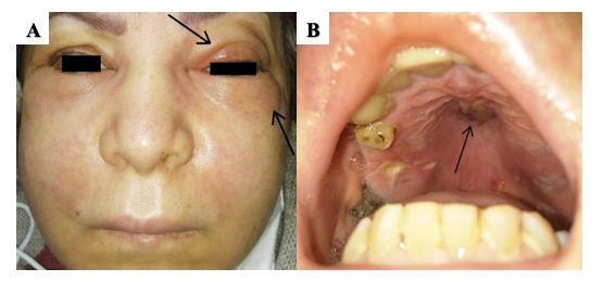
Abstract
Coronavirus disease 2019 (COVID-19) first emerged in Wuhan, China in December 2019, and since then the frequency of bacterial and
fungal coinfections has been continuously rising. While invasive pulmonary aspergillosis is increasingly being recognized in
association with COVID-19, there is limited information with regards to COVID-19 associated mucormycosis. Here, we describe a 50-
year-old woman with uncontrolled diabetes who received systemic corticosteroids and remdesevir during her admission for COVID-19.
Few days after discharge, the patient was readmitted due to facial swelling and numbness, and a diagnosis of COVID-19 associated
rhinosinusitis mucormycosis due to Rhizopus arrhizus (formerly Rhizopus oryzae) was confirmed with sequencing of the internal
transcribed spacer (ITS) region of the ribosomal DNA. This report aims to address the importance of short-term follow-up in COVID-19
patients who have received systemic corticosteroids, particularly those with predisposing conditions, as early detection and prompt,
aggressive treatment is essential for the management of invasive fungal infections.

Introduction
Emerging evidence suggests that patients infected with severe acute respiratory syndrome coronavirus-2 (SARS-CoV-2) may develop
bacterial and fungal secondary infections [1]. While invasive pulmonary aspergillosis (IPA) is increasingly being recognized in
association with coronavirus disease 2019 (COVID-19), especially in critically ill patients hospitalized in the intensive care unit, [2] there
are only a few cases of COVID-19 associated mucormycosis (CAM) available in the literature [3]. Mucormycosis is a rare, opportunistic,
highly fatal fungal infection that typically occurs in individuals with underlying compromising conditions, such as diabetes mellitus,
corticosteroid use, hematologic malignancies, neutropenia, solid organ/allogeneic stem cell transplant, primary immunodeficiency, and
treatment with immunosuppressants. Nevertheless, such infections can be seen in apparently immunocompetent patients on extremely
rare occasions [4]. Rhino-orbito-cerebral mucormycosis is considered as the most common manifestation of mucormycosis that is
thought to be acquired via the inhalation of fungal spores into the paranasal sinuses. Here, we describe a patient with uncontrolled
diabetes who received dexamethasone and remdesevir for COVID-19 treatment, but was readmitted after discharge with a diagnosis of
rhinocerebral mucormycosis.

Case Report
A 50-year-old woman presented with a 3-day history of dry cough, shortness of breath, myalgia, and fatigue. Her past medical history
was positive for type 2 diabetes mellitus and hypertension, which had been diagnosed five years ago. She had also undergone gastric
bypass surgery for weight loss two years before the current admission. The patient did not take any medications for her diabetes as
she assumed that she had controlled blood sugars after her surgery and maybe also due to her poor compliance to antidiabetic
therapy, but used two antihypertensive drugs (diltiazem and losartan) on a daily basis. She had no history of tobacco smoking or
alcohol consumption. The patient was admitted with a presumptive diagnosis of coronavirus disease 2019 (COVID-19). Upon
admission, she was hemodynamically stable with a blood pressure of 160/100 mmHg and a pulse rate of 78 bpm. She had no fever
(oral temperature = 37.2 °C), but had an increased respiratory rate of 32 breaths/min and an oxygen saturation of 88% on room air.
Blood tests revealed normal results and her random plasma glucose level was 224 mg/dl. The patient tested positive for SARS-CoV-2
by reverse transcription polymerase chain reaction (RT-PCR) and a diagnosis of COVID-19 pneumonia was confirmed. During
hospitalization, remdesivir (200 mg on day 1 and 100 mg on days 2-5) and dexamethasone (6 mg once daily for 10 days) were initiated
for the patient. After 21 days, the patient was discharged with significant clinical improvement and an oxygen saturation of 95% on
room air.

Five days later, the patient was readmitted due to facial swelling, facial numbness, periorbital edema and erythema, which were more
prominent on the left side of the face, and headache (Figure 1). A careful and thorough physical examination revealed necrotic eschars
on the palate and nasal turbinates. Subsequently, the patient underwent nasal endoscopy for further investigation; surgical evaluation
was immediately performed for debridement of the necrotic tissues and multiple biopsies were obtained for diagnostic purposes. At
admission, laboratory tests were as follows: random plasma glucose level = 256 mg/dl, hemoglobin A1c = 7.4%, leukocytes = 12.8 ×
103 /µl (neutrophils = 78%), hemoglobin = 11.4 g/dl, c-reactive protein = 53 mg/l, erythrocyte sedimentation rate = 71 mm/h and lactate
dehydrogenase = 402 U/l. All other laboratory tests were within normal limits (Table 1). In addition, paranasal sinuses computed
tomography (PNS CT) showed severe mucosal thickening of left maxillary sinus with erosive changes of maxillary sinus wall and the
left inferior orbital rim, which was in favor of invasive fungal rhinosinusitis.

                                                                  Page 2/9
Histopathological examination of tissue biopsies, which were necrotic and suppurative, showed broad, pauciseptate hyphae with right-
angle branching that were visible within the wall and lumen of blood vessels. Direct smear with 10% KOH revealed hyaline mycelium
with hyphae typical of Mucorales. Culture of the tissue on Sabouraud dextrose agar (SDA) at 25 °C and 35 °C yielded a positive result,
showing growth of grayish white (and later grayish dark brown) colonies with a woolly texture 2 days after incubation. On lactophenol
cotton blue staining of the cultured fungi, hyphae with nodal rhizoids and short sporangiophores with round black sporangia were seen
(Figure 2). Finally, sequencing of the internal transcribed spacer (ITS) region of the ribosomal DNA confirmed the diagnosis of infection
by Rhizopus arrhizus (formerly Rhizopus oryzae). Sequencing was performed by amplification using ITS1 and ITS4 primers.
Sequences were compared to the National Center for Biotechnology Information Genbank database using the BLAST algorithm
(https://blast.ncbi.nlm.nih.gov/Blast.cgi) to confirm the identification of the organism as R. arrhizus.
The patient received intravenous liposomal amphotericin B during her stay at hospital and was finally discharged after 28 days.
Discussion
With more than one year since the emergence of COVID-19 in China, there is still lack of a definitive and specific treatment against
SARS-CoV-2. Several therapeutic agents have been investigated for the management of critically ill patients with COVID-19, such as
corticosteroids, antiviral and immunomodulatory drugs; none have been clinically efficacious though [5, 6].
Administration of systemic corticosteroids has been shown to decrease mortality in particular subgroups of patients with COVID-19,
with the greatest efficacy shown in patients receiving invasive mechanical ventilation [7, 8]. Nevertheless, treatment with systemic
corticosteroids causes immunosuppression, thereby predisposing patients to invasive fungal rhinosinusitis. Corticosteroids may induce
hyperglycemia, leading to activation of immune cells, secretion of proinflammatory cytokines and development of inflammation.
According to the European Organization for Research and Treatment of Cancer and the Mycoses Study Group Education and Research
Consortium (EORTC/MSGERC) consensus, prolonged use of corticosteroids at a therapeutic dose of ≥ 0.3 mg/kg for at least three
weeks in the past 60 days is considered a risk factor for invasive fungal diseases [9]. In a study by Jeong et al., corticosteroid use was
identified as a predisposing factor in about 33% (273/851) of patients with mucormycosis. Also, COVID-19 patients with diabetes are
not only at increased risk of developing severe disease, but are also more prone to invasive fungal infections [10]. Diabetes mellitus can
alter the body’s immunological response to pathogens by enhancing fungal proliferation and diminishing the phagocytic capacity of
host immune cells [11]. In addition, the ketone reductase enzyme in Mucorales including Rhizopus organisms allows them to thrive in
high glucose, acidic conditions. This is the reason for the stimulated growth of these organisms in patients with diabetic ketoacidosis
[12]. The case presented here, however, did not have diabetic ketoacidosis. Furthermore, IL-6-inhibiting drugs such as tocilizumab may
cause immune dysregulation and increase the risk of secondary infections without providing substantial clinical benefit in patients with
COVID-19 [13, 14]. COVID-19 patients with acute respiratory distress might be susceptible to secondary infections as a result of
immune dysregulation [15]. Patients infected with SARS-CoV-2 have declined levels of circulating lymphocytes and T cell subsets,
resulting in suboptimal cell-mediated immune responses [16]. With these in mind, one can anticipate that critically ill patients with
COVID-19 are at increased risk of developing severe invasive fungal infections.
Acute invasive fungal rhinosinusitis is characterized by thrombosis, infarction and necrosis of involved tissues due to vascular
invasion by the fungus, which manifests as black palatal or gingival eschars and/or perforation of the nasal septum. Rhinocerebral
mucormycosis usually presents with an acute onset of fever, facial pain, nasal congestion, headache, perinasal swelling, facial
numbness, and visual changes such as diplopia and proptosis. Facial numbness, as seen in the present case, is caused by fifth cranial
nerve involvement, which indicates that the infection has spread beyond the sinuses. With the rapid spread of the fungal infection to
the brain, obtundation, cranial nerve palsy, cavernous sinus thrombosis, and carotid artery involvement may occur. Cavernous sinus
thrombosis is a complication that is usually seen when the fungal infection enters through direct wound contamination into the oral
cavity, thereby involving the mandible. On the other hand, palatal ulcers are commonly seen in infections originating from the nose and
PNS [11].
The estimated incidence of mucormycosis varies among different continents and/or countries; in 2019, the incidence of mucormycosis
(per million) was reported as follows: Europe (from 0.2 cases in Denmark to 95 cases in Portugal), USA (3.0 cases), Canada (1.2 cases),
Australia (0.6 cases), and India (140 cases). However, the incidence of mucormycosis in patients on dexamethasone without DKA has
not been reported in the literature so far.
Page 3/9Based on the available literature, six studies corresponding to 11 patients, including ours, have reported rhino-orbito-cerebral
mucormycosis in association with COVID-19. Detailed descriptions of these cases are provided in Table 2. Based on these studies, all
patients had diabetes, either previously diagnosed or detected during COVID-19 admission; however, not all patients had received
corticosteroids before initiation of symptoms related to mucormycosis. Importantly, the causative fungus has only been identified in a
few studies. So, the most common species causing invasive rhinocerebral mucormycosis could not be determined among these
patients. It is worthy to note that while few patients developed symptoms during hospital stay, others, such as our case, developed
symptoms after being discharged from the hospital for COVID-19 treatment. Therefore, it is very important to make all
physicians aware of the fact that invasive fungal infections might occur after patients with COVID-19 have been discharged and so,
patients, particularly those with predisposing conditions, should be informed about the red flag symptoms of invasive mucormycosis.
Mucormycosis has been diagnosed postmortem in two patients with COVID-19 [17, 18]; hence, it is rational to assume that a fatal
outcome may possibly be precipitated by invasive fungal infections such as mucormycosis in a number of COVID-19 patients with
predisposing factors, and that this devastating infection might have been underdiagnosed during the pandemic. Therefore, the early
diagnosis of invasive fungal infections, such as rhino-orbito-cerebral mucormycosis, is of critical importance in COVID-19 patients with
sinus complaints, particularly those with underlying diseases and those who have received systemic corticosteroids, since prompt,
aggressive treatment is essential for an optimal outcome. Indeed, early diagnosis and timely management with surgical debridement
plus amphotericin B most probably contributed to the favorable outcome achieved in the patient presented here. However, in four
previously reported case reports of rhino-orbito-cerebral mucormycosis associated with COVID-19, patients died despite receiving
therapy [19-22].
In conclusion, defining the characteristics of patients with invasive mucormycosis associated with COVID-19 may help to better
evaluate the course of fungal infection in patients with COVID-19 and to determine the most appropriate and applicable preventive
measures in highly susceptible COVID-19 patients with the intention to reduce morbidity and mortality. In addition, it is important to
note that corticosteroids may be associated with potentially fatal side effects in COVID-19 patients, acting as a double-edged sword.
Declarations
Author contributions:
Payam Tabarsi, Neda Khalili, and Elham Askari were responsible for study conception and design; Payam Tabarsi, Elham Askari, Mihan
Pourabdollah, Somayeh Sharifynia, Ali Safavi Naeini, Jahangir Ghorbani, Abdolreza Mohamadnia, and Zahra Abtahian were
responsible for acquisition of data and patient management; Neda Khalili and Elham Askari were responsible for drafting and revision
of the manuscript. All authors have read and approved the final version of the manuscript.
Acknowledgments: None
Disclosures
Conflict(s) of interest: The authors declare that they have no conflict of interest.
Financial support: This research did not receive any specific grant from funding agencies in the public, commercial, or not-for-profit
sectors.
Ethical conduct of research statement: This study was approved by the ethics committee of our institution. All procedures followed the
ethical standards of the 1975 Declaration of Helsinki, and as revised in 2000. Informed consent to participate in the study was obtained
from the participant. The participant has consented to the submission of the case report to the journal.
References
1. Clancy CJ, Schwartz IS, Kula B, Nguyen MH. Bacterial superinfections among persons with coronavirus disease 2019:A
comprehensive review of data from postmortem studies. Open Forum Infectious Diseases 8(3), ofab065 (2021).
2. Alanio A, Dellière S, Fodil S, Bretagne S, Mégarbane B. Prevalence of putative invasive pulmonary aspergillosis in critically ill
patients with COVID-19. The Lancet Respiratory Medicine 8(6), e48-e49 (2020).
Page 4/93. Garg D, Muthu V, Sehgal IS et al. Coronavirus Disease (Covid-19) Associated Mucormycosis (CAM): Case Report and Systematic
Review of Literature. Mycopathologia doi:10.1007/s11046-021-00528-2 (2021).
4. Xia ZK, Wang WL, Yang RY. Slowly progressive cutaneous, rhinofacial, and pulmonary mucormycosis caused by Mucor irregularis
in an immunocompetent woman. Clinical infectious diseases : an official publication of the Infectious Diseases Society of America
56(7), 993-995 (2013).
* This study described a very rare case of mucormycosis in an immunocompetent woman.
5. Cascella M, Rajnik M, Cuomo A, Dulebohn SC, Di Napoli R. Features, evaluation and treatment coronavirus (COVID-19). Statpearls
[internet] (2020).
6. Beigel JH, Tomashek KM, Dodd LE et al. Remdesivir for the Treatment of Covid-19 - Final Report. N Engl J Med 383(19), 1813-1826
(2020).
7. The WHO Rapid Evidence Appraisal for COVID-19 Therapies Working Group. Association Between Administration of Systemic
Corticosteroids and Mortality Among Critically Ill Patients With COVID-19: A Meta-analysis. JAMA 324(13), 1330-1341 (2020).
** A interesting article that demonstrated the effect of systemic corticosteroids on mortality of critically ill COVID-19 patients.
8. RECOVERY Collaborative Group, et al. Dexamethasone in Hospitalized Patients with Covid-19. N Engl J Med 384(8), 693-704
(2021).
9. Donnelly JP, Chen SC, Kauffman CA et al. Revision and Update of the Consensus Definitions of Invasive Fungal Disease From the
European Organization for Research and Treatment of Cancer and the Mycoses Study Group Education and Research Consortium.
Clinical infectious diseases : an official publication of the Infectious Diseases Society of America 71(6), 1367-1376 (2020).
10. Huang I, Lim MA, Pranata R. Diabetes mellitus is associated with increased mortality and severity of disease in COVID-19
pneumonia – A systematic review, meta-analysis, and meta-regression. Diabetes & Metabolic Syndrome: Clinical Research &
Reviews 14(4), 395-403 (2020).
11. Afroze SN, Korlepara R, Rao GV, Madala J. Mucormycosis in a Diabetic Patient: A Case Report with an Insight into Its
Pathophysiology. Contemp Clin Dent 8(4), 662-666 (2017).
12. Gale GR, Welch AM. Studies of opportunistic fungi. I. Inhibition of Rhizopus oryzae by human serum. The American journal of the
medical sciences 241 604-612 (1961).
13. Kimmig LM, Wu D, Gold M et al. IL-6 Inhibition in Critically Ill COVID-19 Patients Is Associated With Increased Secondary Infections.
Front Med (Lausanne) 7 583897-583897 (2020).
** This study investigated the association between IL-6 inhibition and secondary infections in COVID-19 patients.
14. Furlow B. COVACTA trial raises questions about tocilizumab's benefit in COVID-19. Lancet Rheumatol 2(10), e592-e592 (2020).
15. Clancy CJ, Nguyen MH. Coronavirus Disease 2019, Superinfections, and Antimicrobial Development: What Can We Expect? Clinical
Infectious Diseases 71(10), 2736-2743 (2020).
** This study discusses the secondary infections and superinfections associated with COVID-19.
16. Chen G, Wu D, Guo W et al. Clinical and immunological features of severe and moderate coronavirus disease 2019. The Journal of
clinical investigation 130(5), 2620-2629 (2020).
** An insightful study about the effects of COVID-19 on the immune system.
17. Monte Junior ESD, Santos MELD, Ribeiro IB et al. Rare and Fatal Gastrointestinal Mucormycosis (Zygomycosis) in a COVID-19
Patient: A Case Report. Clin Endosc 53(6), 746-749 (2020).
18. Hanley B, Naresh KN, Roufosse C et al. Histopathological findings and viral tropism in UK patients with severe fatal COVID-19: a
post-mortem study. The Lancet Microbe 1(6), e245-e253 (2020).
19. Mehta S, Pandey A. Rhino-Orbital Mucormycosis Associated With COVID-19. Cureus 12(9), e10726 (2020).
20. Werthman-Ehrenreich A. Mucormycosis with orbital compartment syndrome in a patient with COVID-19. The American Journal of
Emergency Medicine 42(264), e5-e8 (2020).
Page 5/921. Waizel-Haiat S, Guerrero-Paz JA, Sanchez-Hurtado L, Calleja-Alarcon S, Romero-Gutierrez L. A Case of Fatal Rhino-Orbital
Mucormycosis Associated With New Onset Diabetic Ketoacidosis and COVID-19. Cureus 13(2), e13163 (2021).
22. Mekonnen ZK, Ashraf DC, Jankowski T et al. Acute Invasive Rhino-Orbital Mucormycosis in a Patient With COVID-19-Associated
Acute Respiratory Distress Syndrome. Ophthalmic Plastic & Reconstructive Surgery 37(2), e40-e80 (2021).
23. Sen M, Lahane S, Lahane T, Parekh R, Honavar S. Mucor in a Viral Land: A Tale of Two Pathogens. Indian Journal of
Ophthalmology 69(2), 244-252 (2021).
Tables
Table 1. Laboratory tests of the patient in the second admission
Blood routine
White blood cell, × 103 /µl 12.8
Neutrophil (%) 78
Lymphocyte (%) 18
Hemoglobin, g/dl 11.4
152
Platelet, × 103 /µl
Blood chemistry
Urea, mg/dl 23
Creatinine, mg/dl 0.6
Lactate dehydrogenase, U/l 402
Aspartate aminotransferase, U/l 25
Alanine aminotransferase, U/l 13
Blood sugar, mg/dl 256
Hemoglobin A1c, % 7.4
Arterial blood gas
PH 7.36
PCO2 44
HCO3 23
Infection-related markers
Erythrocyte sedimentation rate, mm/h 71
Ferritin, ng/ml 672
C-reactive protein, mg/l 53
Table 2. Description of previously reported cases of rhino-orbito-cerebral mucormycosis in patients with COVID-19
Page 6/9Outcome Died Alive Died Died Died
Alive
Alive
Alive
Alive
Alive
(All had vision loss)
Causative Not identified Not identified Not identified Lichtheimia Rhizopus spp
fungus spp
Diagnosis Culture Histopathology Culture Culture NA
Culture
Not proved
Culture
Culture
Culture
Initiation of Day 10 Day 0 Day 0 Day 0 Day 7
symptoms †
‡
Day 17
‡
Day 30
‡
Day 14
Day 42 ‡
Day 3
Risk factors Diabetes + ARDS + Diabetes New-onset New-onset Diabetes +
methylprednisolone diabetes + diabetes + systemic
+ dexamethasone Diabetes + diabetic diabetic corticosteroids
+ tocilizumab methylprednisolone ketoacidosis ketoacidosis
+ prednisolone
Diabetes +
prednisolone +
dexamethasone
Diabetes +
prednisolone
Diabetes +
dexamethasone
Diabetes +
dexamethasone
Patient 60-year-old male Patient 1: 46-year- 33-year-old 24-year-old 60-year-old
characteristics with bilateral lid old male with female with female with male with
edema ptosis, periocular altered mental left midface right-sided
swelling and loss status, ptosis, pain, left proptosis,
of vision proptosis, fixed eyelid eyelid
Patient 2: 60-year- pupil and swelling, swelling and
old male with ophthalmoplegia proptosis conjunctival
ptosis, painful and and chemosis
limited eye
movements
Page 7/9maxillary
Patient 3: 73-year- hypoesthesia
old male with
ptosis, painful and
limited eye
movements
Patient 4: 72-year-
old male with
periocular
swelling, fixed
pupil and loss of
vision
Patient 5: 62-year-
old male with
ptosis, fixed pupil
and loss of vision
Patient 6: 47-year-
old male with
ptosis, periocular
swelling and loss
of vision
Study Mehta et al. Sen et al. [23] Werthman- Waizel-Haiat Mekonnen et
[19] Ehrenreich et et al. [21] al. [22]
al. [20]
† Date of admission for COVID-19 is considered as baseline (Day 0).
‡
These patients developed symptoms suggestive of mucormycosis after discharge.
Figures
Figure 1
A. Photograph of the patient showing facial swelling, periorbital edema and erythema (arrows), which are more prominent on the left
side. B. Photograph showing necrotic eschars on the palate (arrow).
Page 8/9Figure 2
A, B. Broad, pauciseptate hyphae within the wall and lumen of blood vessels, hematoxylin and eosin stain. C. Direct smear with KOH
10% reveals hyaline mycelium and hyphae with distinct characteristics, consistent with Mucorales. D. Lactophenol cotton blue staining
of the cultured fungi shows hyphae with nodal rhizoids and short sporangiophores with round black sporangia.
Page 9/9You can also read