Increased expression of Protein S in eyes with diabetic retinopathy and diabetic macular edema
←
→
Page content transcription
If your browser does not render page correctly, please read the page content below
www.nature.com/scientificreports
OPEN Increased expression of Protein S
in eyes with diabetic retinopathy
and diabetic macular edema
Masahiko Sugimoto1*, Mineo Kondo1, Taro Yasuma2, Corina N. D’Alessandro‑Gabazza2,
Masaaki Toda2, Hisanori Imai3, Makoto Nakamura3 & Esteban C. Gabazza2
Protein S (PS) is a multifunctional glycoprotein that ameliorates the detrimental effects of diabetes
mellitus (DM). The aim of this study was to evaluate the distribution of PS in diabetic retinopathy
(DR) and diabetic macular edema (DME). This was a study of 50 eyes with DM (37 with DME, 6 with
proliferative DR, and 7 with no DR) and 19 eyes without DM. The level of PS was measured by enzyme
immunoassay and was compared between eyes with or without DM, with or without DME, and
with severe DME (≥ 350 μm) or mild DME (< 350 μm). We also performed immunohistopathologic
evaluations of post-mortem eyes and the cystoid lesions excised during surgery. The aqueous free PS
was significantly higher with DM (7.9 ± 1.2 ng/ml, P < 0.01) than without DM (6.1 ± 0.7). The aqueous
free PS was significantly elevated with DME (8.2 ± 1.2, P < 0.05) compared to proliferative DR (7.0 ± 1.0)
and no DR (7.0 ± 0.7). Eyes with severe DME had significantly higher aqueous free PS than mild DME
(8.5 ± 1.3 vs. 7.7 ± 1.0, P < 0.05). Immunohistochemistry showed PS in the outer plexiform layer of the
retina and cystoid lesion. The higher expression of PS with DR and DME suggests that PS is involved in
their pathogenesis.
There are approximately 400 million people with diabetes mellitus (DM) worldwide, and it has been estimated
that the number will reach 600 million in 20 years1. DM is characterized by the destruction of the pancreatic β
cells leading to impaired insulin secretion, hyperglycemia, and chronic m icroangiopathy2. Common microvas-
cular complications of DM include peripheral neuropathy, nephropathy, and retinopathy. Diabetic retinopathy
(DR) causes severe visual disturbances due to retinal ischemia in eyes and an increase of retinal permeability
leading to diabetic macular edema (DME)3. The retinal ischemia causes protein leakage and edema because of the
disruption of the blood-retinal barrier between the retinal vascular endothelium and retinal pigment e pithelium4.
The prevalence of DME is different from that of DR and not all DM patients develop DME. An earlier study
showed that among 22,896 DM patients, the overall prevalence of DR was 34.6% and that for DME was 6.81%5.
The angiogenic and inflammatory responses in eyes with DR play a critical role in the progression of DR and
DME. The presence of inflammation has led to a growing interest in developing inhibitors of the inflammation
to treat these retinal disorders. In this context, the pro-inflammatory and angiogenic vascular endothelial growth
factor (VEGF) is an important therapeutic target, and anti-VEGF agents are currently the first-line treatment for
DME. Inhibitors of inflammation, including steroids, e.g., triamcinolone acetonide, dexamethasone, and fluo-
cinolone acetonide, are also used to treat non-responders of anti-VEGF agents. However, no curative therapy is
currently available for DME. Therefore, identifying the mechanism-associated factors and developing biomarkers
for the early detection of these disorers are of critical importance.
Protein S (PS) is a 75-kDa vitamin K-dependent glycoprotein that regulates inflammation by inhibiting the
coagulation system, the expression of inflammatory cytokines from several types of cells, and apoptosis6. PS
regulates inflammation and apoptosis by binding to the Tyro3, Axl, and Mer (TAM) tyrosine kinase receptors7,8.
PS circulates in the plasma in a free form and can complex with C4b-binding protein (C4BP), an inhibitor of the
classic complement pathway. Both the free form of PS (free PS) and complex form of PS exert anti-inflammatory
activity. For example, PS in complex with C4BP inhibits complement-mediated inflammation by localizing C4BP
embrane9. These reported beneficial effects suggest the potential therapeutic application of PS for
to the cell m
inflammatory diseases10.
1
Department of Ophthalmology, Mie University Graduate School of Medicine, 2‑174, Edobashi, Tsu, Mie 514‑8507,
Japan. 2Department of Immunology, Mie University Graduate School of Medicine, Tsu, Mie, Japan. 3Division of
Ophthalmology, Department of Surgery, Kobe University Graduate School of Medicine, Kobe, Japan. *email:
sugmochi@clin.medic.mie-u.ac.jp
Scientific Reports | (2021) 11:10449 | https://doi.org/10.1038/s41598-021-89870-5 1
Vol.:(0123456789)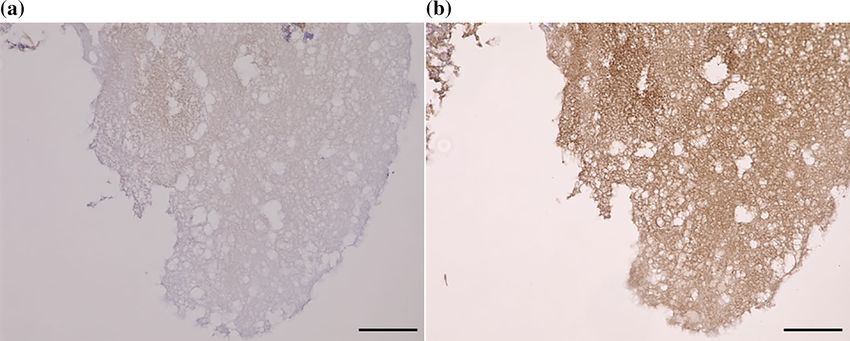
www.nature.com/scientificreports/
N Age HbA1c FBS BUN eGFR DBP SBP
DM( +) (years) (%) (mg/dL) (mg/dL) (mL/min/1.73m2) (mmHg) (mmHg)
DME 37 65.3 ± 12.6 7.8 ± 1.6 173.3 ± 65.9 20.0 ± 12.9 65.9 ± 32.2 141.6 ± 20.5 73.0 ± 10.8
NDR 7 75.0 ± 5.9* 6.5 ± 0.3 135.1 ± 36.9 19.9 ± 6.9 72.2 ± 16.3 139.9 ± 17.6 71.3 ± 13.1
PDR 6 67.5 ± 10.4 7.9 ± 1.9 182.3 ± 34.3 21.2 ± 9.1 52.5 ± 27.5 140.3 ± 29.9 73.0 ± 12.3
Total 50 68.2 ± 10.0 7.6 ± 1.6 168.5 ± 58.9 20.1 ± 11.6 65.1 ± 29.7 141.1 ± 21.0 72.8 ± 11.1
DM( −) 19 73.5 ± 7.5 - 107.1 ± 15.4* 14.9 ± 3.2 70.3 ± 12.5 138.4 ± 15.6 78.1 ± 8.3
P value 0.0003* 0.07 0.0001* 0.79 0.86 0.94 0.56
Table 1. Demographics of all patients. BUN: blood urea nitrogen, DBP: diastolic blood pressure, DM: diabetes
mellitus, DME: diabetic macular edema, eGFR: estimated glomerular filtration rate, FBS: fasting blood sugar,
HbA1c: hemoglobin A1c, NDR: no diabetic retinopathy, PDR: proliferative diabetic retinopathy, SBP: systolic
blood pressure. *: P < 0.05, Kruskal–Wallis test.
We have reported that PS is also protective in eyes of patients with DM11. Increased circulating free PS levels
in transgenic mice inhibit apoptosis of pancreatic β cells and ameliorates DM, and the systemic administration
of PS improves kidney fibrosis and renal dysfunction in diabetic m ice11. The high frequency of vascular events,
including deep vein thrombosis and pulmonary emboli in patients with PS deficiency highlights the clinical
relevance of the anticoagulant function of PS12.
The maintenance of the homeostasis of the retinal vessel is another important role of PS. PS deficiency in a
mouse model is associated with retinal changes resembling retinopathy of prematurity13. PS also regulates the
phagocytosis in the outer layers of the retina and contributes to maintaining a normal retinal architecture14.
However, the role of PS in diabetic retinal complications has not been examined.
Thus, it is important to assess the role played by PS in eyes with DR and DME. The purpose of this study is
to evaluate distribution of PS in diabetic eyes as a first step to accomplish this. We measured the level of PS in
the blood and aqueous humor. We also performed histological examination to determine the site of expression
of PS in a diabetic retina.
Results
Demographics of patients. The demographics of the patients are shown in Table 1. Among the eyes with
DM, 37 eyes had DME, 7 eyes had NDR, and 6 eyes had PDR. In the group with DME, 27 eyes had cystic DME,
9 eyes had sponge DME, and 1 eye had serus DME. The NDR group was significantly older than the other groups
(P = 0.0003, Kruskal–Wallis test). The group with no DM had significantly lower fasting blood glucose than the
other groups (P = 0.0001).
Plasma and aqueous humor levels of PS and related molecules in patients with and without
DM. The concentration of total PS in the plasma of patients with DM was 18.5 ± 5.9 μg/ml which was not
significantly different from that in patients without DM at 20.2 ± 4.5 μg/ml (P > 0.05). The concentration of PS
in the aqueous humor of patients with DM was 38.6 ± 16.3 ng/ml which was not significantly different from that
in patients without DM at 35.6 ± 12.2 ng/ml (P > 0.05; Fig. 1a,d). The plasma concentrations of free PS in patient
with DM was 3.0 ± 0.4 μg/ml which was significantly lower than that in patients without DM at 3.6 ± 0.9 μg/ml
(P = 0.01). The plasma concentration of C4BP was 4.3 ± 0.7 μg/ml in patients with DM which was significantly
lower than the 5.4 ± 1.4 μg/ml in patients without DM (P = 0.01 Fig. 1b,c). The concentration of free PS in the
aqueous humor was 7.9 ± 1.2 ng/ml in patients with DM which was significantly higher than that in non-DM
patients at 6.1 ± 0.7 ng/ml (P = 6.20 × 10⁻⁹; Fig. 1e). There was no significant difference in the concentration of
C4BP in the aqueous humor between the DM group (17.1 ± 8.2 ng/ml) and non-DM group (15.8 ± 11.4 ng/ml;
Fig. 1f).
Plasma and aqueous humor levels of PS and related molecules in DME, non‑DR, and PDR
groups. There was no significant difference in the plasma concentrations of total PS (18.6 ± 6.5 μg/ml vs
17.1 ± 3.2 μg/ml vs 19.9 ± 4.1 μg/ml), free PS (3.1 ± 0.4 μg/ml vs 2.8 ± 0.2 μg/ml vs 2.8 ± 0.4 μg/ml). and C4BP
(4.2 ± 0.4 μg/ml vs 5.0 ± 0.9 μg/ml vs 4.5 ± 1.3 μg/ml) among the DME, NDR and PDR groups (Fig. 2a–c).
There was also no significant difference in the aqueous humor concentrations of total PS (36.1 ± 11.2 ng/ml vs
48.3 ± 33.8 ng/ml vs 42.8 ± 10.0 ng/ml), and C4BP (14.8 ± 4.4 ng/ml vs 22.5 ± 14.7 ng/ml vs 24.1 ± 10.3 ng/ml)
among the three groups (Fig. 2d,f). The concentration of free PS in the aqueous humor was 8.2 ± 1.2 ng/ml in the
DME group which was significantly higher than the 7.0 ± 0.7 ng/ml in the NDR group and the 7.0 ± 1.0 ng/ml in
the PDR group (P = 6.78 × 10⁻⁷; Fig. 2e).
Severity of DME and ocular PS concentration. Because there was a high concentration of free PS in
the aqueous humor in the DME group, we divided the eyes with DME into two groups: a group with severe DME
(CRT ≥ 350 μm, n = 25) and a group with mild DME (CRT < 350 μm, n = 12). The demographics of the patients
are shown in Table 2. The values of the different parameters were not significantly different between the two
groups other than CMT. The aqueous humor level of free PS was significantly higher in the group with severe
DME than the group with mild DME (8.5 ± 1.3 ng/ml vs 7.7 ± 1.0 ng/ml, P = 0.04, Fig. 3a). The aqueous humor
Scientific Reports | (2021) 11:10449 | https://doi.org/10.1038/s41598-021-89870-5 2
Vol:.(1234567890)www.nature.com/scientificreports/
Figure 1. Plasma and aqueous humor concentrations of Protein S and related molecules in patients with or
without diabetes mellitus (DM). The plasma concentration of total PS is not significantly different in the DM
patients and non-DM patients (a). Plasma levels of free PS and C4BP are significantly lower in the DM patients
than in the non-DM patients (b, c). The total PS and C4BP levels are not significantly different among the
different groups (d, f). The free PS level in the aqueous humor is significantly higher in the DM patients than in
the non-DM patients (e). C4BP, C4b-binding protein; DM, diabetes mellitus; PS, protein S. *P < 0.05, **P < 0.01.
Statistical analyses by Mann–Whitney U test.
concentrations of total PS (37.6 ± 12.6 ng/ml vs 32.7 ± 6.6 ng/ml) and C4BP (14.6 ± 4.6 ng/ml vs15.4 ± 4.0 ng/ml)
were not significantly different between patients with severe and mild DME (Fig. 3b,c). The plasma concentra-
tions of free PS (3.2 ± 0.4 μg/ml vs 3.0 ± 0.3 μg/ml), total PS (19.5 ± 7.1 μg/ml vs 16.5 ± 4.8 μg/ml), and C4BP
(4.2 ± 0.5 μg/ml vs 4.1 ± 0.3 μg/ml) were not significantly different (Fig. 3d–f) between the two groups.
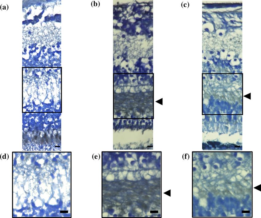
Localization of PS in human retina. We stained the retina of post-mortem human eyes to determine
the expression sites of PS in eyes with DM. The retina of the eyes from a non-DM patient (62-years-old, woman;
Fig. 4a,d) did not have any localized staining for PS. However, PS staining was positive in the outer plexiform
layer (OPL) of the diabetic eyes of a 93-year-old man with NPDR (Fig. 4b,e) and a 60 years-old man with PDR
(Fig. 4c,f).
PS location in cystoid lesion in eyes with DME. We collected specimens of the cystoid lesions for PS
staining in eyes with DME during pars plana vitrectomy. The specimens stained positive for PS (Fig. 5a,b).
Discussion
Macular edema is the accumulation of extravascular fluid in the OPL, the inner nuclear layer, and the Müller cells
leading to localized expansion15. Cystoid macular edema has a perifoveal configuration with cyst-like spaces due
to fluid accumulation16,17. The presence of cystic changes in eyes with DME in the OPL is common by pathologi-
cal examinations18. Byeon et al. also reported cysts within the outer plexiform and OPL that may be related to
leaky vessels when examined by OCT19. The cysts frequently progress and become hard exudates in the outer
plexiform and outer nuclear layers. Deposits of the hard exudates causes irreversible retinal damages leading
to loss of central visual function. Thus, the evidence suggests that the outer plexiform and outer nuclear layers
are major sites of tissue injury in DME. Our findings showed an increase of free PS in DM eyes particularly in
severe DME, localized expression of PS in the OPL, and within cysts in cystic DME. These findings suggest the
close association of PS expression with the DME pathological conditions.
PS inhibits the coagulation system by increasing the activity of activated protein C (APC), an anticoagulant
protein20. PS can accelerate fibrinolysis by suppressing the activation of thrombin-activatable fibrinolysis inhibitor
or by supporting the APC-mediated inhibition of plasminogen activator inhibitor-121. Fibrinogen is a soluble
Scientific Reports | (2021) 11:10449 | https://doi.org/10.1038/s41598-021-89870-5 3
Vol.:(0123456789)www.nature.com/scientificreports/
Figure 2. Plasma and aqueous humor concentrations of protein S and related molecules in patients with
diabetic retinopathy (DR). The plasma levels of total PS (a), free PS (b), and C4BP (c) are not significantly
different among the different groups. The aqueous humor levels of total PS and C4BP are also not significantly
different (d, f). The aqueous humor level of free PS is significantly higher in the DME group than the other
groups (e). C4BP, C4b-binding protein; DM, diabetes mellitus; PS, protein S. *P < 0.05. Statistical analysis by
Kruskal–Wallis test.
Age CMT HbA1c FBS BUN eGFR DBP SBP
(mL/
N (years) (μm) (%) (mg/dL) (mg/dL) min/1.73m2) (mmHg) (mmHg)
High
25 64.1 ± 12.2 473.3 ± 83.6* 7.9 ± 1.8 181.0 ± 35.9 20.7 ± 15.1 69.6 ± 32.5 142.2 ± 21.5 73.7 ± 11.3
(≧350 μm)
Low
12 68.3 ± 13.8 309.6 ± 42.1 7.7 ± 1.2 157.2 ± 66.5 18.2 ± 5.2 57.8 ± 31.6 140.1 ± 18.8 71.6 ± 10.1
(< 350 μm)
P value 0.40 5.80 × 10−5* 0.82 0.36 0.62 0.35 0.78 0.62
Table 2. Demographics of the DME patients. BUN: blood urea nitrogen, CMT: central subfoveal macular
thickness, DBP: diastolic blood pressure, eGFR: estimated glomerular filtration rate, FBS: fasting blood sugar,
HbA1c: hemoglobin A1c, SBP: systolic blood pressure.
macro-molecule that forms an insoluble clot or gel after conversion to fi brin22. The C4BP-PS complex may accel-
erate fibrin degradation during inflammation by interacting with p lasminogen23. This increased interaction of
C4BP and PS with plasminogen accelerates the plasminogen activator-mediated conversion of plasminogen to
plasmin on the fibrin surface which leads to increased fibrinolysis. Mass spectrometry analysis has shown that the
components of the cystoid lesion in eyes with DME is composed of microfibrin wrapped in collagen fibrils and
fibrinogen24. Based on these observations, we suggest that the PS present in the OPL and inside the cystoid lesion
activates the fibrinolysis pathway to reduce the deposition of fibrin. This effect may also prevent hard exudates
deposition that causes irreversible retinal damage. The results of earlier studies have shown that PS protects the
blood–brain barrier from hypoxic/ischemic damage through its Tyro3 receptor, and mice deficient in PS develop
embryonic lethal coagulopathy with disruption of the blood–brain barrier25,26. Based on these observations, we
can speculate that PS protects against blood-retinal barrier disruption which contributes significantly to protein
leakage with DME and it may explain our observation, significant higher the aqueous humor levels of free PS
level in the DME group compared to the NPDR and PDR groups. But because small number of samples were
evaluate PS levels in PDR and NPDR for ELISA which was not balanced, this may affect the results. In addition,
Scientific Reports | (2021) 11:10449 | https://doi.org/10.1038/s41598-021-89870-5 4
Vol:.(1234567890)www.nature.com/scientificreports/
Figure 3. Degree of DME affects ocular protein S concentration. The DME eyes were divided into two groups;
severe DME group (CMT ≥ 350 μm, n = 25) and mild DME group (CMT < 350 μm, n = 12). The aqueous humor
free PS level is significantly higher in the severe DME group than in the mild DME group (a). The aqueous
humor levels of total PS (b) and C4BP (c) are not significantly different between the groups. The plasma levels
of free PS (d), total PS (e), and C4BP (f) were not significantly different between the groups. C4BP, C4b-binding
protein; CMT, central subfoveal macular thickness; DM, diabetes mellitus; DME, diabetic macular edema; PS,
protein S. *P < 0.05. Statistical analysis by Mann–Whitney U test.
we could obtain a few number of post-mortem eye with DM because of the difficulty to obtain post-mortem eye
for research in Japan. We need further investigation with many number of tissue samples.
An interesting observation was the discrepant levels of free PS in plasma and aqueous humor. While there
was decreased levels of free PS in plasma, the free PS level was increased in the aqueous humor. Synthesis and
release of PS by cells in the eyes may explain these findings. However, although vascular endothelial cells can
secrete PS, there is no evidence showing the expression of PS by retinal capillary endothelial cells. The biological
significance of the elevated level of PS in the eyes with DM is also unclear. Although the local increase of PS in
the eyes may be a compensatory response to an enhanced retinal injury, a detrimental role of PS in disease patho-
genesis also deserves consideration. Such dissociation of the molecule between local and systemic distribution
is sometimes observed for other molecules. Though many cytokines are expressed in skeletal muscle following
exercise, some of them are not released into the circulation at least in large amounts because they are produced
locally and sufficient quantities of cytokines to increase their concentration in the systemic circulation may not
be secreted27. But because our histological examination couldnot certify the origin of the PS expression, it is not
cleaer whether similar spiculation can explain dissociation of local and systemic PS distribution. We need much
consideration for this matter in the further study.
On the contraly, there were several reports about unfavorable effects of PS. PS exacerbates acute liver injury
by prolonging the natural killer T cells survival and worsens liver fibrosis by inhibiting apoptosis of extracel-
lular matrix-producing fibroblasts28,29. The expression of PS may also be disease stage-related. Zhong et al. have
reported a high glomerular level of PS in early stages and low glomerular PS level in late stages of diabetic
nephropathy despite the normal circulating level of P S30. There is a possibility that PS distribution may differ
among the stage of disease. However, the occurrence of DME is not related to the clinical stage of DR5. Based
on our present findings, we suggest that PS expression is not related to the stage of DR and that PS expression
only increases in DME. Therefore, detecting PS expression may be a biomarker for the early diagnosis of DME
before the abnormalities are detected by OCT or ophthalmoscopy. However, further investigations are needed
to corroborate these findings.
Though we reported importance of PS in diabetic retina, there are some limitations in this study including
the small number of samples. First, the inclusion of a larger number of eyes with cyst-type DME than sponge- or
serus-type DME are limitations of the present study. There is a possibility that PS behavior differ with other type
of DME like sponge or serus type. The relationship between the retinal morphologic changes and concentrations
of intravitreal cytokines in eyes with DME was reported and the significant association of serus type DME with
Scientific Reports | (2021) 11:10449 | https://doi.org/10.1038/s41598-021-89870-5 5
Vol.:(0123456789)www.nature.com/scientificreports/
Figure 4. Location of protein S in the human retina with and without diabetes. The retina of post-mortem
human eyes was stained with a PS antibody. No staining was observed in the retina from patients without DM
(62-years-old, woman, (a). PS is present in the OPL of patients with DM (b, 93-year-old, man with NPDR;
60-year-old, man with PDR). Black arrow heads shows localization of PS on OPL. Magnified images of each
images were shown (d–f). DM, diabetes mellitus; NPDR, non-proliferative diabetic retinopathy; OPL, outer
plexiform layer; PDR, proliferative diabetic retinopathy; PS, protein S. Bar: 10 μm.
Figure 5. Location of Protein S in the components of a cystoid lesion with diabetic macular edema. The
component of cystoid lesion with DME was stained with anti-hPS antibody. No staining was observed in the
control nonspecific IgG (a). PS was identified inside the component of cystoid lesion (b). DME, diabetic macular
edema; PS, protein-S. Bar: 100 μm.
Scientific Reports | (2021) 11:10449 | https://doi.org/10.1038/s41598-021-89870-5 6
Vol:.(1234567890)www.nature.com/scientificreports/
intravitreal interleukin-6 level was observed and which indicated that inflammation may play an important role
in the development of serus type D ME31. Because there is a possibility PS may only affect the formation of cystic
DME, we also need advanced examination for various DME type.
Second, the circadian rhythm is important for in the progression of arteriosclerosis and t hrombosis32. Undar
et al.33. reported that plasma PS levels were also affected by circadian rhythm, significantly the highest at 6 a.m.
and the lowest at noon. We did not take into account physiological chage of PS due to circadian variation. But
they reported that no significant differences were observed at other hours and intervals. Though we collectted
samples at daily time (from 9 a.m to 5 p.m) when PS levels were not reported to be affected33, we should take
into account the effects of circadian rhythm.
And finally, Here we did not evaluate vitreous level of PS for DR and DME. There is a possibility that PS levels
in vitreous fluid reflects PS levels in retina more sensitive. But some reports concern that there is a significant
relationship between VEGF and interleukin-6 levels in aqueous humor and in vitreous fluid34. Though it is not
clear whether this is also applied for PS, we need consideration for this in the further study.
Our results showed the presence and increased expression of PS in eyes with DR and DME. The expression
of PS was especially high in the OPL and cystoid lesions. These observations suggest that PS is involved in the
pathogenesis of diabetic retinal complications, and it can be used as a biomarker and therapy for DME.
Methods
Subjects. The Mie University Ethics Committee for Clinical Investigations approved the investigation proto-
col (Approval #3087) and Kobe University Ethics Committee for Clinical Investigations approved the investiga-
tion protocol (Approval #B200233). The study was registered at http://www.umin.ac.jp (UMIN ID 000033728).
The procedures conformed to the tenets of the Declaration of Helsinki, and all patients signed an informed
consent form before entry. The patients enrolled were patients in the Department of Ophthalmology and Endo-
crinology, Mie University Hospital and Kobe University. The clinical history of all patients was obtained from
their medical records.
Sample collection. Blood serum samples were collected during regular checkups before receiving any oph-
thalmic treatments. The samples were centrifuged 400 × g for 5 min, the supernatants removed, and immediately
stored at − 80 °C until use. In addition, 50 μl of aqueous humor was collected before the ocular treatments. The
samples were frozen immediately and stored at − 80 °C until use. We did not collect any vitreous fluid because
sampling can cause complications such as retinal detachments.
Inclusion and exclusion criteria. Patients that were ≥ 20-years with type 1 or type 2 DM and those with
DR or DME were included in the DM group. The diagnosis of DME was based on findings of the fundus exami-
nations and spectral-domain optical coherence tomography (OCT). The stage of diabetic nephropathy before
treatment was obtained from the medical charts. Trained retinal specialists (M.S. and M.K.) examined the fun-
dus by indirect ophthalmoscopy and wide field fundus imaging obtained by the Optos ultra-widefield imaging
system (Optos Panoramic 200MA™, Optos PLC. Dunfermline, Scotland, UK). The severity of DR was classified
into three groups from these evaluations: no DR (NDR), non-proliferative DR (NPDR), and proliferative DR
(PDR) according to the International Clinical Diabetic Retinopathy Disease Severity Scale (DRSS)35. DME was
defined as a central subfoveal macular thickness (CMT) of ≥ 250 μm measured as the mean retinal thickness in
the central 1 mm diameter circle in the OCT images. Subjects with NDR or NPDR had no DME.
The exclusion criteria for patients with DM were; history of any pars plana vitrectomy, history of intravitreal
or sub-tenon injections of any drugs including anti-VEGF agents within two months before the beginning
of this study, eyes with any inflammatory disease, drusen, vitreous hemorrhage or retinal hemorrhage which
involved the intra- or subfoveal spaces, glaucoma or intraocular pressure ≥ 21 mmHg, and media opacities that
significantly affected the OCT images. Women with pregnancy, under fertility treatments or use of contraceptives
were also excluded from all the groups.
We also collected aqueous humor from patients without DM during surgical procedures for cataract, macular
hole, and retinal detachment. The inclusion criteria for patients without DM were; patients ≥ 20-years with no
DM and IOP of ≤ 21 mmHg. The exclusion criteria for patients without DM were; patients with severe systemic
disorders, any history of other ocular diseases, history of pars plana vitrectomy, or glaucoma. DME eyes were
also divided into two groups based on CMT measurement: a group with severe DME (higher CMT more than
350 μm) and a group with mild DME (lower CMT less than 350 μm).
The exclusion criteria for both DM patients and non-DM control subjects were; uncontrolled systemic medical
conditions, history of a thromboembolic event or ischemic disease including myocardial infarction or cerebral
infarction, prior treatment with anticoagulants, i.e., aspirin or with systemic anti-VEGF agents, i.e., bevacizumab,
diagnosis of diseases causing hypercoagulability, high refractive errors (spherical equivalent > − 3 or + 3 diopters,
axial lengths longer than 24.0 mm or less than 22.0 mm, and amblyopia.
Hematological analyses were done before the treatment to measure the level of fasting blood sugar (FBS,
normal value 70–110 mg/dL), hemoglobin A1c (HbA1c, NGSP, normal value 4.9–6.0%), blood urea nitrogen
(BUN, normal value 8–20 mg/dL), and estimated glomerular filtration rate (eGFR, normal value 60–120 ml/
min/1.73 m2). The blood pressure was also measured before beginning the treatment.
Immunoassays. To determine the level of free human PS (hPS), a 96-well microplate was coated with C4BP
(ATGen Corp., Sampyeong-dong, Korea), and after appropriate washing and blocking, samples were incubated
to allow free hPS to bind C4BP. Then, biotin-labeled polyclonal rabbit anti-hPS antibody (Dako Cytomation,
Scientific Reports | (2021) 11:10449 | https://doi.org/10.1038/s41598-021-89870-5 7
Vol.:(0123456789)www.nature.com/scientificreports/
Glostrup, Denmark) was added. Complement C4BP was measured using an enzyme immunoassay kit from
Assaypro (St. Charles, MO) and the total hPS as described28.
Immunohistochemical examination of human eyes. Post-mortem human eyes were obtained from
the National Disease Research Interchange (Philadelphia, PA) fixed in formalin and embedded in paraffin. The
postmortem time ranged from 14 to 24 h. The slides were deparaffinized in xylene and rehydrated through an
alcohol series for staining. Endogenous peroxidase was blocked by immersion in 0.3% hydrogen peroxidase
for 30 min. Slides were incubated with goat polyclonal anti-hPS antibody (A0384, DAKO, Glostrup, Denmark)
followed by treatment with horseradish peroxidase. Samples were examined with a fluorescence microscope
(BZ-9000: Keyence, Osaka, Japan).
En bloc removal of the component of cystoid lesion. The components of cystoid lesions in eyes with
DME were collected during pars plana vitrectomy. The surgical procedure for en bloc extraction of cystoid lesion
component combined with pars plana vitrectomy in DME patients was described in d etail24. Briefly, after inci-
sion of the external wall of the subfoveal cystoids with a 27-gauge instruments, the exposed components of the
cystoid lesions were grasped by forceps and excised en bloc. After removal, the cystoid lesion components were
stored at − 80 °C and embedded in paraffin at the time of examination.
Statistical analyses. Data are presented as the means ± standard deviations. The significance of the differ-
ences between two variables was determined by Mann–Whitney U-tests and between three or more variables by
the Kruskal–Wallis test with the Scheffe test. A P < 0.05 was considered significant.
Received: 8 February 2021; Accepted: 4 May 2021
References
1. Wild, S. et al. Global prevalence of diabetes: estimates for the year 2000 and projections for 2030. Diabetes Care 27, 1047–1053
(2004).
2. American Diabetes Association. Classification and diagnosis of diabetes. Diabetes Care 38(Suppl), S8–S16 (2015).
3. Klein, R., Klein, B. E. & Moss, S. E. Visual impairment in diabetes. Ophthalmology 91, 1–9 (1984).
4. Finkelstein, D. Ischemic macular edema. Recognition and favorable natural history in branch vein occlusion. Arch. Ophthalmol.
110, 1427–1434 (1992).
5. Yau, J. W. et al. Global prevalence and major risk factors of diabetic retinopathy. Diabetes Care 35, 556–564 (2012).
6. Ahnstrom, J. et al. Activated protein C cofactor function of protein S: a novel role for a gamma-carboxyglutamic acid residue.
Blood 117, 6685–6693 (2011).
7. Hafizi, S. & Dahlback, B. Gas6 and protein S. Vitamin K-dependent ligands for the Axl receptor tyrosine kinase subfamily. FEBS
J. 273, 5231–5244 (2006).
8. Linger, R. M., Keating, A. K., Earp, H. S. & Graham, D. K. TAM receptor tyrosine kinases: biologic functions, signaling, and
potential therapeutic targeting in human cancer. Adv. Cancer Res. 100, 35–83 (2008).
9. Bouwens, E. A., Stavenuiter, F. & Mosnier, L. O. Mechanisms of anticoagulant and cytoprotective actions of the protein C pathway.
J. Thromb. Haemost. 11(Suppl 1), 242–253 (2013).
10. Rezende, S. M., Simmonds, R. E. & Lane, D. A. Coagulation, inflammation, and apoptosis: different roles for protein S and the
protein S-C4b binding protein complex. Blood 103, 1192–1201 (2004).
11. Yasuma, T. et al. Amelioration of diabetes by protein S. Diabetes 65, 1940–1951 (2016).
12. Comp, P. C., Nixon, R. R., Cooper, M. R. & Esmon, C. T. Familial protein S deficiency is associated with recurrent thrombosis. J.
Clin. Investig. 74, 2082–2088 (1984).
13. Burstyn-Cohen, T., Heeb, M. J. & Lemke, G. Lack of protein S in mice causes embryonic lethal coagulopathy and vascular dysgen-
esis. J. Clin. Investig. 119, 2942–2953 (2009).
14. Hall, M. O. et al. Both protein S and Gas6 stimulate outer segment phagocytosis by cultured rat retinal pigment epithelial cells.
Exp. Eye Res. 81, 581–591 (2005).
15. Scholl, S., Kirchhof, J. & Augustin, A. J. Pathophysiology of macular edema. Ophthalmologica 224(Suppl 1), 8–15 (2010).
16. Wolter, J. R. The histopathology of cystoid macular edema. Albrecht Von Graefes Arch. Klin. Exp. Ophthalmol. 216, 85–101 (1981).
17. Rotsos, T. G. & Moschos, M. M. Cystoid macular edema. Clin. Ophthalmol. 2, 919–930 (2008).
18. Tso, M. O. Pathology of cystoid macular edema. Ophthalmology 89, 902–915 (1982).
19. Byeon, S. H. et al. New insights into the pathoanatomy of diabetic macular edema: angiographic patterns and optical coherence
tomography. Retina 32, 1087–1099 (2012).
20. Dahlback, B. Vitamin K-Dependent Protein S: Beyond the Protein C Pathway. Semin. Thromb. Hemost. 44, 176–184 (2018).
21. Mosnier, L. O., Meijers, J. C. & Bouma, B. N. The role of protein S in the activation of thrombin activatable fibrinolysis inhibitor
(TAFI) and regulation of fibrinolysis. Thromb. Haemost. 86, 1040–1046 (2001).
22. Weisel, J. W. & Litvinov, R. I. Fibrin formation, structure and properties. Subcell Biochem. 82, 405–456 (2017).
23. Agarwal, V., Talens, S., Grandits, A. M. & Blom, A. M. A novel interaction between complement inhibitor C4b-binding protein
and plasminogen that enhances plasminogen activation. J. Biol. Chem. 290, 18333–18342 (2015).
24. Imai, H. et al. Effectiveness of en bloc removal of fibrinogen-rich component of cystoid lesion for the treatment of cystoid macular
edema. Retina 40, 154–159 (2020).
25. Saller, F. et al. Generation and phenotypic analysis of protein S-deficient mice. Blood 114, 2307–2314 (2009).
26. Zhu, D. et al. Protein S controls hypoxic/ischemic blood-brain barrier disruption through the TAM receptor Tyro3 and sphingosine
1-phosphate receptor. Blood 115, 4963–4972 (2010).
27. Peake, J. M., Gatta, P. G., Suzuki, K. & Nieman, D. C. Cytokine expression and secretion by skeletal muscle cells: regulatory mecha-
nisms and exercise effects. Exerc Immunol Rev. 21, 8–25 (2015).
28. Chelakkot-Govindalayathil, A. L. et al. Protein S exacerbates alcoholic hepatitis by stimulating liver natural killer T cells. J. Thromb.
Haemost. 2015(13), 142–154 (2015).
29. Totoki, T. et al. Protein S exacerbates chronic liver injury and fibrosis. Am. J. Pathol. 188, 1195–1203 (2018).
30. Zhong, F. et al. Protein S protects against podocyte injury in diabetic nephropathy. J. Am. Soc. Nephrol. 29, 1397–1410 (2018).
31. Sonoda, S. et al. Retinal morphologic changes and concentrations of cytokines in eyes with diabetic macular edema. Retina 34,
741–748 (2014).
Scientific Reports | (2021) 11:10449 | https://doi.org/10.1038/s41598-021-89870-5 8
Vol:.(1234567890)www.nature.com/scientificreports/
32. Man, A. W. C., Li, H. & Xia, N. Circadian rhythm: potential therapeutic target for atherosclerosis and thrombosis. Int. J. Mol. Sci.
22, 676 (2021).
33. Undar, L., Ertuğrul, C., Altunbaş, H. & Akça, S. Circadian variations in natural coagulation inhibitors protein C, protein S and
antithrombin in healthy men: a possible association with interleukin-6. Thromb. Haemost. 81, 571–575 (1999).
34. Funatsu, H. et al. Aqueous humor levels of cytokines are related to vitreous levels and progression of diabetic retinopathy in diabetic
patients. Graefes Arch. Clin. Exp. Ophthalmol. 243, 3–8 (2005).
35. Wilkinson, C. P. et al. Proposed international clinical diabetic retinopathy and diabetic macular edema disease severity scales.
Ophthalmology 110, 1677–1682 (2003).
Acknowledgements
We thank Professor Emeritus Duco Hamasaki of the Bascom Palmer Eye Institute of the University of Miami
for critical discussion and final manuscript revisions.
Author contributions
M.S., M.K., and E.C.G. conceived and coordinated the study, evaluated the patients, and wrote the paper. T.Y,
C.N.D.G., and M.T. performed the experiments. M.S. take histological images. H.I. and M.N. performed surgery
for the component of the cystoid lesion. All authors reviewed the results and approved the final version of the
manuscript.
Funding
This research was supported in part by a Kakenhi Grant-in-Aid from the Ministry of Education, Culture, Sports,
Science, and Technology of Japan (No. 18K09402) and Okasan-Kato Foundation 2017. The funders had no role
in study design, data analysis, decision to publish, or preparation of the manuscript.
Competing interests
Masahiko Sugimoto have: [F] Alcon Pharma (class III), Bayer (class III). [I/E/C/P] None. [R] Alcon pharma
(class II), Kowa Pharma (class II), Senjyu Pharma (class II), Daiichi Yakuhin Sangyo (class II), Bayer (class II),
Wakamoto Pharma (class II).
Additional information
Correspondence and requests for materials should be addressed to M.S.
Reprints and permissions information is available at www.nature.com/reprints.
Publisher’s note Springer Nature remains neutral with regard to jurisdictional claims in published maps and
institutional affiliations.
Open Access This article is licensed under a Creative Commons Attribution 4.0 International
License, which permits use, sharing, adaptation, distribution and reproduction in any medium or
format, as long as you give appropriate credit to the original author(s) and the source, provide a link to the
Creative Commons licence, and indicate if changes were made. The images or other third party material in this
article are included in the article’s Creative Commons licence, unless indicated otherwise in a credit line to the
material. If material is not included in the article’s Creative Commons licence and your intended use is not
permitted by statutory regulation or exceeds the permitted use, you will need to obtain permission directly from
the copyright holder. To view a copy of this licence, visit http://creativecommons.org/licenses/by/4.0/.
© The Author(s) 2021
Scientific Reports | (2021) 11:10449 | https://doi.org/10.1038/s41598-021-89870-5 9
Vol.:(0123456789)You can also read