Cytomorphological Assessment and Thyroid Function Analysis - A Dual Approach to Diagnose Thyroid Lesions
←
→
Page content transcription
If your browser does not render page correctly, please read the page content below
Original Article                                                                                             DOI: 10.7860/NJLM/2016/20542:2128

     Cytomorphological Assessment

                                                                                                                                                 Pathology Section
     and Thyroid Function Analysis –
     A Dual Approach to Diagnose
     Thyroid Lesions
     M. S. Siddegowda, Jasneet Kaur Sandhu, S. Shivakumar

     ABSTRACT                                                               Results: A total of 350 patients underwent cytomorphological
     Introduction: Thyroid lesions are a commonly encountered               assessment using FNAC, of which most were females with
     clinical problem in all age groups. Accurate preoperative              most patients being in the age group of 21 to 40 years. Non-
     assessment guides the clinical management and improves the             neoplastic lesions were more common, with colloid goitre being
     patient outcome.                                                       the most common diagnosis. Among the neoplastic lesions,
                                                                            follicular adenoma was the most common diagnosis. Thyroid
     Aim: To describe the cytomorphological features of palpable
                                                                            hormone profile was done in 327 patients. Same thyroid
     thyroid nodules using fine needle aspiration cytology (FNAC)
                                                                            disease showed different thyroid status in different patients.
     along with the assessment of thyroid hormonal status of the
                                                                            Cyto-histopathological correlation was done in 28 patients.
     patient. The study also tried to assess the sensitivity, specificity
                                                                            The sensitivity of FNAC in the present study was 87.5%,
     and diagnostic accuracy of FNAC as an initial diagnostic
                                                                            specificity was 91.7%, positive predictive value (PPV) was 93%
     modality for thyroid lesions.
                                                                            and negative predictive value (NPV) was 85%. The diagnostic
     Materials and Methods: The study was conducted in the                  accuracy of FNAC in diagnosing thyroid lesions was 89.3%.
     Department of Pathology of a teaching hospital during July
                                                                            Conclusion: FNAC is a sensitive and specific technique for
     2013 to December 2014 and included 350 patients with thyroid
                                                                            the diagnosis of thyroid lesions. FNAC in conjunction with
     lesions. Cytological assessment was done using FNAC along
                                                                            hormonal analysis helps in proper patient assessment and
     with serological assessment of thyroid hormones and cyto-
                                                                            management.
     histopathological correlation.

                                    Keywords: Cyto-histopathological correlation, Hashimoto’s thyroiditis, Nodular colloid goitre, TSH

     Introduction                                                           hormones, thyroxin (T4) and 3,5,3’-triiodothyronine (T3)
     The prevalence of thyroid swelling ranges from 4% to 7%                through a series of metabolic steps. In circulation, most
     in the general adult population and from 0.2% to 1.8% in               (70%) of the T4 circulates bound to thyroid binding globulin
     children. The clinical importance of thyroid nodule diagnosis          (TBG) followed by transthyretin (20%) and albumin (10%).
     rests with the need to exclude thyroid cancer, which occurs            While most of the circulating T3 is bound to TBG, it does so
     in 7%-15% of the cases depending on age, sex, radiation                with a tenfold-reduced affinity as compared with that of T4.
     exposure, family history and miscellaneous other factors [1].          A small percentage of T4 and T3 remain unbound and are
     Pre-operative distinction of benign lesions is of paramount            metabolically active [1].
     importance to avoid unnecessary surgery.                               Thyroid diseases may be classified on the basis of clinical
     The normal thyroid gland is composed of follicles, which               features of the patient and the hormone profile as hyperthyroid,
     are the site for synthesis and storage of thyroid hormones.            hypothyroid and euthyroid [1]. However, in majority of the
     An intact hypothalamus-pituitary-thyroid axis and a steady             patients, symptoms are subtle in presentation, so the onus
     source of iodide are essential for the normal thyroid hormone          lies on biochemical testing and cytological evaluation to
     synthesis. The hypothalamus secretes thyrotropin-releasing             make a diagnosis [2]. The various thyroid diseases can have
     hormone (TRH), which in turn stimulates the thyrotrophs                either of the above status at different stages of the disease
     of anterior pituitary to secrete thyroid-stimulating hormone           development in the same patient.
     (TSH). Under TSH stimulation, iodine enters the follicular             In the past 5 to 6 decades, FNAC has been increasingly
     cells as inorganic iodide and is transformed into thyroid              utilized for the investigation of thyroid lesions. FNAC by

16                                                                              National Journal of Laboratory Medicine. 2016 Jul, Vol-5(3): PO16-PO21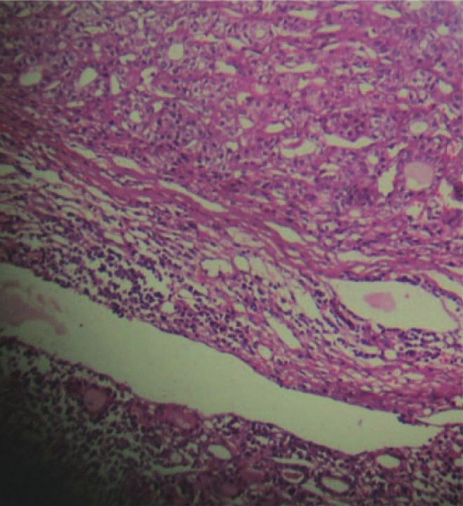
www.njlm.net                                                        M. S. Siddegowda et al., Cytomorphological Assessment and Thyroid Function Analysis

aspiration or non-aspiration technique is a simple outpatient                    • Accuracy = TP + TN / Total No. of Patients x 100
procedure performed on thyroid swellings, which has                              Descriptive statistics were assessed using SPSS 20.
the sensitivity of 93.4%, specificity of 74.9% and positive
predictive value of 98.6% [3].                                                   Results
The use of FNAC in conjunction with thyroid hormonal profile                     The study included 350 patients with palpable thyroid
helps in assessing the stage of the disease and deciding the                     lesions, of which, 334 (95.4%) were females and 16 (4.6%
treatment option for the patient. In this study, we aimed to                     were males) [Table/Fig-1]. The age of the patients in the
assess the cytological findings of palpable thyroid nodules in                   study ranged from 11years to 80 years. Most of the patients
conjunction with thyroid hormonal profile of the patient.                        belonged to the age group of 21years- 40 years.
                                                                                 FNAC was performed by aspiration and non-aspiration
materials and methods                                                            technique on all 350 patients with serological thyroid
A retrospective descriptive study was conducted on 350                           hormone estimation on 327 patients. The non-neoplastic
patients in the Pathology Department of a teaching hospital                      lesions were more common (333/350; 95.1%), with colloid
after obtaining clearance from institutional ethical committee.                  goitre (142/333; 42.6%) being the commonest diagnosis
The study included all the patients, irrespective of age and                     followed by Hashimoto’s thyroiditis (126/333; 37.8%).
gender who presented to the cytology section with a referral                     Among the neoplastic lesions (17/350), follicular neoplasms
for thyroid fine needle aspiration cytology during July 2013                     (13/17; 76.4%) were most common followed by papillary
to December 2014. The patients who presented for a repeat                        carcinoma [Table/Fig-2-7].
FNAC due to any cause were excluded from the study. Out
of 350 patients, histopathological confirmation was available                       Age group (in years)          Male         Female          Total
for 28 patients. At the time of presentation to the Department,                              0-20                  05             035           040
history was obtained and clinical examination of the thyroid
                                                                                            21-40                  05             191           196
lesion was performed.
                                                                                            41-60                  06             087           093
After consent of the patient (or the guardian/parent, in
children below 18 years of age, whichever applicable), using                                61-80                  00             021           021
aseptic precautions FNAC was performed by aspiration                                         Total                 16             334           350
and non-aspiration technique by 23-gauge needle with                              [Table/Fig-1]: Age and gender distribution of patients with palpable
10ml syringe. On aspiration of cystic swellings, the fluid                        thyroid lesions (n=350).
was centrifuged, following which sediment smears were
prepared. These smears were fixed in 95% alcohol (wet
fixation) for haematoxylin and eosin staining and air-dried for
May Graunwald Geimsa staining. The results of serological
estimation of T3, T4, and TSH using enzyme linked
fluorescent assay (ELFA) by VIDAS Biomeriuex, which was
already done on clinician’s directions, were collected from
the patient record at the time of FNAC.
Plan of Data Analysis: True positivity was considered when
the lesion was found to be malignant on both FNAC and
evaluation of post-surgical specimen. False positive were
those cases wherein cytology was reported as malignant
but on evaluation histopathologically the lesion turned out                       [Table/Fig-2]: Hormonal status in various thyroid lesions.
to be of benign nature. True negative (TN) were benign
on both cytology and histopathology. False negative (FN)
were negative on cytology but positive for malignancy on
histopathology. Sensitivity was the detection of disease
when it was actually present and was a measure of detection
of thyroid cancer by FNAC in our study. Similarly specificity
was defined by the ability of FNAC to exclude malignancy,
that is, diagnose benign lesions. Diagnostic accuracy was
calculated by using sensitivity and specificity. The statistical
formulae used were as follows [4]:
• Sensitivity = TP / (TP + FN) x 100
• Specificity = TN / (TN + FP) x 100                                              [Table/Fig-3]: Microphotograph showing abundant thick colloid
                                                                                  with the presence of follicular cell clusters (MAGG, x4).

National Journal of Laboratory Medicine. 2016 Jul, Vol-5(3): PO16-PO21                                                                                     17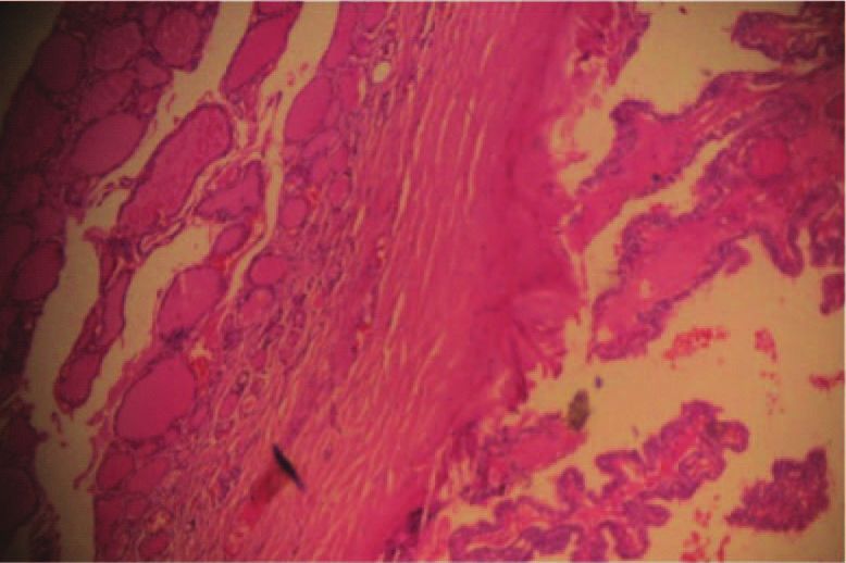
M. S. Siddegowda et al., Cytomorphological Assessment and Thyroid Function Analysis www.njlm.net
[Table/Fig-4]: Microphotograph showing polymorphous population of lymphoid cells with thyroid follicular cells (MGG, x10); Inset: Hurthle cells
in a background of lymphoid cells (H & E, x40). [Table/Fig-5]: Microphotograph showing monomorphic thyroid follicular cells in sheets, clusters
and microfollicular pattern with scant colloid (MGG, x10); Inset: Microfollicular pattern (MGG, x40). [Table/Fig-6]: Microphotograph showing
neoplastic thyroid follicular cells arranged in papillary structures (H & E, x10); Inset: Nuclei showing powdery chromatin, nuclear groove and
intranuclear inclusion (H & E, x40).
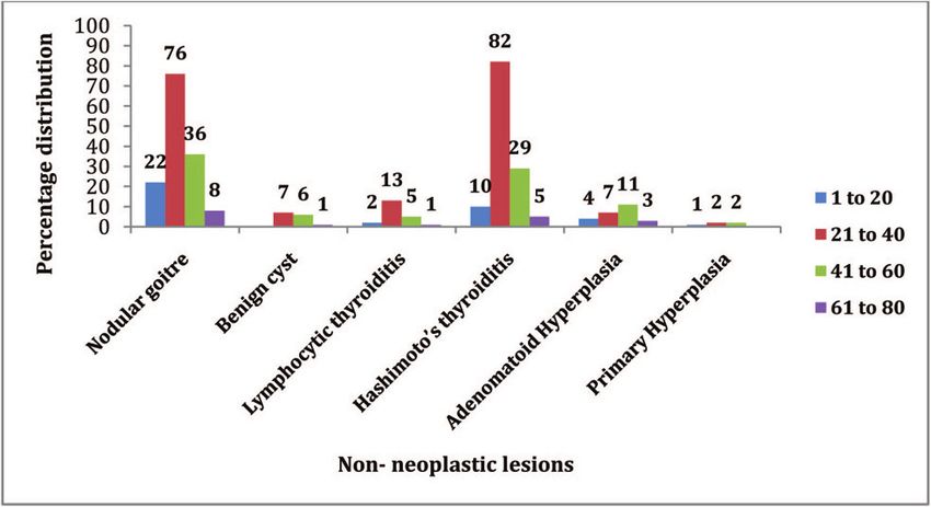
Non-neoplastic lesions Age group (in years)
(n=333)
1-20 21-40 41-60 61-80
Nodular goitre 22 76 36 08
Benign cyst 00 07 06 01
Lymphocytic thyroiditis 02 13 05 01
Hashimoto’s thyroiditis 10 82 29 05
Adenomatoid hyperplasia 04 07 11 03 [Table/Fig-8]: Microphotograph showing papillary carcinoma with
multinodular goiter (H & E, x4). [Table/Fig-9]: Microphotograph
Primary Hyperplasia 01 02 02 00
showing a papilla with fibrovascular core and lined by thyroid follicular
Neoplastic lesions Age group (in years) cells (H & E, x10); Inset: Nuclear features of papillary carcinoma-
(n=17) chromatin clearing with nuclear grooves (H & E, x100).
1-20 21-40 41-60 61-80
Follicular 01 09 02 01
Papillary 00 01 00 01
Anaplastic 00 00 01 01
[Table/Fig-7]: Distribution of FNAC finding of thyroid lesions
(n=350)
Thyroid hormone profile was done in 327 patients, of which
173 were euthyroid, 134 were hypothyroid and 20 were
hyperthyroid. Different thyroid status was observed in the
same disease process in different patients [Table/Fig-2].
In the present study, out of the 350 patients, surgical
intervention was done for 28 patients. In the surgical
specimens, neoplastic lesions (16/28; 57.1%) were more
common than non-neoplastic lesions (12/28; 42.9%).
Among the neoplastic lesions, 12 were of follicular type
(10-follicular adenoma, 02- follicular carcinoma), 02 were [Table/Fig-10]: Microphotograph showing encapsulated follicular
papillary carcinoma, 01 was papillary carcinoma with goitre adenoma (H & E, x10).
[Table/Fig-8,9]. and 01 was follicular adenoma with colloid
whereas 01 case of adenomatoid hyperplasia in multinodular
goitre [Table/Fig-10]. In the neoplastic lesions, cytology
goitre was diagnosed as follicular neoplasm on cytology
correlated positively in all cases of follicular type and 02
[Table/Fig-11].
cases of papillary carcinoma. One case of goitre with papillary
carcinoma was diagnosed as goitre on cytology and another Thus, the sensitivity of FNAC in the present study was
case of goitre with follicular adenoma was diagnosed as 87.5%, specificity was 91.7%, positive predictive value (PPV)
goitre with Adenomatoid hyperplasia on cytology. Cytology was 93% and negative predictive value (NPV) was 85%. The
correlated positively in 11 cases of non-neoplastic lesions diagnostic accuracy of FNAC in diagnosing thyroid lesions
(07-multinodular goitre, 04-goitre with cystic change), was 89.3% [Table/Fig-12].
18 National Journal of Laboratory Medicine. 2016 Jul, Vol-5(3): PO16-PO21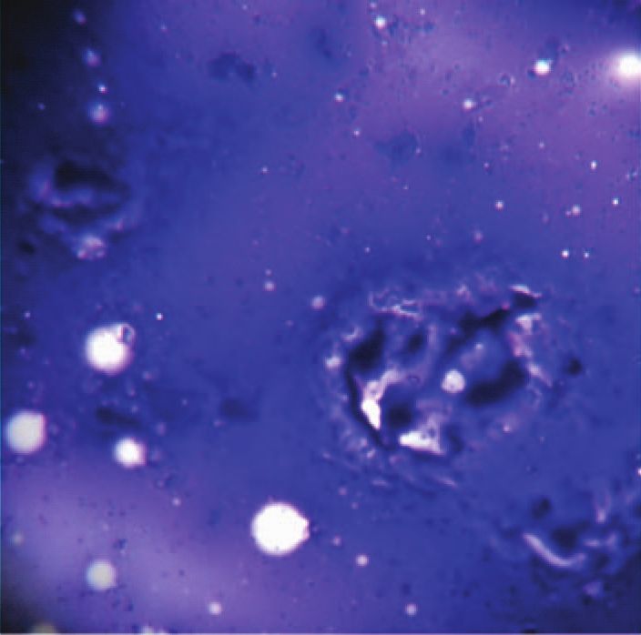
www.njlm.net                                                        M. S. Siddegowda et al., Cytomorphological Assessment and Thyroid Function Analysis

         FNAC                      Histology                     Total           goitre (114/142) had euthyroid status. This finding was
                                                                                 similar to a study done by Junu et al.,[13]. Enlargement of the
                        Neoplastic       Non-neoplastic
                                                                                 thyroid, or goiter is caused by impaired synthesis of thyroid
 Neoplastic                  14                 01                15             hormone, which is most often the result of dietary iodine
 Non-neoplastic              02                 11                13             deficiency leading to increase in TSH levels. Increased TSH
 Total                       16                 12                28             stimulates the thyroid follicular cells causing compensatory
                                                                                 hypertrophy and hyperplasia, which finally culminates in
[Table/Fig-11]: Cytohistological correlation (n=28).
                                                                                 gross enlargement of the gland. Anatomic increase in size
                                                                                 of thyroid leads to increased hormonal production and
Discussion                                                                       achievement of euthyroid status in most of the individuals
Thyroid lesions are a common problem encountered in                              [14].
clinical practise. In our study, during 18 months, 350 cases of
                                                                                 In patients with Hashimoto’s thyroiditis, most of the patients
palpable thyroid nodules underwent FNAC by aspiration and
                                                                                 were hypothyroid (96/126; 76.2%). Minnu Prasannan et
non-aspiration technique and 327 of them had serological
                                                                                 al.,[15] have reported similar findings. Usually Hashimoto’s
estimation of thyroid hormones. Of the 350 patients, most
                                                                                 thyroiditis presents as painless thyromegaly in middle aged
lesions were observed in females (95.4%), which correlate
                                                                                 woman. In most of the cases, it progresses over a period of
with similar studies done by Sheela et al., [5], CK Sang et
                                                                                 time to subclinical and then clinically overt hypothyroidism.
al.,[6], Gupta R et al., [7] and Rupam et al.,[8]. The age of
                                                                                 However, in some cases, it may be preceded by a transient
the patients in the study ranged from 11 years to 80 years.
                                                                                 hyperthyroid state (hashitoxicosis) due to destruction of
Most of the patients were in the age group of 21 to 40 years.
                                                                                 follicles and subsequent rise in the free T3 and T4 along
Sheela et al., [5], CK Sang et al.,[6] and Gupta R et al., [7]
                                                                                 with fall in TSH and diminished radioactive iodine uptake
have reported similar findings.
                                                                                 [14]. Cytologically in our study, oxyphil cells, abundant
Thyroid enlargement needs thorough investigation, mainly to                      polymorphous population of lymphocytes, scant or no colloid
rule out malignancy and thyroiditis. The recommendation of                       were observed. Some cases also showed the presence of
American Thyroid Association (ATC) (2015) state that for any                     multinucleated giant cells and epithelioid cells, similar to the
thyroid nodule > or equal to 2cm, ultrasound should be the                       findings of Gharib et al.,[16]. Hence concurrent FNAC and
initial investigation followed by FNAC [9]. However, in our                      estimation of thyroid hormonal status helps the clinician in
study, the initial investigation following clinical examination                  proper management of such patients.
and history taking was FNAC, which is the scenario in most
                                                                                 Among the non-tested individuals, neoplastic lesions were
of the government hospitals in India. Out of the 350 patients,
                                                                                 more than non-neoplastic lesions. It could be due to the
333 had non-neoplastic lesions and 17 had neoplastic
                                                                                 fact that neoplastic lesions are usually present in euthyroid
pathology, similar to studies done by Silverman JF et al.,[10],
                                                                                 individuals as asymptomatic nodules.
Uma H et al.,[11], Sengupta et al.,[12] and Gupta R et al.,[7].
Among the non-neoplastic lesions, colloid goitre was most                        In the present study, histopathological correlation was
commonly encountered followed by Hashimoto’s thyroiditis.                        available in 28 cases, of which 16 were neoplastic and 12
These findings are in concordance with studies done by                           were non-neoplastic lesions [Table/Fig-11]. In neoplastic
Sheela et al.,[5], CK Sang et al.,[6] and Gupta R et al.,[7].                    lesions, positive cyto-histological correlation was observed
Among the neoplastic lesions, follicular lesions were most                       in 14 cases. One case of goitre with papillary carcinoma
common (13/17; 76.4%) followed by papillary carcinoma,                           was diagnosed on cytology as goitre [Table/Fig-3]. Stromal
similar to studies of Sheela et al.,[5] and CK Sang et al.,[6].                  degenerative fragments can mimic bubble-gum type colloid
                                                                                 and large amounts of loose colloid may also be seen in
The cytological analysis was accompanied with serological
                                                                                 papillary carcinoma, causing confusion in the diagnosis
testing of thyroid hormones. According to the guidelines put
                                                                                 [17]. Another case of nodular colloid goiter with co-existent
forth by ATC, serum estimation of TSH should be part of
                                                                                 follicular adenoma was diagnosed as goiter with adenomatous
initial assessment of thyroid swellings. If overt or subclinical
                                                                                 hyperplasia in our study. Pavithra et al., observed similar
hyperthyroidism is present, additional evaluation is required.
                                                                                 findings in their study [18].This could be explained by the
A higher serum TSH level, even within the upper part of
                                                                                 presence of colloid rich areas in the goitrous nodule and
the reference range, is associated with increased risk of
                                                                                 could have been avoided by aspiration from multiple sites in
malignancy in a thyroid nodule, as well as more advanced
                                                                                 a setting of multinodular lesion. In difficult cases, presence
stage thyroid cancer [9].
                                                                                 of nuclear crowding and overlapping with monomorphic
Thyroid diseases can present with different thyroid function                     population of cells in predominantly microfollicular pattern
status, depending on the stage and extent of the disease                         and absence of colloid favours the diagnosis of malignancy
development. In our study of the 350 patients, serological                       [19].
testing was available in 327 patients and most of the patients
                                                                                 Among the non-neoplastic lesions, positive correlation
were euthyroid (173/327). While correlating the thyroid
                                                                                 was observed in 11 cases and one case of Adenomatous
disease with hormonal status most patients with colloid

National Journal of Laboratory Medicine. 2016 Jul, Vol-5(3): PO16-PO21                                                                                     19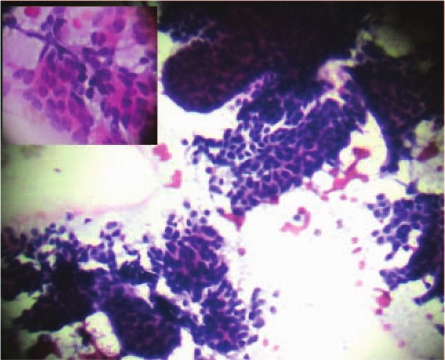
M. S. Siddegowda et al., Cytomorphological Assessment and Thyroid Function Analysis                                                       www.njlm.net

     hyperplasia in multinodular goitre was reported as follicular                a district hospital. Due to the rural setting, patients are not
     neoplasm on cytology. Hyperplastic nodules are characterized                 able to undergo all diagnostic tests owing to economical
     by the presence of variable cellular patterns varying from                   and social reasons. The loss to follow up (due to the same
     honeycomb or follicular arrangement to singly scattered                      reasons or) of patients referred from surrounding areas
     thyroid follicular cells. These cells display scant to abundant              as well as the fact that most of the lesions do not require
     (oncocytic change), mildly vacuolated cytoplasm and                          surgical intervention, also limits the number of cases
     small dark staining nuclei. Watery colloid with cytoplasmic                  wherein histopathological correlation is available. None
     thyroglobulin blebs may also be seen. In certain cases, the                  of our cases underwent a thyroid scan or antibody panel
     presence of features like nuclear grooves, chromatin clearing                analysis. However, in spite of the limited resources we had
     with overlapping of nuclei might create diagnostic dilemma                   a high satisfactory rate of smears and FNAC proved to be
     [17].                                                                        a sensitive and specific diagnostic modality. The ability of
     The most commonly described pitfall in diagnosing papillary                  FNAC to differentiate benign from malignant lesions, with
     carcinoma is the presence of cyst along with papillary                       the exception of follicular neoplasm, aids in deciding the
     carcinoma. However, our study has also highlighted the                       management algorithm with the biochemical tests helping
     presence of co-existent papillary carcinoma and nodular                      in making a decision on the use of combined medical and
     colloid goiter without cystic change causing difficulty in                   surgical modalities in individual cases.
     diagnosis on cytology alone. This is because of the occult
     papillary carcinoma, which was masked by the nodular                         Conclusion
     goiter (which formed the major bulk of the swelling). These                  FNAC is a simple, safe and cost effective diagnostic modality
     kinds of lesions require a high degree of suspicion and                      for thyroid lesions with high specificity and accuracy,
     careful examination of nuclear features in long standing                     especially in developing countries like India. Skillful sampling
     goiter cases. Chen and Rossi have also emphasized the                        of the lesion ensures a satisfactory cytological analysis and
     importance of nuclear features in diagnosing papillary                       in conjunction with thyroid hormone profile, it helps clinicians
     carcinomas as compared to the presence of characteristic                     determine the course of therapy in the management of
     architectural pattern [20].                                                  patients with thyroid nodules.
     In our study, we observed a high sensitivity and specificity
     of FNAC in diagnosing the thyroid lesions, similar to various                REFERENCES
                                                                                   [1] Helena A.Guber, Amal F.Farag, James Lo, James Sharp.
     other studies done previously. The diagnostic accuracy in our
                                                                                         McPherson & Pincus: Henry’s Clinical Diagnosis and
     study was 89.3%. The accuracy of FNAC is distinctly higher                          Management by Laboratory Methods. 21st edition. Philadelphia:
     in centres where both procedure and interpretation are done                         Saunders Elsevier; 2006. Evaluation of endocrine function.In:
     by trained cytopathologists. FNA cytology of thyroid has a                          Richard A.McPherson, Mathew R.Pincus, editors.
     high negative predictive value, which is useful to reassure the               [2]   Hegedus L. The thyroid nodule. New Engl J Med.
     majority of patients presenting with thyroid enlargement and                        2004;35(1):1764-71.
                                                                                   [3]   Gita Jayaram, Svante R.Orell. Orell & Sterrett’s Fine Needle
     can be used to plan definitive surgery. [Table/Fig-12]. Thyroid
                                                                                         Aspiration Cytology, 5th edition. China: Churchill Livingstone
     function tests and FNAC used in conjunction in selected                             Elsevier; 2012. Thyroid. In: Svante R. Orell, Gregory F. Sterrett.
     cases complement one another.                                                       p118-55.
                                                                                   [4]   Basic Epidemiology. 6th edition. Hong Kong: WHO; 2002.
          Study          Sensitivity Specificity   PPV    NPV Accuracy                   Epidemiology and prevention. In: Beaglehole R, Bonita R,
                            (%)         (%)        (%)    (%)   (%)                      Kjellstrom T, editors. p83-105.
     Piromalli et al.,       95          98         95     97        -             [5]   Chaudhari S, Hatwal D, Bhat P, Batra N, Bhat S.
     [21]                                                                                Cytological evaluation of thyroid lesions and its correlation
                                                                                         with histopathology: A prospective study. Int J Sci Stud.
     Sheela et al.,          90          96          -     -        94
                                                                                         2015;3(8):132-35.
     [5]
                                                                                   [6]   Sang CK, Kigondu CS, Muchiri L. Fine needle aspiration cytology
     Inamullah et            75          96         81     95       93                   of thyroid nodules at Kenyatta National Hospital, Nairobi. East
     al., [22]                                                                           Afr Med J. 2007;84(3):117-20.
     Rupam et              82.14        86.8       65.71 94.04     83.6            [7]   Gupta R, Bagde S, Ganguly S, Minj M, Tembhurnikar P.
     al.,[8]                                                                             Fine needle aspiration cytology of thyroid lesions-10 years
                                                                                         retrospective study at Chhattisgarh Institute of Medical Sciences.
     Lewis et                -            -          -     93        -                   J of Evidence Based Med & Hlthcare. 2015;2(41):6911-15.
     al.,[23]                                                                      [8]   Borgohain R, Lal RK, Chatterjee P, Brahma N, Khanna S. A
     Present study          87.5        91.7        93     85      89.3                  study of cyto-histological correlation in the diagnosis of thyroid
                                                                                         swelling. IOSR-JDMS. 2014;13(11):46-49.
     [Table/Fig-12]: Comparison of statistical analysis among various
                                                                                   [9]   Haugen BR, Alexander EK, Bible KC, Doherty GM, Mandel
     studies
                                                                                         SJ, Nikitorov YE, et al. 2015 American thyroid association
                                                                                         management guidelines for adult patients with thyroid nodules
     We did a retrospective audit based analysis of data and
                                                                                         and differentiated thyroid cancer. Thyroid. 2016;26(1):1-133.
     review of slides at a medical college hospital attached to

20                                                                                       National Journal of Laboratory Medicine. 2016 Jul, Vol-5(3): PO16-PO21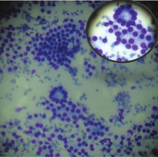
www.njlm.net                                                        M. S. Siddegowda et al., Cytomorphological Assessment and Thyroid Function Analysis

[10] Silverman JF, West RL, Larkin EW, Park HK, Finley JL, Swanson               [17] Canberk S, Firat P, Schmitt F. Pitfalls in the cytological
       MS, Fore WW. The role of fine-needle aspiration biopsy in the                    assessment of thyroid nodules. Turk Patoloji Derg.
       rapid diagnosis and management of thyroid neoplasm. Cancer.                      2015;31(suppl):18-33.
       1986;57(6):1164–70.                                                       [18]   Pavithra P, Rashmi MV. Utility of cytodiagnosis in the management
[11]   Handa U, Garg S, Mohan H, Nagarkar N. Role of fine needle                        of thyroid lesions. Int J Pharm Bio Sci. 2014;5(4):1173-82.
       aspiration cytology in the diagnosis and management of thyroid            [19]   Chandanwale S, Singh N, Kumar H, Pradhan P, Gore C, Rajpal
       lesions: A study on 434 patients. J Cytol. 2008;25(1):13-17.                     M. Clinicopathological correlation of thyroid nodules. Int J Pharm
[12]   Sengupta A, Pal R, Kar S, Zaman FA, Sengupta S, Pal S. Fine                      Biomed Sci. 2012;3(3):97-102.
       needle aspiration cytology as the primary diagnostic tool in              [20]   Manimaran D, Karthikeyan TM, Khan DM, Thulasi R. Follicular
       thyroid enlargement. J Nat SC Biol Med. 2011;2(1):113-18.                        Variant of Papillary Thyroid Carcinoma: Cytological Indicators of
[13]   Devi J, Aziz N. Cytomorphological evaluation and thyroid function                Diagnostic Value. J Clin Diagn Res. 2014; 8(3): 46–48.
       test analysis in various thyroid diseases – our experience at             [21]   Piromalli D, Martelli G, Del Prato I, Collini P, Pilotti S. The role
       tertiary care centre. International Journal of Medical Science                   of fine needle aspiration in the diagnosis of thyroid nodules:
       and Clinical Inventions. 2014;1(8):387-92. Available at http://                  analysis of 795 consecutive cases. J Surg Oncol. 1992;50:247-
       valleyinternational.net/index.php/our-jou/ijmsci.                                50.
[14]   Maitra A. Endocrine system. In: Kumar V, Abbas AK, Fausto                 [22]   Khan I, Naz S, Akhter ZM, Aziz N. Diagnostic accuracy of fine
       N, Astor JC, editors. Robbins and Cotran Pathologic Basis of                     needle aspiration of thyroid nodule verses biopsy in thyroid
       Disease. 9th ed. Philadelphia: Elsevier; 2014. p. 1090-92.                       lesions. J Ayub Med Coll Abbottabad. 2010;22(4):179-81.
[15]   Prasannan M, Kumar SA. Hashimoto’s thyroiditis-a                          [23]   Lewis CM, Chang KP, Pitman M, Faquin WC, Randolph GW.
       cytomorphological study with serological correlation. IJSAR.                     Thyroid fine-needle aspiration biopsy: variability in reporting.
       2015 ;2(8):47-52.                                                                Thyroid. 2009;19(7):717-23.
[16]   Gharib H, Goellner JR, Johnson DA. FNAC of thyroid. Clinics in
       Laboratory Medicine. 1993;13(3):699-709.

   AUTHOR(S):                                                                    NAME, ADDRESS, E-MAIL ID OF THE
   1. Dr. M. S. Siddegowda                                                       CORRESPONDING AUTHOR:
   2. Dr. Jasneet Kaur Sandhu                                                    Dr. Jasneet Kaur Sandhu,
   3. Dr. S. Shivakumar                                                          A007, Ground Floor, Renaissance Prospero Apartments,
                                                                                 Bytryanpura, Bangalore-560092, India.
   PARTICULARS OF CONTRIBUTORS:                                                  E-mail: Drjasneetkaur82@gmail.com
   1. Associate Professor, Department of Pathology,
      Mandya Institute of Medical Sciences, Mandya,                              Financial OR OTHER COMPETING INTERESTS:
      Karnataka, India.                                                          None.
   2. Tutor, Department of Pathology, Mandya Institute of
      Medical Sciences, Mandya, Karnataka, India.
   3. Professor and HOD, Department of Pathology,
      Mandya Institute of Medical Sciences, Mandya,
                                                                                                                          Date of Publishing: Jul 01, 2016
      Karnataka, India.

National Journal of Laboratory Medicine. 2016 Jul, Vol-5(3): PO16-PO21                                                                                         21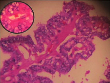
You can also read