Organic Amnesia Arthur P. Shimamura University of California, Berkeley
←
→
Page content transcription
If your browser does not render page correctly, please read the page content below
Shimamura, A. P. (1992), Organic Amnesia, from
L. R. Squire (Ed.), Encyclopedia of Learning and
Memory, pp. 30-35, Macmillan: New York.
Organic Amnesia
Arthur P. Shimamura
University of California, Berkeley
Organic amnesia is a neurological disorder that
affects learning and memory but leaves other
mental abilities relatively preserved. One important
aim of research on this disorder is to understand
how learning and memory are disrupted by brain
dysfunction in order to obtain clues to brain orga-
nization and normal memory processes.
Much of the current interest in memory and
brain function was initiated by Milner and col-
leagues who studied a now-famous patient with
organic amnesia, case H. M., who in 1953 under-
went surgery for relief of severe epileptic seizures.
The surgery involved bilateral excision of the me-
dial temporal region, which reportedly included
removal of the uncus (amygdala), anterior two
thirds of the hippocampus, and hippocampal gyrus
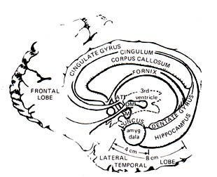
Figure 1. Schematic drawing of the medial surface of the
(see Figure 1). Following surgery, H. M.'s seizure human brain showing structures in the medial temporal
activity was attenuated, but he exhibited a profound lobe (e.g., hippocampus, amygdala) and in the diencephalic
anterograde amnesia—that is, he was unable to midline (e.g., dorsomedial [DM] and anterior [AT] thalamic
remember events and information encountered nuclei, mammillary nucleus [M]). Figure reprinted from
since his operation. Despite this severe impairment Squire (1984).
in new learning ability, there was no detectable
impairment in intellectual or language abilities.
There was some retrograde amnesia, which refers minutes. Thus, the central feature of H. M.'s mem-
to impairment of memory for events that occurred ory disorder is anterograde amnesia or new learn-
before the onset of amnesia. For example, H. M. ing impairment. This impairment affects informa-
could not remember the layout of the hospital ward tion received from all sensory modalities and in-
or recognize members of the medical staff. cludes impairment of both verbal and nonverbal
Moreover, he could not recall the death of a favor- (e.g., spatial) memory. For example, H. M. has
ite uncle who had died three years previously. Yet, failed to acquire new vocabulary words that have
following surgery H.M.'s retrograde amnesia was been added to the dictionary since his surgery. He
not severe, as indicated by the fact that he per- also exhibits severe impairment on laboratory tests
formed as well as control subjects on a test of of word and picture recall, cued-word learning
memory for faces of celebrities who became fa- (e.g., learning word pairs), and recognition mem-
mous prior to 1950. He was also capable of re- ory.
calling well-formed autobiographical episodes Despite the severity of his amnesia, H. M. can
from his adolescence. think and act normally, as indicated by his pre-
H. M. is still alive, and clinical observations served I.Q. Indeed, even some memory functions
indicate that memory for ongoing events is se- are spared, such as short-term memory, which can
verely impaired. For example, 30 minutes after be measured by intact performance on tests of
eating lunch, H. M. could not recall what he had immediate digit span. Nevertheless, as soon as
eaten and could not even recall if he had lunch at information is out of conscious experience, it is
all. H. M. is aware of his disorder and has re- forgotten. The analysis of H. M.'s amnesia stands
flected upon his impairment as always "waking as a milestone in our progress to understand
from a dream." In other words, he seems to lack memory in the brain. He has provided the crucial
continuity in the memory of events across time, evidence for the specific role of the medial tempo-
even when the events are separated by only a few ral region in the process of memory formation and
Page 1storage. Indeed, the analysis of H. M. by Milner show bilateral damage along the diencephalic mid-
and colleagues has provided the impetus for many line, typically involving the dorsomedial thalamic
important animal and human studies on the role of nuclei and a subthalamic nuclei called the mam-
the medial temporal region in learning and mem- millary bodies. In addition, cortical atrophy and
ory. cerebellar damage are often observed.
The Anatomy of Memory Patients with Korsakoff's syndrome exhibit
There are other neurological disorders that severe anterograde amnesia and often extensive
damage the medial temporal region and thus pro- retrograde amnesia. The severity of retrograde
duce an amnesic syndrome similar to that seen in amnesia, however, is variable among these patients,
H. M. For example, tumors, head injuries, or vas- with some showing extensive retrograde amnesia
cular disorders (e.g., strokes) in this region can and others showing little. One factor that compli-
cause organic amnesia. Also, some neurological cates the characterization of the memory impair-
disorders—such as viral infection, ischemia (i.e., ment in Korsakoff's syndrome is widespread cor-
loss of blood flow to the brain), or hypoxia (i.e., tical atrophy, which is presumed to be a conse-
loss of oxygen to the brain), particularly damage quence of chronic alcohol abuse. Indeed, some
the medial temporal region. In these disorders, mental functions, such as stimulus encoding, at-
anterograde amnesia is often the outstanding cog- tention, and problem solving are impaired in pa-
nitive impairment, though retrograde amnesia can tients with Korsakoff's syndrome but not in other
also occur. General intellectual abilities and short- amnesic patients. Moreover, patients with Kor-
term memory are generally intact. sakoff's syndrome are often emotionally flat, apa-
thetic, and without insight about their deficit. These
An amnesic patient studied by Zola-Morgan et additional cognitive and personality disorders may
al. has provided additional clues concerning the occur as a result of extensive cortical damage, in
prominent role of the hippocampus in memory. particular damage to the prefrontal cortex.
Patient R. B. became amnesic in 1978, when he
experienced an ischemic episode that occurred Not all amnesic syndromes are permanent. For
during open-heart surgery. R. B. was given exten- example, head injury can cause a transient and
sive neuropsychological assessment and was selective memory impairment. Following initial
found to exhibit anterograde amnesia but little if stages of unconsciousness or confusion, an-
any retrograde amnesia. In 1983, R. B. suffered a terograde and retrograde amnesia occurs, and the
fatal cardiac arrest, and, with the encouragement of severity of anterograde amnesia is often correlated
his family, a comprehensive examination of his with the temporal extent of retrograde amnesia.
brain was performed. This examination revealed a Retrograde amnesia tends to follow Ribot's Law,
discrete bilateral lesion restricted to a portion of which states that memory for the recent past is
the hippocampus called the CA1 subfield. R. B. affected more severely than memory for the distant
represents the first extensively studied case of past. Amnesia following head trauma can last for
amnesia that occurred as a result of damage re- minutes, days, or even weeks. In mild trauma
stricted to the hippocampus. cases, new learning ability recovers to premorbid
levels. In more severe cases, both amnesia and
Another area of the brain, the diencephalic mid- other cognitive impairment can be long-lasting and
line, can also produce organic amnesia (see figure sometimes permanent.
1). This area includes various midline thalamic
nuclei (nuclei are bundles of neurons) as well as Amnesia can also occur after electroconvulsive
subthalamic nuclei. These nuclei receive and send therapy (ECT), which is sometimes prescribed for
projections to various areas in the brain, including severe depressive illness. Anterograde amnesia can
the medial temporal region. Patients with neuro- be quite severe, particularly in patients who receive
logical damage due to cerebrovascular stroke or bilateral ECT. Retrograde amnesia is often tempo-
head injury in this area often exhibit organic am- rally-graded, following Ribot's Law. By several
nesia. months after ECT treatment, there is extensive
recovery of new learning capacity. Retrograde
The best-studied cases of amnesia resulting amnesia also resolves considerably when testing
from damage to the diencephalic midline are pa- occurs 6-9 months after ECT. Although the bio-
tients with Korsakoff's syndrome. As reviewed by logical factors that cause the transient amnesic
Butters and Cermak, Korsakoff's syndrome is an disorder following ECT are not well understood, it
amnesic disorder that develops after many years of is known that the hippocampus has one of the
chronic alcohol abuse and nutritional deficiency. lowest seizure thresholds of all brain structures.
Studies by Victor et al. of postmortem brain tissueThus, hippocampal functioning may be particularly trials and good retention when the same puzzle
compromised following ECT. was given 1 week later.
Advances in neuroimaging techniques, such as Cohen and Squire observed preserved skill
computed tomography (CT), magnetic resonance learning by amnesic patients on a mirror reading
(MR) imaging and positron emission tomography task. In this task, subjects were asked to read mir-
(PET), have allowed more detailed analysis of the ror-reversed words. Patients with Korsakoff's
brain areas that are damaged in neurological pa- syndrome, patients prescribed ECT, and patient N.
tients. For example, analyses of computed tomo- A. improved their reading speed of mirror-reversed
graphy (CT) brain scans of patients with Kor- words across training sessions to the same extent
sakoff's syndrome corroborated postmortem as control subjects. Moreover, amnesic patients
findings by identifying signs of increased fluid exhibited normal retention of the mirror-reading
and low neural density in the midline diencephalic skill even when they were tested one month after
region. A new technique by Press et al. has been learning. Despite this intact skill learning perform-
developed for MR imaging of the hippocampus. ance, patients failed to recognize the the words
This technique produces a clear cross-sectional used in the task. Moreover, the patients often did
image of the hippocampal formation and has al- not even recognize the testing apparatus nor did
ready provided remarkable images of the extent of they have conscious recollection of having en-
hippocampal damage in amnesic patients. For gaged in the task before. Performance by amnesic
example, it was shown that, compared to control patients in these tasks indicates that skill learning
subjects, amnesic patients exhibited an average can be preserved even when the patient has little or
loss of 49% of tissue in the area of the hippocam- no recollection of having acquired the skill. These
pal formation. Despite this tissue loss in the hip- findings suggest that amnesic patients can exhibit
pocampal area, the average area of the temporal a certain "unconscious" form of knowledge
lobe in these patients was nearly identical to that of ("knowing how") in the absence of explicit or
control subjects. Although PET analyses of amne- declarative knowledge ("knowing that").
sic patients have not been thoroughly studied, they There are several other forms of preserved
offer another approach to the physiological dys- memory function in amnesia. One form is illus-
function associated with organic amnesia. trated by an early anecdote of "unconscious"
memory that was reported by Claparede. During
Preserved Memory Functions in Amnesia an interview with an amnesic patient, Claparede hid
One of the most striking findings is that amne- a pin between his fingers and surreptitiously
sic patients can perform in an entirely normal pricked the patient on the hand. At a later time
fashion on certain "implicit" or "nondeclarative" during the interview, he once again reached for the
memory tests. These tests involve habit or auto- patient's hand, but the patient quickly withdrew her
matic learning, such as the kind of memory ex- hand. The patient did not acknowledge the previ-
pressed on tests of skill learning, classical condi- ous incident, and, when asked why she withdrew
tioning, and "priming." For example, H. M. her hand, she simply stated, "...sometimes pins are
showed considerable retention of perceptual-motor hidden in people's hands." This anecdote is an
skill on a mirror drawing task in which he was example of stimulus-response learning without
required to trace the outline of a star while viewing awareness. Another form of such learning was
the star in a mirror. The task is difficult at first but demonstrated by Weiskrantz & Warrington, who
then becomes easier and easier with practice. H. assessed Pavlovian classical conditioning of the
M. also exhibited skill learning on a pursuit-rotor eyeblink response in two amnesic patients. These
task in which a stylus must be kept on a rotating patients retained the eyeblink response for as long
target. In these tests, H. M. performed as a skilled as 24 hours, even though they did not recognize
individual but did not have conscious knowledge the test apparatus.
of having performed the task before. Preserved A memory phenomenon known as priming is
skill learning has been observed in other cases of also preserved in amnesia. Priming is an automatic
amnesia as well. For example, normal pursuit-rotor facilitation or bias in performance as a result of
skill learning and one-week retention in three pa- recently encountered information. The seminal
tients with Korsakoff's syndrome and two patients evidence for preservation of priming in amnesia
with amnesia due to viral encephalitis. Also, in a came from Warrington and Weiskrantz. Amnesic
jig-saw puzzle assembly task, these amnesic pa- patients were asked to identify words or pictures
tients exhibited faster completion times across six that were presented in a degraded form. If the
subject could not identify the stimulus, a succes-sion of less degraded versions of the stimulus scious" recollection from unconscious or auto-
were shown until identification was successful. matic memory. Squire and colleagues suggested
When amnesic patients were asked to identify the that amnesia impairs declarative memory and
same degraded words or pictures at a later time, spares procedural or nondeclarative memory.
their performance was facilitated by the previous Others have used related terms such as explicit and
experience; that is, they were able to identify the implicit memory or memory and habit. Such de-
stimuli more quickly. This priming effect occurred scriptions provide a framework for theoretical
despite failure to discriminate previously presented views about the organization of memory in the
stimuli from new ones in a recognition memory brain.
test. Memory Systems in the Brain
Graf et al. used a word completion task to study Findings from amnesic patients have led to the
priming effects. In this task, words are presented conclusion that there are multiple memory systems
(e.g., MOTEL) to the subject and later cued by in the brain, such that some forms exist entirely
three-letter word stems (e.g., MOT). Subjects are outside the brain regions that are damaged in or-
asked to say the first word that comes to mind for ganic amnesia. Amnesic patients apparently cannot
each word stem. In both amnesic patients and explicitly or consciously recollect information
control subjects, the tendency to use previously learned since the onset of amnesia. The impair-
presented words in the word completion test was ment is often thought to affect the ability to store
increased by 100-200% over baseline levels. In and also to retrieve newly learned information.
this test, words appear to "pop" into mind, and Amnesic patients, however, can often perform in a
amnesic patients exhibited this effect to the same normal fashion on certain "indirect" tests of mem-
level as control subjects. However, when subjects ory—tests that do not require conscious recollec-
were asked to use the same word stems as aids to tion of past learning sessions. Tests of skill learn-
recollect words from the study session, the control ing, classical conditioning, and priming can be
subjects exhibited better performance than amnesic characterized as indirect or implicit tests.
patients.
Various theories have been proposed to de-
A variety of priming paradigms have since been scribe the amnesic disorder. Squire and colleagues
used to demonstrate preserved priming in amnesia. have specified a neurological basis for declarative
For example, in one task subjects were presented memory—the form of memory that is impaired in
words (e.g., BABY) and later asked to "free asso- amnesia. It is hypothesized that declarative mem-
ciate" to related words (e.g., CHILD). Amnesic ory involves a storage or consolidation process
patients exhibited a normal bias to use recently that depends critically on the interaction of the
presented words in this word association task. hippocampus with areas in neocortex. The hippo-
This finding suggests that semantic associations campal "system" receives projections from many
can also be used to prime information in memory. neocortical areas. Thus this region may be in-
This priming effect, as well as others, are short- volved in relating or connecting information be-
lasting, and decline to baseline levels after a 2-hour tween various neocortical areas so that memory
delay. Although patients with circumscribed dien- storage and retrieval can be accomplished quickly
cephalic or medial temporal lesions exhibit normal and efficiently. Warrington and Weiskrantz have
priming effects, patients with the clinical diagnosis suggested that amnesia is due to a disruption of
of Alzheimer's disease do not. For example, im- diencephalic midline projections that connect the
paired word completion and word association medial temporal region to the prefrontal cortex.
priming has been observed in patients with senile Similarly, Mishkin has suggested that amnesia is
dementia of the Alzheimer type. These findings caused by disruption of the interaction of the hip-
suggest that priming effects may depend critically pocampus and amygdala with structures in neo-
on neocortical areas that are damaged in Alz- cortex and in the diencephalic midline (mediodor-
heimer's disease. sal and anterior nuclei of the thalamus).
Demonstrations of preserved memory functions In summary, neuropsychological studies of
in amnesic patients suggest that some memory memory functions in amnesic patients have pro-
processes can be dissociated from the brain re- vided useful information about the organization of
gions that are damaged in organic amnesia. As memory systems in the brain. Damage to the me-
reviewed by Squire, various taxonomies have been dial temporal region or diencephalic midline
used to distinguish the memory forms that are causes an amnesic syndrome in which conscious
impaired in amnesia from those that are preserved. or declarative memory is severely impaired. Inter-
For example, many distinguish between "con- estingly, other implicit or procedural functions(e.g., skills, habits) are entirely preserved. These Shimamura AP: Priming in amnesia: Evidence for
findings suggest that there are multiple memory a dissociable memory function. Q J Exper
systems in the brain and that one system can be Psychol 38, A: 619-644, 1986
dissociated from other memory and cognitive sys- Squire LR: The neuropsychology of memory. In
tems. These findings may offer important avenues Marler P, Terrace H (Editors), The Biology of
for both rehabilitative and pharmocological inter- Learning, Dahlem Konferenzen, Berlin:
ventions. That is, it may be possible to develop Springer-Verlag, 1984.
more efficient and more specific diagnoses and Squire LR: Memory and Brain. New York: Ox-
therapies for neurological patients as well as for ford University Press, 1987
individuals with more subtle forms of memory Victor M, Adams R.D, Collins GH: The Wer-
dysfunction, such as that observed in aging. nicke-Korsakoff Syndrome. Philadelphia:
Davis Company, 1971
Warrington EK, Weiskrantz L: Amnesia: A dis-
Bibliography connection syndrome? Neuropsychologia 20:
233-248, 1982
Andreason NC: Brain imaging: Applications in Warrington EK, Weiskrantz L: New method of
psychiatry. Science: 239: 1381-1388, 1988. testing long-term retention with special refer-
Butters N, Cermak LS: Alcoholic Korsakoff's ence to amnesic patients. Nature 217: 972-
Syndrome: An Information Processing Ap- 974, 1968
proach. New York: Academic Press, 1980 Weiskrantz L, Warrington EK: Conditioning in
Cohen NJ, Squire LR: Preserved learning and amnesic patients. Neuropsychologia 17: 187-
retention of pattern analyzing skill in amnesia: 194, 1979
Association of knowing how and knowing Zola-Morgan S, Squire LR, Amaral DG: Human
that. Science. 2l0: 207-209, 1980 amnesia and the medial temporal region: En-
Corkin S: Lasting consequences of bilateral medial during memory impairment following a bilat-
temporal lobectomy: Clinical course and ex- eral lesion limited to field CA1 of the hippo-
perimental findings in H. M. Sem Neurol 4: campus. J Neurosci 6: 2950-2967, 1986
249-259, 1984
Graf P, Squire LR, Mandler G: The information
that amnesic patients do not forget. J Exper
Psychol: Learn Mem Cognit 10: 164-178,
1984
Graff-Radford NR, Tranel D, VanHoesen GW,
Brandt J.P: Diencephalic amnesia, Brain, 113,
1-25, 1990
Milner B, Corkin S, Teuber H: Further analysis of
the hippocampal amnesic syndrome: 14-year
follow-up study of H.M. Neuropsychologia
6: 215-234, 1968
Mishkin, M. A memory system in the monkey. In
D. E. Broadbent, & L. Weiskrantz, Eds., The
Neuropsychology of Cognitive Function, pp.
85-95 London: The Royal Society, 1982
Press, GA, Amaral DG, Squire LR: Hippocampal
abnormalities in amnesic patients revealed by
high-resolution magnetic resonance imaging.
Nature 341: 54-57, 1989
Schacter DL: Implicit memory: History and cur-
rent status. J Exper Psychol: Learn Mem
Cognit 13: 501-518, 1987
Shimamura AP: Disorders of memory: The cogni-
tive science perspective. Boller F Grafman J.
Handbook of NeuropsychologyAmsterdam,
The Netherlands: Elsevier Sciences Publish-
ers, 1989: 35-73You can also read