Association of 2D and 3D transvaginal ultrasound findings with adenomyosis in symptomatic women of reproductive age: a prospective study - SciELO
←
→
Page content transcription
If your browser does not render page correctly, please read the page content below
ORIGINAL ARTICLE

Association of 2D and 3D transvaginal ultrasound
findings with adenomyosis in symptomatic women
of reproductive age: a prospective study
Ana Luiza Santos Marques0 0 -0 0 -0 0 -0 0 ,I Marina Paula Andres0 0 -0 0 -0 0 -0 0 ,I,II Leandro A. Mattos0 0 -0 0 -0 0 -0 0 ,III Manoel O. Gonc¸alves0 0 -0 0 -0 0 -0 0 ,IV
Edmund Chada Baracat0 0 -0 0 -0 0 -0 0 ,I Mauricio Simões Abrão0 0 -0 0 -0 0 -0 0 I,II,*
I
 Departamento de Obstetricia e Ginecologia, Hospital das Clinicas HCFMUSP, Faculdade de Medicina, Universidade de Sao Paulo, Sao Paulo, SP, BR.
II
 Divisao de Ginecologia, Hospital Beneficencia Portuguesa de Sao Paulo, Sao Paulo, SP, BR. III Departamento de Diagnostico por Imagem, Escola Paulista
de Medicina da Universidade Federal de Sao Paulo (EPM-UNIFESP), Sao Paulo, SP, BR. IV Secao de Diagnosticos Pelvicos Femininos, Alta Excelencia
Diagnostica, Sao Paulo, SP, BR.
Marques ALS, Andres MP, Mattos LA, Gonc¸alves MO, Baracat EC, Abrão MS. Association of 2D and 3D transvaginal ultrasound findings with adenomyosis
in symptomatic women of reproductive age: a prospective study. Clinics (Sao Paulo). 2021;76:e2981
*Corresponding author. E-mail: msabrao@mac.com

     OBJECTIVE: To evaluate the association of two-dimensional (2D) and three-dimensional (3D) transvaginal
     ultrasound (TVUS) findings with adenomyosis symptoms.
     METHODS: This prospective study conducted between January and December 2018 enrolled 78 women aged 18
     to 40 years with abnormal uterine bleeding (AUB), infertility, and/or pelvic pain. All patients underwent 2D and
     3D TVUS. Signs of adenomyosis on TVUS were identified according to the consensus of the Morphological
     Uterus Sonographic Assessment group.
     RESULTS: The prevalence of adenomyosis on TVUS was 55.12%. Patients with adenomyosis were older (p=0.002)
     and had more dysmenorrhea, AUB, and endometriosis than those without adenomyosis. When comparing the
     presence of symptoms with each adenomyosis feature, on 2D TVUS, severe dyspareunia was significantly
     associated with the presence of a poorly defined junctional zone (JZ) (p=0.023) and on 3D TVUS, patients with
     AUB had a more irregular (p=0.003), poorly defined (p=0.028), and interrupted JZ (p=0.011). After logistic
     regression analysis, signs of adenomyosis on TVUS remained significantly associated only with age over 30 years
     (OR: 1.2; 95% CI: 1.0-1.2) and AUB (OR: 7.65; 95% CI: 2-29). Patients with diffuse adenomyosis were older and
     presented with more infertility and AUB than patients with focal or no adenomyosis.
     CONCLUSION: The findings of adenomyosis by 2D and 3D TVUS showed association with age and AUB. 3D TVUS
     alterations in the JZ were associated with AUB and dyspareunia. Diffuse adenomyosis was associated with older
     age, a greater prevalence of infertility, and AUB.

     KEYWORDS: Adenomyosis; Abnormal Uterine Bleeding; Dysmenorrhea; Pelvic Pain; Transvaginal Ultrasound.

’ INTRODUCTION                                                                           with gynecological comorbidities such as endometriosis (4,5)
                                                                                         and uterine fibroids (6).
   Adenomyosis was once considered a disease of multi-                                      Studies have shown that imaging tools such as magnetic
parous women in late reproductive age, usually diagnosed                                 resonance imaging and two-dimensional (2D) and three-
after hysterectomies (1). However, it has been gaining                                   dimensional (3D) transvaginal ultrasound (TVUS) can accu-
worldwide interest as it has become a multifaceted disease                               rately diagnose women with diffuse adenomyosis with a
diagnosed by noninvasive imaging techniques in young                                     sensitivity of 80-86% and 50-96% (7,8), respectively. How-
women (2) with abnormal uterine bleeding (AUB), infertility,                             ever, few studies have investigated the early stages of
and pelvic pain and even in asymptomatic women (3).                                      adenomyosis, when patients are diagnosed with focal adeno-
In addition, adenomyosis is often diagnosed in association                               myosis with mild or no symptoms (9–12). Therefore, clinical
                                                                                         criteria and shared imaging criteria are still lacking, and data
                                                                                         from previous studies are heterogeneous and not fully
Copyright & 2021 CLINICS – This is an Open Access article distributed under the          comparable (13). It is important to remember that there
terms of the Creative Commons License (http://creativecommons.org/licenses/by/           are controversies about pathogenic theories, classifications,
4.0/) which permits unrestricted use, distribution, and reproduction in any
medium or format, provided the original work is properly cited.                          and diagnostic imaging criteria that prevent a shared defi-
                                                                                         nition of adenomyosis, even after histopathological exam-
No potential conflict of interest was reported.
                                                                                         ination (14).
Received for publication on March 24, 2021. Accepted for publi-                             Although the diagnostic process of adenomyosis is
cation on June 17, 2021                                                                  currently challenging, this study, given the importance of
DOI: 10.6061/clinics/2021/e2981                                                          TVUS as a first-line diagnostic examination in the patient’s

                                                                                     1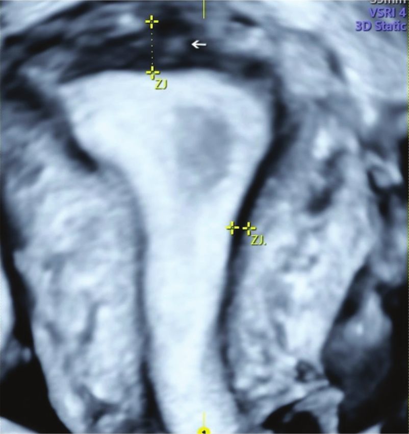
Transvaginal ultrasound and adenomyosis                                                                      CLINICS 2021;76:e2981
Marques ALS et al.

gynecological investigation, aims to contribute by evaluating         All included women underwent 2D TVUS and 3D TVUS
the association of 2D and 3D TVUS findings with adeno-              performed by an experienced sonographer (ALSM) with
myosis symptoms.                                                    more than 18 years of experience in gynecological ultra-
                                                                    sound. A total of 88 patients were eligible and underwent
’ METHODS                                                           the TVUS protocol with bowel preparation (16). Of the 88
                                                                    recruited patients, 10 were excluded from the study (seven
Study design                                                        for pregnancy and three for refusing participation), resulting
   This prospective study was conducted at the Getúlio              in 78 patients included in the final analysis (Figure 1).
Vargas University Hospital (Federal University of Amazo-
nas) in collaboration with the Endometriosis Section, Divisão
de Clinica Ginecologica, Hospital das Clinicas Faculdade de         Two- and three-dimensional transvaginal
Medicina, Universidade de São Paulo, São Paulo, Brazil.             ultrasound
The study was approved by the Internal Review Board of                 All TVUS examinations were performed using Voluson E8
both hospitals (IRB: 2.131.291/2017) and was conducted in           Expert or Voluson E10 BT18 (GE Healthcare Ultrasound;
accordance with the Helsinki Declaration. All participants          Zipf, Austria) equipped with a multifrequency endovaginal
provided written informed consent to participate in the             probe (6-12 MHz) and a multifrequency 3D endovaginal
study.                                                              probe (2.8-10 MHz) after bowel preparation.
   From January 2018 to December 2018, women aged 18-40                The 2D TVUS evaluation included evaluation and mea-
years with AUB, pelvic pain, and/or infertility, were recrui-       surement of the pelvic organs. The uterus and endometrium
ted from the outpatient clinic of the Getúlio Vargas Univer-        were measured, and the uterine volume was calculated
sity Hospital. Patients who were pregnant, had cervical             using the ellipsoidal formula (longitudinal uterine diameter
cancer, were in current use of hormonal treatment, had              transverse diameter  anteroposterior diameter  0.532). Any
no previous vaginal intercourse, or had fibroids 48.0 cm            myometrial lesions were described and measured and if there
(maximum diameter) or more than three fibroids 4 5.0 cm             were findings of pelvic endometriosis (ovaries, tubas, recto-
(maximum diameter) were excluded (9). Infertility was               sigmoid, sigmoid, bladder, distal ureters, retrocervical region,
defined as failure to achieve a clinical pregnancy after            rectovaginal septum and vagina), the extent of the disease
12 months or more of regular unprotected sexual intercourse.        was evaluated. Power Doppler was performed using prein-
   The clinical history of each patient was retrieved through       stalled fixed settings (Frequency: 6-9 MHz; pulse repeti-
an interview using a structured questionnaire by one                tion frequency: 0.6-0.3 kHz; gain: 4.0; wall motion filter:
researcher (ALSM). The questionnaire included age, height,          40 Hz).
weight, AUB, personal history of endometriosis or myoma,               In the 3D TVUS evaluation, two to four uterus gray-scale
and history of infertility and pain (dysmenorrhea, dyspar-          static volumes were obtained from the sagittal and trans-
eunia, and chronic pelvic pain). For each painful symptom,          verse planes. The volume acquisition technique was stan-
the intensity was assessed using a visual analog scale from         dardized, with frequency set to 6-9 MHz, uterus enlarged up
0 to 10 (15).                                                       to half the screen, sweep angle at 120o, scan speed adjusted

Figure 1 - Flowchart of included patients.

                                                                2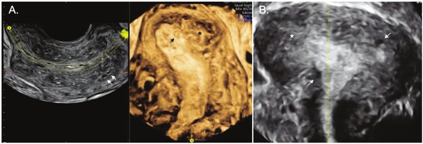
CLINICS 2021;76:e2981                                                                      Transvaginal ultrasound and adenomyosis
                                                                                                                 Marques ALS et al.

from medium to maximum quality, and the 3D volume                        The 3D TVUS (Figure 3) evaluated the characteristics of
box exceeding uterine borders by 1 cm on each side. The               the JZ. If it was found to be poorly defined, irregular, and
rendering mode, or Omni View, was used for the reconstruc-            interrupted and if its thickness was X8 mm or the difference
tion of the coronal plane; this consisted of placing a straight       between the maximum and minimum thickness was X4 mm
or curved line along the endometrial strip in the sagittal and        (JZ Dif) (17), it was considered suggestive of adenomyosis.
transverse planes (Panels A and B in multiplanar mode).               We considered focal and diffuse adenomyosis of the outer
Multiplanar vision was then manipulated until a satisfactory          and inner myometrium separately and classified adenomyo-
coronal plane image of the uterine external profile and cavity        sis types according to TVUS features as assessed in the
was obtained, with bilateral visualization of the interstitial        coronal plane. The focal of the inner myometrium, or JZ type,
portion of the fallopian tube. The volume contrast image              was a JZ interrupted with one hyperechogenic island (bud,
(slice thickness 2-4 mm) with volume rendering (mixed light           Figure 4). The focal of the outer myometrium type was a
surface and gradient light) was then applied. After acquisi-          heterogeneous uterus with irregular JZ and hyperechogenic
tion, the ultrasound volumes were stored on the hard disk of          subendometrial lines or buds, heterogeneous uterus with one
the machine.                                                          cystic area within the myometrium, or heterogeneous uterus
   Adenomyosis was identified by TVUS using the consensus             with adenomyoma (Figure 5). Diffuse type was defined by
of the Morphological Uterus Sonographic Assessment group              three or more findings evaluated by 2D-TVUS or 3D-TVUS
(10). The 2D TVUS features were a heterogeneous uterus,               or the presence of a question mark sign (18) (Figure 6). The
myometrial cysts, hypoechoic linear striae (when a radiating          ‘‘Question Mark Sign’’ was defined when the uterus was
pattern of thin acoustic shadows not arising from echogenic           flexed backward, with the uterus fundus facing the posterior
foci or leiomyoma, fan-shaped shadowing was present),                 pelvic compartment with the cervix directed frontally toward
hyperechogenic islands (buds), a poorly defined junctional            the urinary bladder (19).
zone (JZ), and posterior and anterior wall asymmetry.
Uterine wall asymmetry was assessed by measuring in the
                                                                      Statistical analysis
sagittal plane, perpendicular to the endometrium, from the
                                                                         For the comparison of the means, Student’s t-test or
uterine serosa external to the external endometrial contour;
                                                                      analysis of variance was used; however, for the comparison
measurements included the JZ but not the endometrium.
                                                                      of frequencies, the nonparametric Fisher’s exact test or chi-
Both measurements were recorded from the same image and
                                                                      square test with Yates correction was used. For the com-
ideally obtained from the thickest point of the myometrial
                                                                      parison of TVUS characteristics and clinical symptoms, the
wall. The ratio between the anterior and posterior wall
                                                                      latter were considered as independent variables and the
thickness was calculated. A ratio of approximately 1 indi-
                                                                      former as dependent variables. Multivariate analysis was
cates that the myometrial walls are symmetrical and a ratio
                                                                      performed using a logistic regression analysis. Variables with
well above or below 1 indicates asymmetry, although this
                                                                      po0.02 were included in the model.
may also be estimated subjectively (Figure 2). Power Dop-
                                                                         A significance level of 5% was considered statistically
pler was used in 2D mode to differentiate adenomyomas
                                                                      significant. Data were analyzed using the Epi Info software
from leiomyomas and myometrial cysts from gaps or
                                                                      (version 7.2 for Windows program; US Centers for Disease
vascular components. The vascular pattern within the myo-
                                                                      Prevention and Control, www.cdc.gov/epiinfo) and the SPSS
metrium can be uniform or non-uniform, and the vascular
                                                                      (v25) software.
pattern of a myometrial lesion can be circumferential,
translesional, or both. The type of translesional vasculariza-
tion, which is characterized by the presence of vessels per-          ’ RESULTS
pendicular to the uterine/serous cavity crossing the lesion,
is more frequent in adenomyoma, while the circumferential               Of the 78 patients included in the study, findings of
type is typical of fibroids (10,17).                                  adenomyosis were observed by 2D TVUS only in 1 patient,

Figure 2 - Two-dimensional ultrasound imaging of a uterus in a longitudinal section showing typical sonographic features of
adenomyosis. A. Note the asymmetry of the myometrial wall (posterior wall thicker than anterior wall) and the heterogeneous
myometrium with hypoechoic linear striae (arrows). B. Poorly defined junctional zone (arrows).

                                                                  3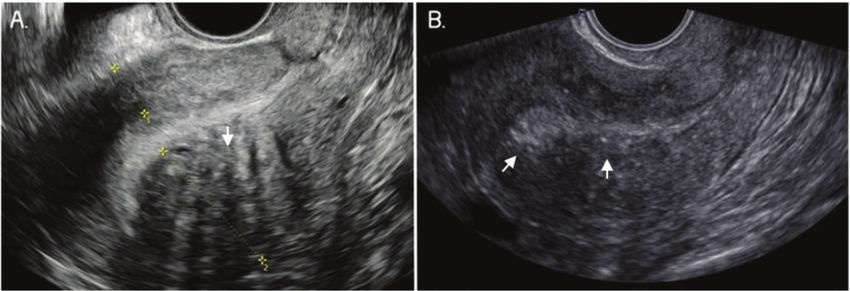
Transvaginal ultrasound and adenomyosis CLINICS 2021;76:e2981
Marques ALS et al.
Figure 3 - Three-dimensional ultrasound imaging of a uterus with adenomyosis in the coronal plane. A. Protrusions of the
endometrium into the junctional zone (JZ) (arrows). B. Junctional zone thickened, interrupted, and infiltrated by subendometrial
echogenic nodules (buds) (arrows). In this image, the JZ difference is 6.8 mm.
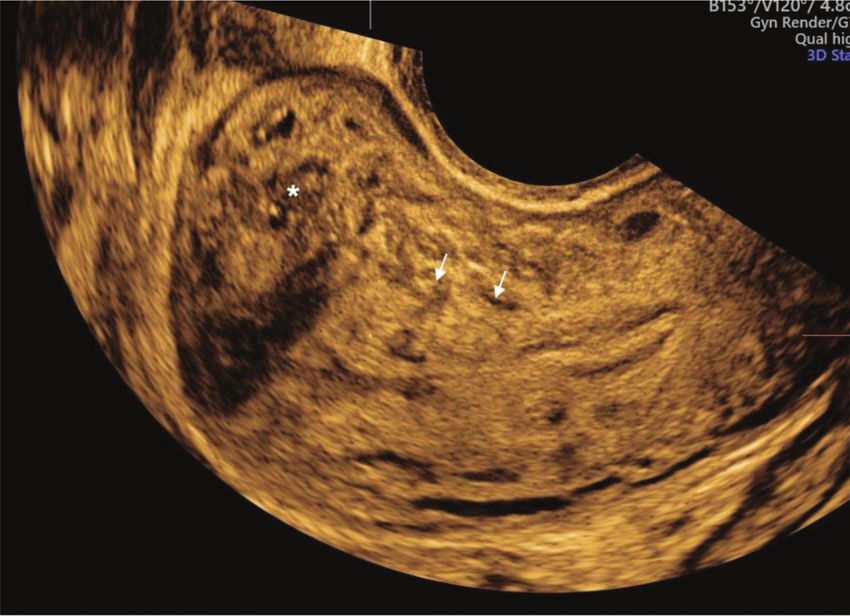
Figure 4 - Three-dimensional ultrasound imaging of a uterus in the coronal plane showing focal adenomyosis of the inner myometrium:
junctional zone interrupted with one hyperechogenic island (arrows).
4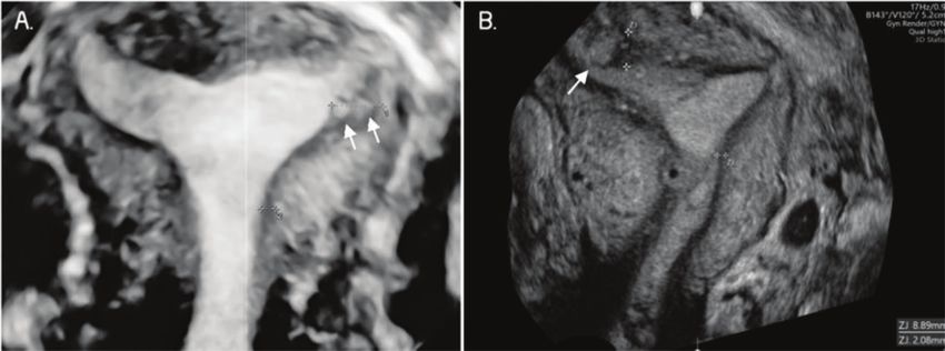
CLINICS 2021;76:e2981                                                                       Transvaginal ultrasound and adenomyosis
                                                                                                                  Marques ALS et al.

Figure 5 - Two-dimensional ultrasound imaging of a uterus in the sagittal plane showing a focal adenomyosis of the outer
myometrium: a heterogeneous uterus with adenomyoma and an irregular junctional zone (arrows and asterisks).

Figure 6 - Two and three-dimensional ultrasound imaging of a uterus with diffuse type adenomyosis. A. Heterogeneous uterus with an
irregular and interrupted junctional zone associated with myometrial cysts (arrows). B. Severe diffuse adenomyosis. Globular, enlarged,
and heterogeneous uterus with myometrial cysts and an ill-defined junctional zone, completely infiltrated by adenomyosis (arrows and
asterisks).

by 3D TVUS only in 2 patients, and by both 2D and 3D TVUS             years; p=0.002; Table 1). Patients with any sonographic
in 40 (55.1%) patients, resulting in 43 patients with any             finding of adenomyosis presented with significantly more
features of adenomyosis. Adenomyosis was classified as                AUB (23.3% vs. 0%; p=0.002) and significantly more dys-
focal of the inner myometrium in 22 (51.1%) cases, focal of           menorrhea (97.7% versus 82.9%; p=0.041; Table 1) than those
the outer myometrium in 11 (25.6%) cases, and diffuse in              without any sonographic finding of adenomyosis. 2D TVUS
10 (23.3%) cases. Fibroids were present in 11.4% (4 out of 35)        findings of adenomyosis were significantly associated with
of patients without adenomyosis and in 30.2% (13 out of 43)           endometriosis (55.8% versus 25.7%; p=0.011, Table 1), with
of patients with adenomyosis.                                         45% deeply infiltrating endometriosis of the posterior
   The mean age of patients with TVUS findings of adeno-              compartment and 10% ovarian endometriomas associated
myosis was greater than that of patients without TVUS                 with posterior compartment endometriosis. We did not find
findings of adenomyosis (31.6±5.8 years versus 27.4±5.7               any endometriosis in the anterior compartment.

                                                                  5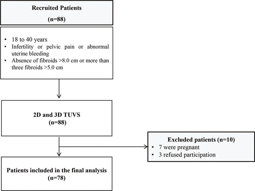
Transvaginal ultrasound and adenomyosis                                                                                        CLINICS 2021;76:e2981
Marques ALS et al.

Table 1 - Characteristics of included patients evaluated by TVUS.
                                          With TVUS signs of adenomyosis             Without TVUS signs of adenomyosis
                                                      (n=43)                                      (n=35)

Characteristics                               n                    %                       n                        %                 Total         p

Age                                                     31.6±5.8                                    27.4±5.7                                     0.002a
BMI (kg/m2)                                             25.7±4.0                                    24.2±5.2                                     0.158a
Previous pregnancy                             3                    7.0                    0                        0                   3        0.248b
AUB                                           10                   23.3                    0                        0                  10        0.002b
Severe Dysmenorrhea (VAS X 7)                 42                   97.7                   29                       82.9                71        0.041b
Severe Dyspareunia (VAS X 7)                  33                   76.7                   21                       60.0                54        0.178c
Severe Acyclic pain (VAS X 7)                 32                   74.4                   20                       57.1                52        0.171c
Myoma                                         13                   30.2                    4                       11.4                17        0.084c
Endometriosis                                 24                   55.8                    9                       25.7                33        0.011c

TVUS: transvaginal ultrasound; BMI: body mass index; AUB: abnormal uterine bleeding; VAS: visual analog scale of pain; aStudent’s t-test; bFisher’s exact
test; cChi-square test with Yates correction. P-values in bold indicate statistical differences at the 5% significance level.

Table 2 - Prevalence of 2D and 3D transvaginal ultrasound                       over 30 years (OR: 1.2; 95% CI: 1.0-1.2; p=0.0375; Table 3) and
features in patients with adenomyosis.                                          AUB (OR: 7.65; 95% CI: 2-29; p=0.0029; Table 4).
                                                                                  When comparing the type of adenomyosis, patients with
Adenomyosis findings                               n                  %
                                                                                diffuse adenomyosis were older (36.7±2.6 years) than
2D TVUS                                            42                55.0       patients in all other groups (focal adenomyosis of the inner
  Heterogeneous myometrium                         36                83.7       myometrium, 30.5±5.6 years; focal adenomyosis of the
  Buds                                             27                62.8       outer myometrium, 29.4±5.7 years; without adenomyosis,
  Linear striae                                    19                44.2
                                                                                27.4±5.7 years; po0.001; Table 5). In addition, all patients
  Globular uterus                                  17                39.5
  Uterine wall asymmetry                           15                34.9
                                                                                with diffuse adenomyosis presented with infertility and
  Myometrial cysts                                 14                32.6       AUB, and these frequencies were significantly higher than
  Poorly defined JZ                                 5                11.6       those in patients with focal or without adenomyosis
  Question Mark sign                                4                 9.3       (Table 5).
3D TVUS                                            41
  Interrupted JZ                                   38                88.4
  JZ Dif (X4.0 mm)                                 22                56.4
  Irregular JZ                                     21                48.8       ’ DISCUSSION
  Defined JZ                                        3                 7.0
  Poorly defined JZ                                 4                 9.3
                                                                                   The aim of this study was to evaluate the association of
                                                                                adenomyosis symptoms (infertility, pelvic pain, and AUB)
2D: two dimensions; 3D: three dimensions; TVUS: transvaginal ultrasound;        with 2D and 3D TVUS findings in young women of
JZ: junctional zone; JZ Dif: difference between the maximum and                 reproductive age. Although 2D and 3D TVUS features have
minimum thickness of the junctional zone.
                                                                                been shown to be associated with adenomyosis (14), the
                                                                                association of each feature or its combination with clinical
                                                                                symptoms such as pain, bleeding, and pregnancy needs to be
   The most frequently observed ultrasonographic features of                    defined.
adenomyosis by 2D TVUS are summarized in Table 2 and                               In this study, in 55.8% of cases, an association between
included a heterogeneous myometrium (n=36; 83.7%), sub-                         adenomyosis and endometriosis was observed, as assessed
endometrial echogenic nodules (n=27; 62.8%), hypoechoic                         by TVUS with bowel preparation. Both diseases share com-
linear striae (n=19; 44.2%), and a globular uterus (n=17,                       mon symptoms such as dysmenorrhea, infertility, dyspar-
39.5%). The most frequent 3D TVUS features observed were                        eunia, and chronic pelvic pain and are reported to be
interrupted JZ (n=38; 88.4%) and JZ thickness X4 mm (n=22;                      associated in 13.2–89.4% of cases (20-23). It was also
56.4%).                                                                         previously reported (18) that women with diffuse adeno-
                                                                                myosis showed a higher association with endometriosis than
                                                                                those with mild adenomyosis.
Comparison of clinical characteristics with TVUS                                   One study (23) evaluated the TVUS findings of adeno-
findings                                                                        myosis and clinical symptoms in 718 patients. The research-
  When comparing the presence of symptoms with each                             ers observed that 2D TVUS features of adenomyosis were
adenomyosis feature on 2D TVUS, patients with severe                            present in 21.9% of symptomatic women requiring consulta-
dyspareunia had more poorly defined JZ (81.3% vs. 56.5%;                        tion and that they were significantly associated with the
p=0.023; Table 3) than those without severe dyspareunia.                        severity of menstrual pain. In our study, which included
When comparing symptoms with each adenomyosis feature                           patients with infertility, AUB, and/or pelvic pain, 55% of
on 3D TVUS, patients with AUB showed more irregular JZ                          women presented with sonographic signs of adenomyosis.
(86.5% vs. 56.1%; p=0.003), more poorly defined JZ (100%                        Moreover, we observed that patients with severe dysmenor-
versus 65.7%; p=0.028), and more interrupted JZ (77.8% versus                   rhea and AUB had a higher prevalence of TVUS features of
57.5%; p=0.011) than those without AUB. After logistic                          adenomyosis than those without these symptoms (97.7%
regression analysis, the presence of sonographic signs of                       versus 82.9%; 23.3% versus 0%, respectively). This is in
adenomyosis remained significantly associated only with age                     accordance with a previous study (18), which showed a

                                                                            6
CLINICS 2021;76:e2981                                                                                 Transvaginal ultrasound and adenomyosis
                                                                                                                            Marques ALS et al.

Table 3 - Comparison of the presence of clinical symptoms and TVUS signs of adenomyosis.
                                                 Severe Dyspareunia            Severe Dysmenorrhea            Severe Acyclic pain
Characteristic               Infertility              (VASX7)                        (VASX7)                       (VASX7)                   AUB

2D TVUS
Globular uterus
   No                        19 (79.2)                27 (64.3)                      28 (66.7)                     26 (61.9)               30 (71.4)
   Yes                       28 (90.3)                25 (69.4)                      20 (55.6)                     24 (66.7)               25 (69.4)
   p                          0.276a                   0.630b                         0.315b                        0.662b                  0.848b
Asymmetry
   No                        22 (75.9)                33 (66.0)                      29 (58.0)                     32 (64.0)               34 (68.0)
   Yes                       25 (96.2)                19 (67.9)                      19 (67.9)                     18 (64.3)               21 (75.0)
   p                          0.054a                   0.867b                         0.391b                        0.980b                  0.515b
Myometrial cysts
   No                        36 (81.8)                44 (66.7)                      40 (60.6)                     43 (65.2)               45 (68.2)
   Yes                       11 (100)                  8 (66.7)                      8 (66.7)                      7 (58.3)                10 (83.3)
   p                          0.188a                    0.999a                        0.758a                        0.747a                  0.492a
Buds
   No                        42 (85.7)                48 (66.7)                      45 (62.5)                     45 (62.5)               49 (68.1)
   Yes                        5 (83.3)                 4 (66.7)                       3 (50.0)                      5 (83.3)                6 (100)
   p                           0.999a                   0.999a                         0.670a                        0.411a                 0.172a
Linear striae
   No                        39 (83.0)                46 (65.7)                      41 (58.6)                     45 (64.3)               48 (68.6)
   Yes                        8 (100)                  6 (75)                        7 (87.5)                      5 (62.5)                7 (87.5)
   p                          0.587a                   0.712a                         0.143a                        0.999a                  0.424a
Poorly defined JZ
   No                        20 (83.3)                26 (56.5)                      26 (56.5)                     27 (58.7)               29 (63.0)
   Yes                       27 (87.1)                26 (81.3)                      22 (68.8)                     23 (71.9)               26 (81.3)
   p                          0.718a                   0.023b                         0.275b                        0.233b                  0.083b
3D TVUS
Irregular JZ
   No                        17 (89.5)                24 (58.5)                      24 (58.5)                     26 (63.4)               23 (56.1)
   Yes                       30 (83.3)                28 (75.7)                      24 (64.9)                     24 (64.9)               32 (86.5)
   p                          0.700a                   0.109b                         0.566b                        0.894b                  0.003b
Poorly defined JZ
   No                        38 (86.4)                45 (67.2)                      42 (62.7)                     44 (65.7)               44 (65.7)
   Yes                        9 (81.8)                 7 (63.6)                       6 (54.5)                      6 (54.5)               11 (100)
   p                           0.654a                   0.999a                         0.741a                        0.511a                 0.028a
Interrupted JZ
   No                        16 (88.9)                23 (57.5)                      23 (57.5)                     25 (62.5)               23 (57.5)
   Yes                       22 (84.6)                22 (81.5)                      19 (70.4)                     19 (70.4)               21 (77.8)
   p                          0.890a                   0.127a                         0.557a                        0.610a                  0.011a

2D: two dimensions; 3D: three dimensions; TVUS: transvaginal ultrasound; AUB: abnormal uterine bleeding; VAS: visual analog scale for pain; aFisher’s
exact test; bChi-square test. Significant differences are highlighted in boldface.

Table 4 - Comparison of clinical symptoms in patients with TVUS                reported a significant association between the number of 2D
findings of adenomyosis.                                                       TVUS adenomyosis features with dysmenorrhea intensity
                                                                               and heavy menstrual bleeding in a cohort of nulliparous
Characteristics                          OR       CI 95%           pa          women, aged 18-30 years.
Age 430 years                          1.12     1.01–1.24         0.0375          A recent study (18) included 108 patients with sonographic
Endometriosis                          2.28     0.65–7.95         0.1966       signs of adenomyosis. The study reported that patients with
Severe dysmenorrhea (VASX7)            9.48     0.52–170.64       0.1274       older age and heavy menstrual bleeding were more
Severe dyspareunia (VASX7)             0.67     0.17–2.66         0.5744       associated with diffuse adenomyosis than those with focal
Leiomyomas                             6.86     0.99–47.46        0.0511
                                                                               disease with no difference in the presence of dysmenorrhea
Severe acyclic pain (VASX7)            2.37     0.61–9.23         0.2128
AUB                                    7.64     2.01–29.09        0.0029
                                                                               and dyspareunia. The study (18) also showed that in patients
                                                                               trying to conceive, the presence of ultrasound findings of
TVUS: transvaginal ultrasound; VAS: visual analog scale; OR: odds ratio;       focal disease was associated with a higher percentage of
CI 95%: confidence interval 95%; aLogistic regression. Significant             infertility (82% versus 52%; po0.005) and miscarriage (69%
differences are highlighted in boldface.
                                                                               versus 36%; po0.05) than that of diffuse disease. Similarly, in
                                                                               our study, after logistic regression analysis, clinical variables
prevalence of 72.2% of TVUS signs in patients with severe                      associated with the presence of adenomyosis were age 430
dysmenorrhea.                                                                  years and AUB. In contrast, we observed a higher frequency
   The authors previously reported that when adenomyosis                       of infertility in patients with diffuse adenomyosis (100%)
is detected late, a greater number of ultrasound features are                  than in those with focal adenomyosis of the inner (72.7%)
observed, and patients present with worse symptoms and                         and outer (81.8%; po0.001) myometrium.
more severe disease. One study (23) showed that the greater                       The limitations of this study include the small number of
the number of adenomyosis features identified on 2D TVUS,                      patients and the absence of histological analysis to evaluate
the greater the pain score. Another similar study (2) also                     the accuracy of TVUS as we included only women with

                                                                           7
Transvaginal ultrasound and adenomyosis CLINICS 2021;76:e2981
Marques ALS et al.
Table 5 - Comparison of adenomyosis severity and symptoms.
Adenomyosis
Characteristic No Focal (inner myometrium) Focal (outer myometrium) Diffuse p
N 35 22 11 10
Age 27. 4±5.7 30.5±5.6 29.4±5.7 36.7±2.6 o0.001a
Infertility 14 (40.0) 16 (72.7) 9 (81.8) 10 (100) o0.001b
Severe Dyspareunia (VASX7) 17 (48.6) 14 (63.6) 8 (72.7) 8 (80.0) 0.21b
Severe Dysmenorrhea (VASX7) 29 (82.9) 22 (100) 10 (90.9) 10 (100) 0.11b
Severe Acyclic pain (VASX7) 20 (57.1) 15 (68.2) 8 (72.7) 8 (80.0) 0.506b
AUB 16 (45.7) 18 (81.8) 8 (72.7) 10 (100) 0.002b
AUB: abnormal uterine bleeding; VAS: visual analog scale; aANOVA: analysis of variance.
b
Chi-square test. Significant differences are highlighted in boldface.
reproductive desire. The main strengths of our study were 8. Andres MP, Borrelli GM, Ribeiro J, Baracat EC, Abrão MS, Kho RM.
the inclusion criteria, the prospective design, and the fact Transvaginal Ultrasound for the Diagnosis of Adenomyosis: Systematic
Review and Meta-Analysis. J Minim Invasive Gynecol. 2018;25(2):257-64.
that all TVUS were performed by only one ultrasonographer, https://doi.org/10.1016/j.jmig.2017.08.653
minimizing interobserver variability. 9. Van den Bosch T, de Bruijn AM, de Leeuw RA, Dueholm M, Exacoustos
C, Valentin L, et al. Sonographic classification and reporting system for
’ CONCLUSION diagnosing adenomyosis. Ultrasound Obstet Gynecol. 2019;53(5):576-82.
https://doi.org/10.1002/uog.19096
10. Van den Bosch T, Dueholm M, Leone FP, Valentin L, Rasmussen CK,
Findings of adenomyosis by 2D and 3D TVUS were Votino A, et al. Terms, definitions and measurements to describe sono-
associated with age and AUB. 3D TVUS alterations of the JZ graphic features of myometrium and uterine masses: a consensus opinion
were associated with AUB and dyspareunia. Diffuse adeno- from the Morphological Uterus Sonographic Assessment (MUSA) group.
myosis was associated with older age, greater prevalence of Ultrasound Obstet Gynecol. 2015;46(3):284-98. https://doi.org/10.1002/
uog.14806
infertility, and AUB. 11. Bazot M, Cortez A, Darai E, Rouger J, Chopier J, Antoine JM, et al.
Ultrasonography compared with magnetic resonance imaging for the
’ ACKNOWLEDGMENTS diagnosis of adenomyosis: correlation with histopathology. Hum Reprod.
2001;16(11):2427-33. https://doi.org/10.1093/humrep/16.11.2427
The authors gratefully acknowledge the contribution of Maria Riselda 12. Reinhold C, Tafazoli F, Mehio A, Wang L, Atri M, Siegelman ES, et al.
Uterine adenomyosis: endovaginal US and MR imaging features with
Vinhote da Silva, MD, for her professional clinical attention to the patients
histopathologic correlation. Radiographics. 1999;19 Spec No:S147-60.
in this study. https://doi.org/10.1148/radiographics.19.suppl_1.g99oc13s147
13. da Silva JR, Andres MP, Leite APK, Gomes MTNA, Neto JS, Baracat EC,
’ AUTHOR CONTRIBUTIONS et al. Comparison of Sensitivity and Specificity of Structured and Nar-
rative Reports of Transvaginal Ultrasonography for Adenomyosis.
Marques ALS was responsible for the study design, collection of data, J Minim Invasive Gynecol. 2021;28(6):1216-24. https://doi.org/10.1016/
j.jmig.2020.11.001
analysis of results and manuscript drafting. Andres MP and Abrão MS were 14. Chapron C, Vannuccini S, Santulli P, Abrão MS, Carmona F, Fraser IS,
responsible for the study design, analysis of results and manuscript drafting. et al. Diagnosing adenomyosis: an integrated clinical and imaging
Mattos LA and Gonc¸alves MO were responsible for the study design and approach. Hum Reprod Update. 2020;26(3):392-411. https://doi.org/
collection of data. Baracat EC was responsible for the study design and 10.1093/humupd/dmz049
manuscript drafting. 15. Price DD, McGrath PA, Rafii A, Buckingham B. The validation of visual
analogue scales as ratio scale measures for chronic and experimental pain.
Pain. 1983;17(1):45-56. https://doi.org/10.1016/0304-3959(83)90126-4
’ REFERENCES 16. Goncalves MO, Dias JÁ Jr, Podgaec S, Averbach M, Abrão MS. Transva-
ginal ultrasound for diagnosis of deeply infiltrating endometriosis. Int J
1. Dueholm M, Lundorf E, Hansen ES, Sørensen JS, Ledertoug S, Olesen F. Gynaecol Obstet. 2009;104(2):156-60. https://doi.org/10.1016/j.ijgo.2008.
Magnetic resonance imaging and transvaginal ultrasonography for the 10.005
diagnosis of adenomyosis. Fertil Steril. 2001;76(3):588-94. https://doi. 17. Exacoustos C, Luciano D, Corbett B, De Felice G, Di Feliciantonio M,
org/10.1016/S0015-0282(01)01962-8 Luciano A, et al. The uterine junctional zone: a 3-dimensional ultrasound
2. Pinzauti S, Lazzeri L, Tosti C, Centini G, Orlandini C, Luisi S, et al. study of patients with endometriosis. Am J Obstet Gynecol. 2013;
Transvaginal sonographic features of diffuse adenomyosis in 18-30-year- 209(3):248.e1-7. https://doi.org/10.1016/j.ajog.2013.06.006
old nulligravid women without endometriosis: association with symp- 18. Exacoustos C, Morosetti G, Conway F, Camilli S, Martire FG, Lazzeri L, et al.
toms. Ultrasound Obstet Gynecol. 2015;46(6):730-6. https://doi.org/ New Sonographic Classification of Adenomyosis: Do Type and Degree of
10.1002/uog.14834 Adenomyosis Correlate to Severity of Symptoms? J Minim Invasive Gynecol.
3. Abbott JA. Adenomyosis and Abnormal Uterine Bleeding (AUB-A)- 2020;27(6):1308-15. https://doi.org/10.1016/j.jmig.2019.09.788
Pathogenesis, diagnosis, and management. Best Pract Res Clin Obstet 19. Di Donato N, Bertoldo V, Montanari G, Zannoni L, Caprara G, Seracchioli
Gynaecol. 2017;40:68-81. https://doi.org/10.1016/j.bpobgyn.2016.09.006 R. Question mark form of uterus: a simple sonographic sign associated
4. Di Donato N, Montanari G, Benfenati A, Leonardi D, Bertoldo V, Monti G, with the presence of adenomyosis. Ultrasound Obstet Gynecol. 2015;
et al. Prevalence of adenomyosis in women undergoing surgery for 46(1):126-7. https://doi.org/10.1002/uog.14750
endometriosis. Eur J Obstet Gynecol Reprod Biol. 2014;181:289-93. 20. Bordonné C, Puntonet J, Maitrot-Mantelet L, Bourdon M, Marcellin L,
https://doi.org/10.1016/j.ejogrb.2014.08.016 Dion E, et al. Imaging for evaluation of endometriosis and adenomyosis.
5. Eisenberg VH, Arbib N, Schiff E, Goldenberg M, Seidman DS, Soriano D. Minerva Obstet Gynecol. 2021;73(3):290-303.
Sonographic Signs of Adenomyosis Are Prevalent in Women Undergoing 21. Decter D, Arbib N, Markovitz H, Seidman DS, Eisenberg VH. Sono-
Surgery for Endometriosis and May Suggest a Higher Risk of Infertility. graphic Signs of Adenomyosis in Women with Endometriosis Are Asso-
Biomed Res Int. 2017;2017:8967803. https://doi.org/10.1155/2017/ ciated with Infertility. J Clin Med. 2021;10(11):2355. https://doi.org/
8967803 10.3390/jcm10112355
6. Brucker SY, Huebner M, Wallwiener M, Stewart EA, Ebersoll S, Schoen- 22. Rasmussen CK, Hansen ES, Ernst E, Dueholm M. Two- and three-
fisch B, et al. Clinical characteristics indicating adenomyosis coexisting dimensional transvaginal ultrasonography for diagnosis of adenomyosis
with leiomyomas: a retrospective, questionnaire-based study. Fertil Steril. of the inner myometrium. Reprod Biomed Online. 2019;38(5):750-60.
2014;101(1):237-241.e1. https://doi.org/10.1016/j.fertnstert.2013.09.038 https://doi.org/10.1016/j.rbmo.2018.12.033
7. Tellum T, Nygaard S, Lieng M. Noninvasive Diagnosis of Adenomyosis: 23. Naftalin J, Hoo W, Nunes N, Holland T, Mavrelos D, Jurkovic D. Asso-
A Structured Review and Meta-analysis of Diagnostic Accuracy in Ima- ciation between ultrasound features of adenomyosis and severity of
ging. J Minim Invasive Gynecol. 2020;27(2):408-418.e3. https://doi.org/ menstrual pain. Ultrasound Obstet Gynecol. 2016;47(6):779-83. https://
10.1016/j.jmig.2019.11.001 doi.org/10.1002/uog.15798
8You can also read