Sonoelastographic evaluation of plantar fascia after shock wave therapy for recalcitrant plantar fasciitis: A 12-month longitudinal follow-up ...
←
→
Page content transcription
If your browser does not render page correctly, please read the page content below
www.nature.com/scientificreports

            OPEN           Sonoelastographic evaluation of
                           plantar fascia after shock wave
                           therapy for recalcitrant plantar
                           fasciitis: A 12-month longitudinal
                           follow-up study
                                                   1
                           Chueh-Hung Wu               , Yun-Yi Lin1,2,3, Wen-Shiang Chen      1
                                                                                                   & Tyng-Guey Wang        1*

                           Extracorporeal shockwave therapy (ESWT) is proposed to be effective in reducing pain and improving
                           functional outcome in chronic plantar fasciitis. However, no long-term reports exist on the changes in
                           plantar fascia (PF) elasticity after ESWT. We aimed to evaluate the changes in PF stiffness in patients
                           with plantar fasciitis undergoing ESWT. The visual analogue scale (VAS, 0–100) was used for evaluating
                           heel pain severity. B-mode sonography and strain sonoelastography were used for evaluating the PF
                           thickness and stiffness. The sonoelastogram was analyzed using hue histogram analysis (value: 0–255,
                           from stiffer to softer). All evaluations were recorded before ESWT, and 1 week, 1 month, 3 months,
                           6 months, and 12 months after ESWT. Repeated measures ANOVA was used to compare pain VAS,
                           PF thickness, and PF hue value at different follow-up time-points. Twenty-two participants (8 men,
                           14 women) completed all measurements for 12 months. The VAS of heel pain, PF thickness, and PF
                           hue values at pre-ESWT, and 1-week, 1-month, 3-month, 6-month, and 12-month evaluations after
                           ESWT were 62.4 ± 4.2, 49.3 ± 5.8, 38.3 ± 5.7, 27.9 ± 5.3, 18.9 ± 4.7, and 13.2 ± 3.0 (p < 0.01 in all
                           measurements post ESWT versus pre-ESWT); 5.57 ± 0.22 mm, 5.64 ± 0.18 mm, 5.45 ± 0.24 mm,
                           5.37 ± 0.20 mm, 5.08 ± 0.20 mm, and 4.62 ± 0.15 mm (p < 0.01 at 6-month; otherwise p > 0.05);
                           and 24.5 ± 2.4, 35.2 ± 3.1, 31.0 ± 4.1, 30.5 ± 3.9, 21.4 ± 2.1, and 15.9 ± 1.6 (p < 0.01 at 1-week and
                           6-month; otherwise p > 0.05), respectively. In conclusion, the heel pain intensity and PF thickness
                           reduced gradually over 12 months after ESWT. The PF stiffness decreased during the first week and
                           increased thereafter; at the 12-month follow-up, stiffness was more than at pre-ESWT.

                           Plantar fasciitis is the most common cause of inferior heel pain in adults1. If a patient’s heel pain lasts over 6
                           months despite conservative treatment, it is considered chronic recalcitrant plantar fasciitis2. In such patients,
                           low-intensity extracorporeal shockwave therapy (ESWT) has been proposed to be effective in reducing pain and
                           improving functional outcome3. While the exact mechanism of ESWT remains unknown, animal experiments
                           have demonstrated that ESWT induces neovascularization at the tendon-bone junction, which might imply
                           improvement of blood supply and tissue regeneration4,5. Through this regeneration process, there might be cer-
                           tain material property changes of the plantar fascia (PF), but such changes have not been described.
                               Morphologically, a significant decrease in PF thickness after ESWT was revealed by using B-mode sonogra-
                           phy6,7. In addition to morphology changes, regeneration of the PF may be also associated with changes of its elas-
                           ticity. However, there has been no report on long-term follow-up of PF elasticity changes after ESWT. To achieve
                           such evaluation, an ultrasound-based technique, strain sonoelastography provides an estimation of soft tissue
                           elasticity. Previous studies using this technology have shown softening of the PF in plantar fasciitis patients8–13.
                           Therefore, in this prospective assessor-blind longitudinal follow-up study, we aimed to evaluate elasticity changes

                           1
                            Department of Physical Medicine and Rehabilitation, National Taiwan University Hospital, College of Medicine,
                           National Taiwan University, Taipei, Taiwan. 2Institute of Health Policy & Management, College of Public Health,
                           National Taiwan University, Taipei, Taiwan. 3Department of Rehabilitation Medicine, Da-Chien Hospital, Miao-Li,
                           Taiwan. *email: tgw@ntu.edu.tw

Scientific Reports |   (2020) 10:2571 | https://doi.org/10.1038/s41598-020-59464-8                                                           1
www.nature.com/scientificreports/ www.nature.com/scientificreports
of the PF after ESWT by using strain sonoelastography. We hypothesized that softened PF would become stiffer
after ESWT.
Materials and Methods
Participants. This prospective longitudinal follow-up study was approved by the Research Ethics Committee,
National Taiwan University Hospital (Ref. No.: 201007055R). Each participant provided written informed consent
before evaluation, and the work was completed in accordance with the principles laid down by the Declaration of
Helsinki. The participants were recruited between September 7, 2010 and January 28, 2014.
Inclusion criteria of the participants were (1) age 20–80 years old; (2) unilateral heel pain at the insertion of
the PF on the medial tubercle of the calcaneus; (3) worse pain when waking up in the morning or after rest; (4)
pain duration longer than 6 months despite conservative treatments including shoe modification, arch support,
medication and physiotherapy; (5) visual analogue scale (VAS) of worst heel pain in previous one week >40 on
a scale of 100; and (6) B-mode ultrasound (US) examination revealing a thickened (>4 mm) and hypoechoic PF.
Exclusion criteria were (1) having received steroid or hyaluronic acid injections to the heel; (2) having undergone
shock wave therapy or operations on the heel; (3) a history of systemic inflammatory diseases.
Extracorporeal shock wave therapy. Piezoson100 (Richard Wolf, Knittlingen, Germany), a
piezoelectric-type device, was used by one physiatrist for ESWT. All participants received three sessions of ESWT
(3000 shock waves per session of 0.08–0.2 mJ/mm2) at weekly intervals. No local anesthesia was applied during
treatment. The target of treatment was determined by the self-reported tender area. A sonographic examination
was performed only before each session of ESWT to confirm the depth of the PF. The shock waves were applied
to the maximum pain sites and to the surrounding area within a 1-cm radius.
Ultrasound and sonoelastographic examination. The examination of each PF included B-mode US
scanning and strain sonoelastography using an Acuson S2000 US system (Siemens, Munich, Germany) with a
6–14-MHz linear transducer (14L5; Siemens). US imaging settings (transducer frequency: 11 MHz, total gain:
+20 dB, focus: 1.5 cm in depth, dynamic range: 70, SieClear (SC): 2, map: B, space/time (ST): 0) were set to
standardize each measurement. All examinations were conducted by one physiatrist, with a 6-year experience of
musculoskeletal ultrasound.
Each participant was examined in a prone position with 90° of knee flexion in the neutral ankle position8,14.
The ultrasound transducer can be hold more steadily in this posture, which is important for obtain high-quality
strain sonoelastography images. The entire width of the PF was examined to localize the thickest area. In a lon-
gitudinal view, the PF thickness was measured from the anterior edge of the inferior calcaneal border vertically
to the inferior border of the PF. Sonoelastograms of the PF were obtained three times in the same position with a
quality factor exceeding 60, indicating stable transducer positioning.
The stiffness color scale used in the sonoelastogram expresses differing degrees of tissue stiffness with corre-
sponding colors15. For the Siemens system, the scale indicates the relative stiffness of the examined tissues within
the region of interest (ROI) and ranges from red (hardest), yellow (relatively hard), green (intermediate stiffness),
blue (relatively soft), to purple (softest).
Hue histogram analysis of sonoelastogram. Image J software (version 1.43 u; National Institutes of
Health, Bethesda, USA) was used to acquire an RGB color histogram and a hue histogram of a manually-selected
standardized rectangular ROI for each PF sonoelastogram. Since the site of abnormality in plantar fasciitis mostly
occurs near the PF origin at the medial tuberosity of the calcaneus16, the longer side of the ROI was set 1 cm distal
to the proximal PF insertion on the distal calcaneus border, and the shorter side was set 2 mm toward the heel
from the point of the inferior calcaneus border. This was done to avoid including the area outside the PF since
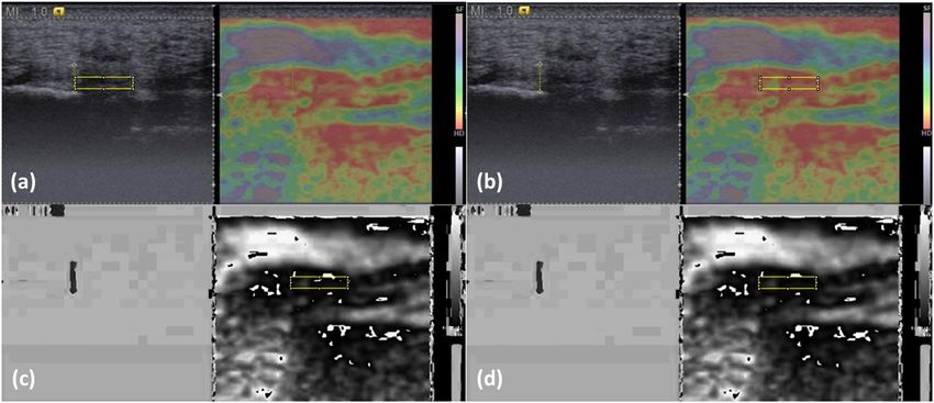
most normal PFs are thicker than 2 mm17 (see Fig. 1a, the yellow rectangular delineated area).
In RGB color histogram analysis, we used the Image J “Color Histogram” function to analyze all pixels within
the ROI. The red, green, and blue color intensities (ranging from 0 to 255) of each pixel were calculated separately
and averaged as mean values. Such analysis may be superior to dichotomizing the PF stiffness into hard and soft
solely based on the percentage of blue area18.
In hue histogram analysis, sonoelastograms were converted from the original RGB coded images (Fig. 1b) to
the hue component of hue, saturation, and brightness (HSB) coded images using the “HSB Stack” function. The
originally-colored sonoelastograms and stiffness color scale were thus converted to monotonic gray scale images
(Fig. 1c) in which each hue value (0–255) represented a different level of relative stiffness within the ROI, with
0 representing the hardest and 255 representing the softest. Following conversion, images contained some black
or white noise signals at the softest and hardest parts (Fig. 1c). These noise signals resulted from violations of
the monotonically increasing rule during conversion from RGB to HSB color-coding systems according to the
conversion equations19. We corrected these noise signals by assigning those pixels to hue value of 0 in the hard-
est region and value 255 in the softest region. The post-processing image is shown in Fig. 1d. We then used the
“Histogram” function to obtain the average hue value of all pixels within the ROI to represent the average relative
stiffness. A higher hue value indicated a relatively lower stiffness. The results of the hue histogram and RGB color
histogram analyses of the three images from the same patient were averaged for statistical analysis.
Outcome measurements and follow-up. VAS of heel pain, PF thickness, and PF elasticity (hue value)
were recorded before ESWT, and 1 week, 1 month, 3 months, 6 months, and 12 months after ESWT.
Statistical analysis. MedCalc Statistical Software version 19.0.5 (MedCalc Software bvba, Ostend, Belgium;
https://www.medcalc.org; 2019) was used for all statistical analysis. Because a preliminary Kolmogorov–Smirnov
test demonstrated that all samples followed a normal distribution, repeated measures ANOVA was used to
Scientific Reports | (2020) 10:2571 | https://doi.org/10.1038/s41598-020-59464-8 2www.nature.com/scientificreports/ www.nature.com/scientificreports
Figure 1. Image processing for RGB color histogram and hue histogram analyses. (a) Select a region of interest
(ROI) in the B-mode image, (b) move the ROI to the same place on the sonoelastogram for color histogram
processing, (c) convert the color-coded sonoelastogram into hue component of the HSB system, and
(d) following noise signal correction, obtain the hue histogram of all pixels within the ROI.
Before ESWT 1 week 1 month 3 months 6 months 12 months
VAS (0–100) 62.4 ± 4.2 49.3 ± 5.8* 38.3 ± 5.7* 27.9 ± 5.3* 18.9 ± 4.7* 13.2 ± 3.0*
PF thickness (mm) 5.57 ± 0.22 5.64 ± 0.18 5.45 ± 0.24 5.37 ± 0.20 5.08 ± 0.20 4.62 ± 0.15*
PF hue values 24.5 ± 2.4 35.2 ± 3.1* 31.0 ± 4.1 30.5 ± 3.9 21.4 ± 2.1 15.9 ± 1.6*
Table 1. Heel pain and ultrasonographic evaluations before and after ESWT. *p < 0.01 versus before ESWT
(repeated measures ANOVA). ESWT: extracorporeal shock wave therapy; VAS: visual analogue scale of heel
pain; PF: plantar fascia. Note: a higher hue value indicates a softer plantar fascia.
compare pain VAS, PF thickness, and PF hue value among different timings of follow-ups. A p value ofwww.nature.com/scientificreports/ www.nature.com/scientificreports
Figure 2. Outcome measurements before and after extracorporeal shock wave therapy (ESWT). (Pre: before
ESWT; 1w: 1 week after ESWT; 1 m: 1 month after ESWT; 3 m: 3 months after ESWT; 6 m: 6 months after
ESWT; 12 m: 12 months after ESWT) (*p < 0.01 versus before ESWT). (A) Visual analogue scale score for heel
pain (0–100). (B) Plantar fascia thickness (mm). (C) Plantar fascia elasticity (hue value).
tissue composed primarily of type I collagen, similar to tendons. In a study on rats with the collagenase-induced
Achilles tendinitis, biomechanical and biochemical characteristics of healing tendons were restored 12 weeks
after ESWT22. Increasing TGF-beta 1 expression in the early stage of tendon repair and elevated IGF-I expression
persisting throughout the healing period were also observed22. In another rat study, the associated tenocyte pro-
liferation was reported to be mediated by early up-regulation of PCNA and TGF-beta 1 gene expression, endog-
enous NO release, synthesis of TGF-beta 1 protein, and collagen synthesis (at the 7th day)23. The above findings
Scientific Reports | (2020) 10:2571 | https://doi.org/10.1038/s41598-020-59464-8 4www.nature.com/scientificreports/ www.nature.com/scientificreports
may indicate that ESWT metabolically “activated” these tenocytes and significantly induced them to proliferate24.
Unlike animals, our study showed that it may take a longer time for the human PF to regain stiffness and even
become stiffer.
The initially softer PF after ESWT may have important clinical implications. In the current study, we found
that heel pain intensity decreased gradually, which was consistent with the effectiveness of ESWT on plantar fas-
ciitis reported previously25. Despite the decrease in symptoms in the first few weeks after ESWT, the PF may be
structurally weaker in this period. If the patients return to previous activity level, exerting too much pressure on
the PF without adequate protection, the symptoms may quickly recur. Although we did not evaluate the effective-
ness of an in-sole, we still recommend that after ESWT the patients should be instructed to wear an in-sole and
to avoid excessive pressure on the heel for at least for one month, based on the results that the PF may be softer in
this period. Such a protection strategy may prevent recurrence of plantar fasciitis.
US evaluation of morphological changes of PF after ESWT has been reported6,26. An ultrasonographic study
of plantar fasciitis at 6 months follow-up after extracorporeal shock wave therapy showed that the PF thickness
decreased from 5.2 ± 1.5 mm to 4.4 ± 1.0 mm at 24 weeks of follow-up6. In the current study, there was no statisti-
cally significant difference in PF thickness between pre-ESWT and 6 months post-ESWT, despite a trend towards
a thinner PF at the 6-month follow-up. However, at the 12-month follow-up the decrease in thickness of the PF
was significant. The longer time for the PF to become thinner in this study may be related to the thicker PF before
ESWT, and/or a longer symptom duration (18.6 months in our study; not mentioned in the ultrasonographic
study6).
Based on the above findings, we believe that ESWT is effective in treating chronic recalcitrant plantar fasciitis
and may modify the PF morphology, with the effect lasting for 12 months. In addition to morphological changes,
our study further showed elasticity changes of PF after ESWT. A sonoelastographic study revealed hardening of
the PF 3 months after collagen injection for plantar fasciitis27. In this longitudinal follow-up study, we found an
initially softer PF which became stiffer at one-year follow-up, despite a gradually decreased heel pain intensity.
Several limitations need to be addressed. First, this was a single-center, unblinded study with no control group,
making it difficult to clarify whether elasticity changes were associated with natural recovery. However, in this
study we included those patients with symptoms for at least 6 months despite conservative treatment. It is unlikely
that the thickness and elasticity remained unchanged for 18.6 months (the mean symptom duration in this study)
and then naturally recovered in the following 12 months. Second, unlike a manually marked ROI, a rectangular
ROI for elastogram analysis in this study may not fit the PF well because it was smaller than the PF. Such ROI
may not represent the entire PF. However, we actually conducted comparisons within the same subject at dif-
ferent timings. This standardized ROI may minimize bias due to hand-draw errors. Third, we did not perform
pathohistological correlation in this study. Surgery is rarely necessary in plantar fasciitis, so it is difficult to obtain
PF specimens. Because in previous studies a softer PF was observed in plantar fasciitis patients. In this study, the
change of elasticity after ESWT was our major aim for investigation. Fourth, we utilized strain sonoelastography
with image analysis, which was a semi-quantitative evaluation of tissue elasticity. Shear wave ultrasound elas-
tography (SWUE) can provide objective evaluation of tissue elasticity by providing quantification of shear wave
velocity within the tissue28,29. Investigation of PF stiffness with SWUE should be conducted in future studies.
Because evaluation with SWUE requires the certain ultrasonography system with a particular probe, we thought
that few clinicians or researchers had access to such machines. This study may provide an alternative for follow-up
of tissue elasticity with utilizing a more easily accessible system.
Conclusion
After ESWT for plantar fasciitis, heel pain intensity decreased gradually, while the PF thickness became thinner
at the 12-month follow-up. The PF became softer at 1 week of follow-up and regained stiffness thereafter, finally
becoming stiffer than pre-ESWT at 12 months of follow-up.
Received: 2 November 2019; Accepted: 23 January 2020;
Published: xx xx xxxx
References
1. Goff, J. D. & Crawford, R. Diagnosis and treatment of plantar fasciitis. Am. Fam. Physician 84, 676–682 (2011).
2. Gerdesmeyer, L. et al. Radial extracorporeal shock wave therapy is safe and effective in the treatment of chronic recalcitrant plantar
fasciitis: results of a confirmatory randomized placebo-controlled multicenter study. Am. J. Sports Med. 36, 2100–2109, https://doi.
org/10.1177/0363546508324176 (2008).
3. Yin, M. C. et al. Is extracorporeal shock wave therapy clinical efficacy for relief of chronic, recalcitrant plantar fasciitis? A systematic
review and meta-analysis of randomized placebo or active-treatment controlled trials. Arch. Phys. Med. Rehabil. 95, 1585–1593,
https://doi.org/10.1016/j.apmr.2014.01.033 (2014).
4. Wang, F. S. et al. Physical shock wave mediates membrane hyperpolarization and Ras activation for osteogenesis in human bone
marrow stromal cells. Biochem. Biophys. Res. Commun. 287, 648–655 (2001).
5. Wang, C. J., Huang, H. Y. & Pai, C. H. Shock wave-enhanced neovascularization at the tendon-bone junction: an experiment in dogs.
J. Foot Ankle Surg. 41, 16–22 (2002).
6. Hammer, D. S. et al. Ultrasonographic evaluation at 6-month follow-up of plantar fasciitis after extracorporeal shock wave therapy.
Arch. Orthop. Trauma. Surg. 125, 6–9, https://doi.org/10.1007/s00402-003-0591-z (2005).
7. Vahdatpour, B., Sajadieh, S., Bateni, V., Karami, M. & Sajjadieh, H. Extracorporeal shock wave therapy in patients with plantar
fasciitis. A randomized, placebo-controlled trial with ultrasonographic and subjective outcome assessment. J. Res. Med. Sci. 17,
834–838 (2012).
8. Wu, C. H., Chang, K. V., Mio, S., Chen, W. S. & Wang, T. G. Sonoelastography of the plantar fascia. Radiology 259, 502–507 (2011).
9. Wu, C. H., Chen, W. S., Wang, T. G. & Lew, H. L. Can sonoelastography detect plantar fasciitis earlier than traditional B-mode
ultrasonography? Am. J. Phys. Med. Rehabil. 91, 185 (2012).
10. Sconfienza, L. M. et al. Real-time sonoelastography of the plantar fascia: comparison between patients with plantar fasciitis and
healthy control subjects. Radiology 267, 195–200 (2013).
Scientific Reports | (2020) 10:2571 | https://doi.org/10.1038/s41598-020-59464-8 5www.nature.com/scientificreports/ www.nature.com/scientificreports
11. Wu, C. H., Chen, W. S. & Wang, T. G. Plantar fascia softening in plantar fasciitis with normal B-mode sonography. Skelet. Radiol. 44,
1603–1607 (2015).
12. Rios-Diaz, J. et al. Sonoelastography of Plantar Fascia: Reproducibility and Pattern Description in Healthy Subjects and Symptomatic
Subjects. Ultrasound Med. Biol. 41, 2605–2613, https://doi.org/10.1016/j.ultrasmedbio.2015.05.024 (2015).
13. Wu, C. H. & Wang, T. G. Author reply to “color scaling in sonoelastography”. Am. J. Phys. Med. Rehabil. 94, e85, https://doi.
org/10.1097/PHM.0000000000000332 (2015).
14. Pascual Huerta, J. & Alarcon Garcia, J. M. Effect of gender, age and anthropometric variables on plantar fascia thickness at different
locations in asymptomatic subjects. Eur. J. Radiol. 62, 449–453, https://doi.org/10.1016/j.ejrad.2007.01.002 (2007).
15. Park, G. Y. & Kwon, D. R. Application of Real-Time Sonoelastography in Musculoskeletal Diseases Related to Physical Medicine and
Rehabilitation. American journal of physical medicine & rehabilitation/Association of Academic Physiatrists, https://doi.org/10.1097/
PHM.0b013e31821a6f8d (2011).
16. Rompe, J. D. Plantar fasciopathy. Sports Med. Arthrosc. Rev. 17, 100–104, https://doi.org/10.1097/JSA.0b013e3181a3d60e (2009).
17. McMillan, A. M., Landorf, K. B., Barrett, J. T., Menz, H. B. & Bird, A. R. Diagnostic imaging for chronic plantar heel pain: a
systematic review and meta-analysis. J. foot ankle Res. 2, 32, https://doi.org/10.1186/1757-1146-2-32 (2009).
18. Wu, C. H. A few considerations on “Ultrasound elastography in the early diagnosis of plantar fasciitis”. Clin. Imaging 39, 165, https://
doi.org/10.1016/j.clinimag.2014.09.001 (2015).
19. Ding, J., Cheng, H., Ning, C., Huang, J. & Zhang, Y. Quantitative measurement for thyroid cancer characterization based on
elastography. J. ultrasound medicine: Off. J. Am. Inst. Ultrasound Med. 30, 1259–1266 (2011).
20. Zhu, F., Johnson, J. E., Hlrose, C. B. & Bae, K. T. Chronic plantar fasciitis acute changes in the heel after extracorporeal high-energy
shock wave therapy–observations at MR imaging. Radiology 234, 206–210 (2005).
21. Bosch, G. et al. The effect of focused extracorporeal shock wave therapy on collagen matrix and gene expression in normal tendons
and ligaments. Equine Vet. J. 41, 335–341 (2009).
22. Chen, Y. J. et al. Extracorporeal shock waves promote healing of collagenase-induced Achilles tendinitis and increase TGF-beta1 and
IGF-I expression. J. Orthop. Res. 22, 854–861, https://doi.org/10.1016/j.orthres.2003.10.013 (2004).
23. Chao, Y. H. et al. Effects of shock waves on tenocyte proliferation and extracellular matrix metabolism. Ultrasound Med. Biol. 34,
841–852, https://doi.org/10.1016/j.ultrasmedbio.2007.11.002 (2008).
24. Vetrano, M. et al. Extracorporeal shock wave therapy promotes cell proliferation and collagen synthesis of primary cultured human
tenocytes. Knee Surg. Sports Traumatol. Arthrosc. 19, 2159–2168, https://doi.org/10.1007/s00167-011-1534-9 (2011).
25. Aqil, A. et al. Extracorporeal shock wave therapy is effective in treating chronic plantar fasciitis a meta-analysis of RCTs. Clin.
Orthop. Relat. Res. 471, 3645–3652 (2013).
26. Lai, T. W., Ma, H. L., Lee, M. S., Chen, P. M. & Ku, M. C. Ultrasonography and clinical outcome comparison of extracorporeal shock
wave therapy and corticosteroid injections for chronic plantar fasciitis: A randomized controlled trial. J. Musculoskelet. Neuronal
Interact. 18, 47–54 (2018).
27. Kim, M., Choi, Y. S., You, M. W., Kim, J. S. & Young, K. W. Sonoelastography in the Evaluation of Plantar Fasciitis Treatment:
3-Month Follow-Up After Collagen Injection. Ultrasound Q. 32, 327–332, https://doi.org/10.1097/RUQ.0000000000000233 (2016).
28. Wu, C. H., Ho, Y. C., Hsiao, M. Y., Chen, W. S. & Wang, T. G. Evaluation of Post-Stroke Spastic Muscle Stiffness Using Shear Wave
Ultrasound Elastography. Ultrasound Med. Biol. 43, 1105–1111, https://doi.org/10.1016/j.ultrasmedbio.2016.12.008 (2017).
29. Wu, C. H. et al. Altered stiffness of microchamber and macrochamber layers in the aged heel pad: Shear wave ultrasound
elastography evaluation. J. Formos. Med. Assoc. 117, 434–439, https://doi.org/10.1016/j.jfma.2017.05.006 (2018).
Acknowledgements
This study is supported by the Grants from NTUH 108-M4366 and MOST 107-2314-B-002-045-MY3.
Author contributions
Chueh-Hung Wu: design of the work; acquisition, analysis, and interpretation of data; drafted and substantively
revised the work. Yun-Yi Lin: acquisition, analysis, and interpretation of data; drafted the work. Wen-Shiang
Chen: conception of the work; interpretation of data; substantively revised the work. Tyng-Guey Wang:
conception of the work; interpretation of data; substantively revised the work. All authors have approved the
submitted version and have agreed both to be personally accountable for the author’s own contributions and to
ensure that questions related to the accuracy or integrity of any part of the work.
Competing interests
The authors declare no competing interests.
Additional information
Correspondence and requests for materials should be addressed to T.-G.W.
Reprints and permissions information is available at www.nature.com/reprints.
Publisher’s note Springer Nature remains neutral with regard to jurisdictional claims in published maps and
institutional affiliations.
Open Access This article is licensed under a Creative Commons Attribution 4.0 International
License, which permits use, sharing, adaptation, distribution and reproduction in any medium or
format, as long as you give appropriate credit to the original author(s) and the source, provide a link to the Cre-
ative Commons license, and indicate if changes were made. The images or other third party material in this
article are included in the article’s Creative Commons license, unless indicated otherwise in a credit line to the
material. If material is not included in the article’s Creative Commons license and your intended use is not per-
mitted by statutory regulation or exceeds the permitted use, you will need to obtain permission directly from the
copyright holder. To view a copy of this license, visit http://creativecommons.org/licenses/by/4.0/.
© The Author(s) 2020
Scientific Reports | (2020) 10:2571 | https://doi.org/10.1038/s41598-020-59464-8 6You can also read