Smart glasses display device for fluoroscopically guided minimally invasive spinal instrumentation surgery: a preliminary study
←
→
Page content transcription
If your browser does not render page correctly, please read the page content below
TECHNICAL NOTE
                                                                                                                          J Neurosurg Spine 34:150–155, 2021

                           Smart glasses display device for fluoroscopically guided
                           minimally invasive spinal instrumentation surgery:
                           a preliminary study
                           Keitaro Matsukawa, MD, PhD, and Yoshiyuki Yato, MD, PhD

                           Department of Orthopaedic Surgery, National Hospital Organization, Murayama Medical Center, Musashimurayama, Tokyo,
                           Japan

                           OBJECTIVE Most surgeons are forced to turn their heads away from the surgical field to see various intraoperative
                           support monitors. These movements may result in inconvenience to surgeons and lead to technical difficulties and
                           potential errors. Wearable devices that can be attached to smart glasses or any glasses are novel visualization tools
                           providing an alternative screen in front of the user’s eyes, allowing surgeons to keep their attention focused on the op-
                           erative task without taking their eyes off the surgical field. The aim of the present study was to examine the feasibility of
                           using glasses equipped with a wearable display device that transmits display monitor data during fluoroscopically guided
                           minimally invasive spinal instrumentation surgery.
                           METHODS In this pilot prospective randomized study, 20 consecutively enrolled patients who underwent single-
                           segment posterior lumbar interbody fusion (PLIF) at L5–S1 performed using the percutaneous pedicle screw technique
                           were randomly divided into two groups, a group for which the surgeon used a wearable display device attached to regu-
                           lar glasses while performing surgery (smart glasses group) and a group for which the surgeon did not use such a device
                           (nonglasses group). Real-time intraoperative fluoroscopic images were wirelessly transmitted to the display device at-
                           tached to the surgeon’s glasses. The number of head turns performed by the surgeon to view the standard fluoroscopic
                           monitor during procedures and the operative time, estimated blood loss, radiation exposure time, screw placement ac-
                           curacy, and intraoperative complication rate were evaluated for comparison between the two groups.
                           RESULTS The number of surgeon head turns to view the fluoroscopic monitor in the smart glasses group was 0.10 ±
                           0.31 times, which was significantly fewer than the head turns in the nonglasses group (82.4 ± 32.5 times; p < 0.001). The
                           operative and radiation exposure times in the smart glasses group were shorter than those in the nonglasses group (op-
                           erative time 100.2 ± 10.4 vs 105.5 ± 14.6 minutes, radiation exposure time 38.6 ± 6.6 vs 41.8 ± 16.1 seconds, respec-
                           tively), although the differences were not significant. Postoperative CT showed one screw perforation in the nonglasses
                           group, and no intraoperative complications were observed in either group.
                           CONCLUSIONS This is, to the authors’ knowledge, the first report on the feasibility of using this wearable display
                           device attached to glasses for fluoroscopically guided minimally invasive spinal instrumentation surgery. Smart glasses
                           display devices such as this one may be a valid option to facilitate better concentration on operative tasks by improving
                           ergonomic efficiency during surgery.
                           https://thejns.org/doi/abs/10.3171/2020.6.SPINE20644
                           KEYWORDS smart glasses; head-mounted display; fluoroscopy-guided; minimally invasive; spine instrumentation;
                           surgical technique

V
        arious intraoperative support devices, such as vital                         successful completion of the surgery. This trend has be-
        sign monitors, fluoroscopic guidance, navigation                             come more pronounced in recent years with the rapidly
        systems, and neurophysiological monitoring, are                              increasing use of minimally invasive spine surgery. In
essential to ensure patient safety and avoid complications                           order to achieve the same accuracy as conventional open
during spine surgery. Surgeons are required to collectively                          surgery in a narrow and limited surgical field, surgeons
consider information from these multiple audiovisual de-                             use various intraoperative support devices that can replace
vices, which assists them in making judgments leading to                             or enhance the operator’s eyes and are now indispensable.

ABBREVIATIONS HDMI = high-definition multimedia interface; PLIF = posterior lumbar interbody fusion.
SUBMITTED April 21, 2020. ACCEPTED June 1, 2020.
INCLUDE WHEN CITING Published online October 13, 2020; DOI: 10.3171/2020.6.SPINE20644.

150      J Neurosurg Spine Volume 34 • January 2021                                                    ©AANS 2021, except where prohibited by US copyright law

                                                                                                           Unauthenticated | Downloaded 10/07/21 11:13 PM UTC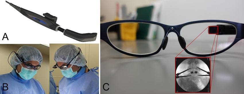
Matsukawa and Yato
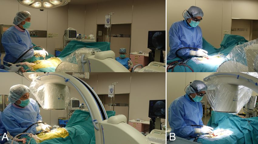
FIG. 1. Comparison of fluoroscopy-guided procedures. A: The surgeon views the standard fluoroscopic monitor by turning the
head away from the surgical field. B: The surgeon uses the smart glasses display device as an alternative method to display fluo-
roscopic images, and so the surgeon’s focus can be maintained on the surgical field. Figure is available in color online only.
There are two major problems with this surgical en- showing information overlaid onto the user’s field of view.
vironment. First, these monitors are not always located Compact, lightweight, and comfortable head-mounted dis-
in positions where they can be easily viewed by surgeons plays are now available that can be attached to any type of
or assistants, because of cumbersome equipment within glasses. By superimposing various kinds of intraoperative
limited-capacity operating rooms. Second, to check the information projected on the display within the user’s visu-
monitors during a delicate spinal procedure, surgeons are al field, device-assisted “smart” glasses allow surgeons to
forced to turn their head away and take their eyes off the keep their attention focused on the operative task without
surgical field, in some cases twisting their body or remov- taking their eyes away from the surgical field (Fig. 1B). In
ing it from the surgical field (Fig. 1A). These unnecessary the literature, some authors have reported the advantages
movements may result in inconvenience to surgeons and of using smart glasses in terms of improved accuracy and
cause technical difficulties and errors. For example, dur- reduced time during various surgical procedures.2–6 Al-
ing percutaneous pedicle screw placement, surgeons de- though minimally invasive surgery has become a popular
termine the insertion points, place needles at the appropri- technique and offers many benefits to both surgeons and
ate depth, and insert cannulated pedicle screws while fre- patients, these techniques depend heavily on indirect vi-
quently checking an intraoperative fluoroscopic monitor sualization and fluoroscopic navigation guidance. We hy-
to avoid displacing wires. Simultaneously, surgeons can pothesized that the utilization of device-assisted glasses for
confirm the correctness of screw placement by checking a the placement of minimally invasive spinal instrumenta-
neuromonitor. Such frequent eye movements between the tion might improve the safety of the surgery. However, to
surgical field and monitors interfere with the smooth pro- the best of our knowledge, little has been reported on the
gression of surgery, potentially leading to unintentional use of a device attached to surgical glasses in spine surgery
for monitoring surgical data projected in the surgeon’s field
hand deviation and critical misplacement, a longer opera-
of view. The aim of the present study was to examine the
tive time, and unnecessary radiation exposure. This ten- feasibility of using a smart glasses display device for fluo-
dency is marked in inexperienced surgeons. roscopy-guided posterior lumbar interbody fusion (PLIF)
Portable head-mounted display devices that present in- involving the percutaneous pedicle screw technique.
formation directly in the user’s visual field and promote
hands-free capabilities have been applied in medical and
healthcare fields in recent years.1 These devices are di- Methods
vided into two major categories, non–see-through displays This was a pilot prospective randomized study conduct-
obscuring the user’s field of view and see-through displays ed from April 2019 to March 2020. The study participants
J Neurosurg Spine Volume 34 • January 2021 151
Unauthenticated | Downloaded 10/07/21 11:13 PM UTCMatsukawa and Yato
FIG. 2. Image capture and streaming system using the smart glasses display device. Intraoperative information (only fluoroscopic
images in this study) is output to a transmission box using an HDMI signal, which is transmitted to the picoLinker device via WiFi.
consisted of 20 consecutively enrolled patients (15 men display device attached to the surgeon’s glasses, which dis-
and 5 women; mean age 61.9 ± 12.5 years, range 42–82 played information in the surgeon’s field of view, using an
years) who underwent single-segment PLIF at L5–S1. All encrypted connection via WiFi (Miracast). The time need-
20 operations were performed by the same surgeon, who ed for image transmission was about 0.2 seconds, which
used the percutaneous pedicle screw technique. There was fast enough for clinical use. A commercially available
were 15 patients with spondylolytic spondylolisthesis, four monocular see-through head-mounted display was used
with recurrent disc herniation, and one with foraminal in this study (picoLinker, Westunitis) (Fig. 3). The pico-
stenosis. Patients were randomly divided into two groups Linker is lightweight (30 g) and its flexible arm and con-
in a 1:1 allocation, one group in which the surgeon used nector clip can be easily attached to any type of glasses,
device-assisted smart glasses to perform the PLIF (smart including a surgical loupe,7 which allows free adjustment
glasses group) and another group in which the surgeon did to the user’s preferred position and quick removal if neces-
not use a smart glasses device (nonglasses group). Ran- sary due to discomfort during surgery. The picoLinker has
domization was achieved by an independent observer who more than 10 hours of battery life and a transparent prism
used the sealed envelope method. screen with resolution of 640 × 360 pixels. The display is
usually positioned in the right upper quadrant of the user’s
Image Capture and Streaming System Using the right eye and renders images equivalent to viewing a 23-
picoLinker Device inch screen at a distance of 2 m in front of the user.
Intraoperative fluoroscopically guided procedures were
conducted using an Arcadis Orbic 3D fluoroscopy device Surgical Procedure
(Siemens). First, real-time intraoperative fluoroscopic im- The same PLIF procedures were performed in all pa-
ages were output to a transmission box using a high-defi- tients, including bilateral total facetectomy, intervertebral
nition multimedia interface (HDMI) signal (Fig. 2). Then, discectomy, local bone grafting, and placement of two
this information was wirelessly transmitted to a wearable rectangular interbody cages, followed by bilateral percu-
FIG. 3. The picoLinker device used in this study. A: The device can be easily attached to any type of glasses using its flexible arm
and connector clip. Copyright Keitaro Matsukawa. Used with permission. B: The surgeon wearing the smart glasses device during
surgery. C: The smart glasses display device and a projected intraoperative fluoroscopic image. Figure is available in color online
only.
152 J Neurosurg Spine Volume 34 • January 2021
Unauthenticated | Downloaded 10/07/21 11:13 PM UTCMatsukawa and Yato
TABLE 1. Summary of patient characteristics (n = 20) TABLE 2. Summary of overall results
Patient Group Patient Group
Smart Glasses Nonglasses p Value Smart Glasses Nonglasses p Value
Mean age, yrs 60.8 ± 15.9 63.1 ± 8.4 0.69 No. of head turns 0.10 ± 0.31 82.4 ± 32.5 0.99 Operative time, mins 100.2 ± 10.4 105.5 ± 14.6 0.36
Mean BMI, kg/m2 24.5 ± 2.5 24.3 ± 2.5 0.73 Estimated blood loss, mL 55.1 ± 35.1 66.3 ± 38.3 0.51
Diagnosis >0.99 Radiation exposure time, sec 38.6 ± 6.6 41.8 ± 16.1 0.57
Spondylolytic 8 7 Screw perforation, no. (%) 0/40 (0%) 1/40 (2.5%) >0.99
spondylolisthesis
Values are presented as mean ± SD unless otherwise indicated.
Recurrent disc herniation 2 2
Foraminal stenosis 0 1
Values are presented as number of patients or mean ± SD. plications, such as intraoperative dural injury, postopera-
tive hematoma, or infection, in any of the other patients
in both groups.
taneous pedicle screw fixation. Fluoroscopic guidance was
typically used for the following procedures: preoperative Discussion
skin marking, cage placement, needle placement, tapping The present study is, to our knowledge, the first to in-
and screw insertion, bilateral rod placement, and reduction vestigate the feasibility of using display device–assisted
maneuvers as needed. In the glasses group, the surgeon uti- smart glasses during fluoroscopy-guided minimally in-
lized the picoLinker smart glasses system during all pro- vasive spine instrumentation surgery. The device-assisted
cess mentioned above, whereas in the nonglasses group, visualization technology, which can be attached to regular
the surgeon utilized a standard fluoroscopic monitor. glasses to display information in the user’s field of view,
allowed surgeons to proceed with the surgery without hav-
Evaluation ing to take their focus away from procedures, and thus this
The number of times the surgeon’s head turned to method has the potential to facilitate surgeon viewing of
view the fluoroscopic monitor during procedures and the monitoring processes and reduce radiation exposure.
operative time, estimated blood loss, radiation exposure Head-mounted display technology with medical appli-
time, accuracy of screw placement, and any intraopera- cations has been expanding in the current decade. Kaneko
tive complications were evaluated for comparison be- et al. and Lim et al. examined the merits of using smart
tween the two groups. Screw positions were assessed in glasses during an ultrasound-guided procedure in their
CT images obtained within 1 week postoperatively, and randomized control studies, and these investigators con-
screw perforation was defined as a screw with any corti- cluded that smart glasses improved ergonomic efficiency
cal breach. by reducing head movements.4,6 In the field of neurospine
surgery, several attempts to use smart glasses displays as
Statistical Analysis an intraoperative neuromonitor,8 endoscopic monitor,9
All results are shown as the mean ± standard devia- or 3D navigation screen have been reported. Yoon et al.
tion. Student and Welch t-tests were used for continuous investigated the feasibility of streaming navigation im-
variables, and Fisher exact tests were used for categorized ages onto smart glasses during pedicle screw placement.3
variables. JMP version 13 (SAS Institute) was used for all According to their report, time per screw placement was
analyses, and the significance was set at p < 0.05. 15.1% shorter when using smart glasses, with a user posi-
tive response rate of 79%. The results of the present study
showed similar trends in terms of shorter operative and
Results radiation exposure times (not significant). The small
There were no significant differences in patient demo- sample size and operator’s familiarity with the minimally
graphic or medical characteristics, such as age, sex, body invasive spine procedure could have a marked impact on
mass index, and diagnosis, between the two groups (Table the results to determine whether smart glasses display de-
1). The number of surgeon head turns to view the fluoro- vice–assisted visualization technology is of merit for use
scopic monitor in the smart glasses group was 0.10 ± 0.31 in pedicle screw placement.
times (range 0–1 times), which was significantly fewer In the lumbosacral region, due to the large volume of
than in the nonglasses group (82.4 ± 32.5 times, range the paraspinal muscles, deeper screw entry points, and
54–132 times; p < 0.001) (Table 2). The operative and ra- large medialized pedicle axis, the percutaneous pedicle
diation exposure times in the smart glasses group were screw technique is particularly effective compared with
shorter than those in the nonglasses group (operative time the conventional technique, which requires extensive
100.2 ± 10.4 vs 105.5 ± 14.6 minutes, radiation exposure muscle dissection for screw insertion. In addition, from
time 38.6 ± 6.6 vs 41.8 ± 16.1 seconds, respectively), al- anatomical standpoints, the sacral vertebrae have few
though the differences were not significant. Postoperative prominent structures, unlike the transverse process of the
CT showed one screw in the nonglasses group with a me- lumbar vertebrae that can be used to identify entry points,
dial breach of less than 2 mm. There were no other com- and S1 screw insertion into the sacral promontory requires
J Neurosurg Spine Volume 34 • January 2021 153
Unauthenticated | Downloaded 10/07/21 11:13 PM UTCMatsukawa and Yato
skillful use of fluoroscopic assistance. Consequently, par- was not investigated. Different conclusions may be drawn
ticularly in this region, frequent fluoroscopic checks are depending on the degree of the surgeon’s familiarity with
required to appropriately place screws. Therefore, the the use of glasses with an additional visual screen pro-
present study included only patients who underwent PLIF vided by an attached device. Second, we investigated only
at the L5–S1 segment. the radiation exposure time and lacked data on radiation
The results of the present study have practical implica- dose to surgeons and the number of images taken during
tions in terms of ergonomic improvements for surgeons fluoroscopy. Because the radiation dose is affected not
during procedures, because intraoperative support moni- only by the time of exposure, but also by the distance from
tors are not in line with the procedural site or the surgeons’ the radiation source and shielding devices, further studies
natural line of sight. Movement of the surgeon’s head away to assess the effective dose of fluoroscopic radiation are
from the surgical field could lead to hand deviation, result- warranted. Third, we evaluated the total operative time in-
ing in changes in position, direction, or depth within the stead of the time taken for each individual procedure, such
surgical field. However, using the smart glasses system as as skin marking, cage placement, and screw placement.
an alternative screen in the normal field of view allows The placement of percutaneous pedicle screws is one of
surgeons to see both the surgical field and the information the procedures that heavily depends on fluoroscopic as-
displayed on monitors simultaneously without moving sistance; therefore, an assessment of the time taken for
their eyes and head. In addition to fluoroscopic images, a screw placement may be useful to elucidate in detail the
variety of images, such as images obtained before surgery true benefits of using smart glasses.
and those for monitoring vital signs10 and neurophysiologi-
cal indicators,8 conducting computer navigation,3 and en- Conclusions
doscopic viewing, can be displayed on the wearable screen
(Fig. 2).9 When using a wireless connection, surgeons can To the best of our knowledge, this is the first report on
perform procedures more comfortably with full-body mo- the feasibility of using device-assisted smart glasses for
bility. fluoroscopy-guided minimally invasive spinal instrumen-
Furthermore, this novel visualization approach provides tation placement. The use of a smart glasses display device
two potential advantages alongside facilitating better con- such as this one may be a valid option to enhance the sur-
centration on the operative task. First, the device-assisted geon’s ability to concentrate on operative tasks by improv-
smart glasses system could contribute to shortening the ing ergonomic efficiency during surgery.
operative time and reducing intraoperative radiation ex-
posure. Chimenti and Mitten2 and Tsubosaka et al.5 per- References
formed a study involving fluoroscopy-guided pin insertion 1. Rahman R, Wood ME, Qian L, et al. Head-mounted display
for hand and femoral neck fracture and demonstrated that use in surgery:a systematic review. Surg Innov. 2020;27(1):
the use of smart glasses reduced the operative time by 88–100.
15%–33% and intraoperative radiation exposure time by 2. Chimenti PC, Mitten DJ. Google Glass as an alternative to
44%–59% compared with a control. In the present study, standard fluoroscopic visualization for percutaneous fixation
of hand fractures:a pilot study. Plast Reconstr Surg. 2015;
the use of a smart glasses display device (picoLinker) 136(2):328–330.
also seemed to reduce unintentional hand deviation dur- 3. Yoon JW, Chen RE, Han PK, et al. Technical feasibility and
ing procedures by eliminating the need to shift the focus safety of an intraoperative head-up display device during
between a standard monitor and the surgical field, leading spine instrumentation. Int J Med Robot. 2017;13(3):e1770.
to shorter operative and radiation exposure times. Second, 4. Kaneko N, Tsunoda M, Mitsuhashi M, et al. Ultrasound-
equipped with a camera and video-recording capabilities, guided fine-needle aspiration in the neck region using an
this new device could become a more useful adjunct tool optical see-through head-mounted display:a randomized
controlled trial. J Ultrasound Med. 2017;36(10):2071–2077.
in surgical education, information sharing, real-time con- 5. Tsubosaka M, Hiranaka T, Okimura K, et al. Additional visu-
sultation, and remote instruction and monitoring.1,11,12 alization via smart glasses improves accuracy of wire inser-
There are some concerns to be noted, such as the wear- tion in fracture surgery. Surg Innov. 2017;24(6):611–615.
ability of and fatigue caused by using a smart glasses de- 6. Lim H, Kim MJ, Park JM, et al. Use of smart glasses for ul-
vice. We did not conduct a survey to assess feedback from trasound-guided peripheral venous access:a randomized con-
multiple users on the usability and comfort of wearing trolled pilot study. Clin Exp Emerg Med. 2019;6(4):356–361.
such a device attached to glasses. However, the surgeon 7. Diaz R, Yoon J, Chen R, et al. Real-time video-streaming to
surgical loupe mounted head-up display for navigated menin-
in this study experienced no discomfort or adverse effects gioma resection. Turk Neurosurg. 2017;28(4):682–688.
and there was no significant increase in the operative time, 8. Golab MR, Breedon PJ, Vloeberghs M. A wearable headset
possibly owing to the lightweight body construction of the for monitoring electromyography responses within spinal
device, adjustability to the user’s preferred position, and surgery. Eur Spine J. 2016;25(10):3214–3219.
transparent monocular display that did not interfere with 9. Liounakos JI, Urakov T, Wang MY. Head-up display assisted
performing tasks. Our study included only patients under- endoscopic lumbar discectomy—a technical note. Int J Med
going single-segment fusion; thus, further research on the Robot. 2020;16(3):e2089.
effects of prolonged use in more complex cases is needed. 10. Iqbal MH, Aydin A, Lowdon A, et al. The effectiveness of
Google GLASS as a vital signs monitor in surgery:a simula-
Several limitations of this study should be acknowl- tion study. Int J Surg. 2016;36(Pt A):293–297.
edged. First, this study presented the initial experience of 11. Guze PA. Using technology to meet the challenges of medi-
using the display device–assisted smart glasses; however, cal education. Trans Am Clin Climatol Assoc. 2015;126:
the presence of a learning curve for the use of this system 260–270.
154 J Neurosurg Spine Volume 34 • January 2021
Unauthenticated | Downloaded 10/07/21 11:13 PM UTCMatsukawa and Yato
12. Rojas-Muñoz E, Cabrera ME, Lin C, et al. The System for the final version of the manuscript on behalf of both authors:
Telementoring with Augmented Reality (STAR):a head- Matsukawa. Statistical analysis: Matsukawa. Administrative/
mounted display to improve surgical coaching and confi- technical/material support: Matsukawa. Study supervision: both
dence in remote areas. Surgery. 2020;167(4):724–731. authors.
Correspondence
Disclosures Keitaro Matsukawa: National Hospital Organization, Murayama
Keitaro Matsukawa is a consultant for Westunitis. Medical Center, Tokyo, Japan. keitaro197897@hotmail.com.
Author Contributions
Conception and design: Matsukawa. Acquisition of data:
Matsukawa. Analysis and interpretation of data: Matsukawa.
Drafting the article: Matsukawa. Critically revising the article:
Yato. Reviewed submitted version of manuscript: Yato. Approved
J Neurosurg Spine Volume 34 • January 2021 155
Unauthenticated | Downloaded 10/07/21 11:13 PM UTCYou can also read