The importance of serum neutrophil gelatinase-associated lipocalin level in patients with lupus nephritis
←
→
Page content transcription
If your browser does not render page correctly, please read the page content below
J Renal Inj Prev. 2019; 8(2): 133-139.

                          http://journalrip.com                                                       DOI: 10.15171/jrip.2019.25

                                          Journal of Renal Injury Prevention

The importance of serum neutrophil gelatinase-
associated lipocalin level in patients with lupus nephritis
                                              ID
Mohammad Reza Jafari Nakhjavani1 , Sima Abediazar2* ID , Amir Ghorbanihaghjo3, Behnaz
Hanafizadeh1, Sepideh Zununi Vahed2, Tala Pourlak1,2
1
 Department of Rheumatology, Connective Tissue Diseases Research Center, Tabriz University of Medical Sciences, Tabriz, Iran
2
 Kidney Research Center, Tabriz University of Medical Sciences, Tabriz, Iran
3
 Biotechnology Research Center, Tabriz University of Medical Sciences, Tabriz, Iran

ARTICLE INFO                               ABSTRACT

Article Type:                              Introduction: The neutrophil gelatinase-associated lipocalin (NGAL) has emerged as a
Original                                   biomarker of renal damage.
                                           Objectives: The aim of this study was to assess the serum levels of NGAL (sNGAL) as a marker
Article History:                           of disease activity in individuals with lupus nephritis (LN).
Received: 12 November 2018                 Patients and Methods: This study contained 50 systemic lupus erythematosus (SLE)
Accepted: 2 January 2019                   individuals with (n = 25) and without (n = 25) nephritis, and 39 healthy controls. The sNGAL

                                                                                                                                              Original
Published online: 26 January 2019          levels were measured by ELISA. Renal function test, urinary parameters, lupus serology
                                           activity, and also calculated SLE disease activity index (SLEDAI) were analyzed to determine
Keywords:                                  their associations with sNGAL.
Systemic lupus erythematosus,              Results: The results revealed that the SLE individuals with or without nephritis had a raised
Biomarker, Lupus nephritis,                serum NGAL levels as compared to control subjects (P < 0.001). Additionally, sNGAL levels in
End-stage renal disease,                   LN individuals were meaningfully higher compared to those in non-LN patients (P < 0.001).
Chronic kidney diseases,                   Serum NGAL showed a significant correlation with the SLEDAI, serum creatinine, and 24-h
Neutrophil gelatinase-                     urinary protein (P < 0.05). More importantly, sNGAL had a significant positive correlation
associated lipocalin,                      with the activity index of LN (r = 0.616, P = 0.001). In the ROC curve analysis, the measurement
                                           of sNGAL level showed a good diagnostic performance for distinguishing individuals with
                                           LN from SLE patients without renal involvement with AUC = 0.902 (P < 0.001), 72% sensitivity,
                                           and 99% specificity. Moreover, sNGAL could identify all of SLE patients from controls with
                                           high accuracy, AUC= 0.99, P < 0.001, with 99% sensitivity, and 97% specificity.
                                           Conclusion: Serum NGAL had an association with clinical parameters and could discriminate
                                           LN from SLE patients without renal involvement. Our result suggests that serum NGAL can
                                           be used for early diagnosis of LN and identifying active LN.

    Implication for health policy/practice/research/medical education:
    The knowledge of LN activity and the disease condition presents significant role in the treatment and prognosis of lupus patients.
    In the present study, we evaluated the diagnostic values of serum NGAL in serum samples of lupus patients with and without
    nephritis and healthy controls. In summary, the increased urinary NGAL but not its serum levels have been confirmed as a
    predictor of renal involvement in SLE and LN activity. We found that serum NGAL can also be a useful biomarker for the
    identification of renal involvement in SLE patients, LN activity and the discrimination of LN among SLE patients. Further studies
    should be conducted to support the results.
    Please cite this paper as: Jafari Nakhjavani MR, Abediazar S, Ghorbanihaghjo A, Hanafizadeh B, Zununi Vahed S, Pourlak
    T. The importance of serum neutrophil gelatinase-associated lipocalin level in patients with lupus nephritis. J Renal Inj Prev.
    2019;8(2):133-139. doi: 10.15171/jrip.2019.25.

Introduction                                                                 Asians in comparison to other races (1). Within 15 years
Systemic lupus erythematosus (SLE) is a chronic                              after diagnosis, LN patients (10%–30%) progress the end-
inflammatory autoimmune disease with several clinical                        stage renal disease (ESRD) (2).
manifestations. Lupus nephritis (LN) is a serious renal                        Renal biopsy is a useful tool for diagnosis, prognosis,
involvement in SLE and is more prevalent in blacks and                       and monitoring therapy of LN. Yet, it is an inconvenient

*Corresponding author: Prof. Sima Abediazar, Email: sima_abedi@yahoo.com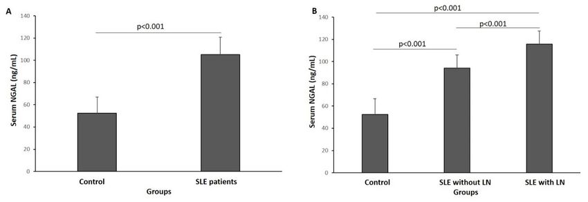
Jafari Nakhjavani MR et al

and invasive process, thus, the American College of                          hemodialysis or renal replacement therapy. All samples
Rheumatology (ACR) criteria is applied to diagnose LN                        were collected from September 2016 to June 2018. Disease
(3) using the quantity of 24-h urinary protein. This test                    activity was determined by the SLE disease activity index
has also often resulted in inaccurate results. Additionally,                 (SLEDAI) (32), the disease was considered active when
the urine sediment, glomerular filtration rate (GFR),                        the SLEDAI score was ≥6.
and complement components (like C3 and C4) are
considered to be too inaccurate to differentiate the acute                   Clinical and laboratory measurement
inflammatory and the chronic degenerative changes (4).                       For each patient, fresh blood sample was collected. Serum
Thus, alternative accurate biomarkers have been required                     samples were frozen within 2 hours after collection and
for LN diagnosis or monitoring (5). Presently, numerous                      stored at −8°C until further analysis. Serum creatinine
studies have been carried out to discover potential                          levels, serum chemistry, complement (C3 and C4), 24-h
biomarkers including urinary monocyte chemoattractant                        urinary protein, antinuclear antibodies (ANA), and anti-
protein-1 (uMCP-1)(6), transforming growth factor beta                       dsDNA antibody (anti-dsDNA) were analyzed. NGAL
1 (uTGF-β1), clusterin (7), and interleukin-17 (uIL-17).                     levels were assessed by enzyme-linked immunosorbent
Neutrophil gelatinase-associated lipocalin (NGAL), an                        assay (ELISA) benefitting a commercially available
iron-transporting protein (8), is a predictor of the acute                   kit (ZellBio GmbH, Cat No: ZB-11719c-H9648,
kidney injury (AKI) while it has also been related to the                    Germany) following the manufacturer’s instructions. All
LN.                                                                          measurements were conducted in triplicate. On the day
   As an innate antimicrobial agent, NGAL was originally                     of the sampling, information about patient demographic
identified in activated neutrophils (9,10). Following                        characteristics, medications, and disease activity was
ischemia, nephrotoxic, or AKI, NGAL is expressed and                         recorded.
accumulated in the kidney tubules and urine (11) in both
animal models and humans (12,13). Moreover, it may be                        Ethical issues
involved in the pathophysiology of chronic renal failure                     The protocol of the study was institutionally approved
such as chronic glomerulonephritis and polycystic kidney                     by the Clinical Research Ethics Committee of the
disease (14). NGAL is correlated with GFR and tubular                        Tabriz University of Medical Sciences, Tabriz, Iran (#
atrophy(15) and its high levels in serum/plasma and urine                    IR.TBZMED.REC.1396.598) and has therefore been
have been observed in patients with SLE (4,16), diabetic                     performed in accordance with the ethical standards laid
nephropathy (17), IgA nephropathy (18), and nephrotic                        down in the 1964 Declaration of Helsinki and its later
syndrome (19).                                                               amendments. The protocol of the study was clarified to all
   In a mouse model of LN, NGAL level in the kidney,                         participants and written informed consent was achieved
urine, and serum is elevated in connection with disease                      from the patients. This study was extracted from Behnaz
severity (20). Likewise, human studies have displayed that                   Hanafizadeh residency thesis in Tabriz University of
urinary NGAL (uNGAL) levels can reflect the decline                          Medical Sciences, Tabriz, Iran (Thesis# 58580).
of renal function and the disease activity in LN (5,21-
30). Furthermore, it may predict poor response after                         Statistical analysis
induction therapy (31). However, the value of serum                          Data were given as mean ± SD for normally or median
NGAL (sNGAL) as a predictor of renal flare is less clear                     with interquartile range (IQR) for non-parametric
because of contradictory results.                                            distributed variables. Categorical variables were compared
                                                                             by Chi-square test. For parametric and nonparametric
Objectives                                                                   data, the differences between groups were analyzed by
The aim of our study was to investigate the association of                   the student’s t test or ANOVA and Mann–Whitney U or
serum NGAL with renal involvement and LN activity in                         Kruskal-Wallis tests, respectively. Spearman’s correlation
SLE patients with and without nephritis.                                     coefficient was used to test correlations between sNGAL
                                                                             and other variables. Areas under ROC curves (AUC) and
Patients and Methods                                                         95% confidence intervals (CI) were calculated. Statistical
Patients’ selection                                                          analysis was performed using SPSS statistical software,
A cross-sectional study was conducted in Imam Reza                           version 16.0 (SPSS, Chicago, IL). P values less than 0.05
hospital, Tabriz, Iran and included 50 SLE patients with                     was considered significant.
and without nephritis. Patients suffering from any of
the following conditions were excluded; active infection                     Results
at the time of sampling, diabetes mellitus, history of                       This study comprised 50 individuals with SLE, 10 males
HIV and hepatitis C virus, patients with other type                          and 40 females, with a mean age of 36.7 ± 11 years old.
of glomerulopathy, overlap syndrome, malignancies,                           The individuals were divided into three groups: group 1;
urinary tract infection, and those with ESRD undergoing                      25 SLE individuals without nephritis and group 2; 25 SLE

134       Journal of Renal Injury Prevention, Volume 8, Issue 2, June 2019                                           http://journalrip.com
Serum NGAL in lupus nephritis
individuals with biopsy-proven LN. Group 3 included 39 LN) had a higher level of sNGAL as compared to controls
age/gender matched healthy subjects with a mean age of with mean of 105±15.9 versus 52±14.3 ng/mL (P < 0.001;
38.4 ± 9.7 years old and considered as controls. The mean Figure 1A). Additionally, level of sNGAL meaningfully
age of onset of LN was 35.4 ± 11.7 years old and their enhanced in cases with LN as compared to those without
disease diagnosed at the time of sampling. The severity LN, with mean of 115.67±11.92 ng/mL and 94.33±11.75
of lupus was evaluated among the patients. Eighty-eight ng/mL, respectively (P < 0001; Figure 1B). In patients who
percent of patients had an active disease (SLEDAI scores had CNS, skin, and kidney involvement, significantly
≥ 6), the median (IQR) score of LN patients was 10 (8- raised values of serum NGAL were observed (P < 0.003;
14) versus SLE without nephritis 6 (5.5 to 9). As expected, Table 2).
all patients with LN had significantly higher levels of In the LN group, significantly positive correlations
proteinuria (Median [IGR] of 1368 [926 to 2000] versus were detected between sNGAL (ng/mL) and SLEDAI (r=
125 [92-170]), lower levels of the complement, and 40% of 0.427, P = 0.033) and anti-dsDNA (r = 0.487, P = 0.014).
them had hematuria (n = 10) (Table 1). However, in terms Moreover, there was a strong positive correlation between
of mean age and gender no significant differences were serum NGAL levels and renal parameters such as 24 hours
observed (P > 0.05). urinary proteins (r = 0.793), serum creatinine (r = 0.756),
Twenty-two LN cases (92%) had active disease (rSLEDAI and LN activity index (r = 0.616), (P < 0.001). Likewise,
≥8). Cases had normal serum creatinine levels. Twenty- in SLE patients without LN, significant correlations were
six SLE patients (52%) had inflammatory arthritis, 52% found between sNGAL and SLEDAI (r= 0.450, P = 0.024),
of the cases had rash, and eight (16%) had a prevalence and proteinuria (r = 0.453, P = 0.023) (Figures 2C and D).
of serositis. Some of SLE patients had leucopenia Moreover, there were correlations between sNGAL and
(24%), hematuria (20%), and thrombocytopenia (12%). other clinical variables in SLE patients with and without
Moreover, five cases (10%) had central nervous system LN, however, they were not statistically significant.
(CNS) involvement. The results showed that the incidence Additionally, correlations between sNGAL and clinical
of hematuria and rash in the two groups was significantly manifestations of the cases are listed in Table 3.
different. The ROC curve for sNGAL was calculated to
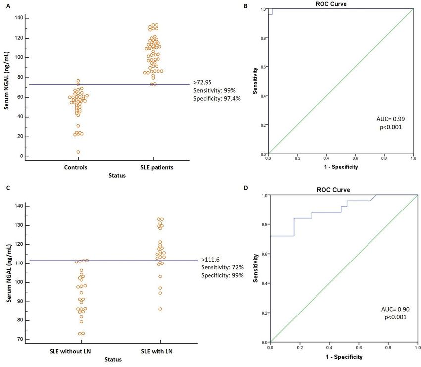
Our results showed that the SLE group (with or without discriminate SLE cases from healthy controls and also SLE
Table 1. Demographic and baseline clinical data
Characteristics/Groups Total SLE patients SLE without LN SLE with LN P Valuea
Demographic characteristics
No. of cases 50 25 25
Gender, No. (%)
10 (20) 5 (20) 5 (20) 1
Male
40 (80) 20 (80) 20 (80)
Female
Age, mean ± SD (y) 36.7 ± 11.3 38 ± 10.9 35.4 ± 11.7 0.429
Biomarkers
C3 (mg/dL) 47.3±31.27 72.28±24.0 22.32 ±10.98Jafari Nakhjavani MR et al
Figure 1. Serum level of NGAL in the studied groups. (A) Serum levels of NGAL between healthy controls and SLE patients and (B)
between SLE patients with and without nephritis. Values are mean± standard deviation. NGAL: neutrophil gelatinase associated lipocalin.
patients with LN from those with non-renal flare (Figure with a cutoff of 72.95 ng/mL and AUC = 0.99 (P < 0.001)
3). The diagnostic performance of sNGAL presented that (Figure 3A and B). Moreover, diagnostic reliability study
with high sensitivity (99%) and specificity (97%), sNGAL in the present study detected a good efficacy for sNGAL
could discriminate SLE patients from healthy controls to distinguish SLE individuals with nephritis from those
without nephritis with the best cutoff of 111.6 (AUC=
Table 2. Serum levels of NGAL based on organ involvement in SLE 0.90) (P < 0.001), sensitivity = 72%, and specificity = 99%
patients (N = 50)
(Figure 3C and D).
Organs Not-involved Involved P value
CNS 102.83±15.0 124.56±9.02 0.003 Discussion
Skin 111.67±15.73 98.84±13.66 0.003
In the current investigation, we studied the NGAL and
traditional measures of LN in serum samples of SLE
Arteritis 107.74±15.94 102.47±15.77 0.246
patients. We found that the serum NGAL was significantly
Leucopenia 104.46±16.50 106.71±14.42 0.674
higher in SLE individuals in comparison to the control
Serositis 103.94±15.6 110.55±17.22 0.278 subjects. Furthermore, sNGAL was even more elevated
Thrombocytopenia 105.33±16.02 102.58±16.33 0.696 in SLE patients with LN when compared to those without
Kidney 94.33±11.75 115.67±11.92Serum NGAL in lupus nephritis
Figure 2. Correlation of serum NGAL with clinical parameters. Correlation
between serum NGAL and (A) SLEDAI, (B) anti-double strand DNA
antibodies, (C) proteinuria, and (D) LN activity index in SLE patients with Figure 3. Receiver operating characteristic curves of serum NGAL.
nephritis. Graph ROC curves analysis showing AUC of serum NGAL to discriminate
(A, B) SLE patients from healthy controls and (C and D) SLE patients with
LN from those without LN. AUC: Area under the Curve, NGAL: neutrophil
gelatinase associated lipocalin.
The biologic function of NGAL is under active
investigation (20). Pawar et al concluded that renal
binding of pathogenic antibodies induces expression disease activity in the clinic, the present study did not find
of NGAL. Increased level of NGAL has a critical role in a statistically significant association between sNGAL and
the pathogenesis of nephritis through the promotion of these clinical parameters. Our results are in line with the
inflammatory gene expression and apoptosis in kidney study of Pitashny et al (34) who showed a highly significant
cells both in vitro and in vivo (20). NGAL is a predictive association between a renal parameter of SLEDAI score.
and early urinary marker that is induced rapidly by active However, they could not find an association between the
inflammation in LN and declined with therapy (16). Data complement or anti-dsDNA antibodies and uNGAL levels
indicated that high uNGAL levels reflect the activity and in adult patients.
severity of kidney disease in SLE patients (4,16,21,27,28, Recently, a meta-analysis performed on fourteen studies
33,34). has confirmed that uNGAL relatively has reasonable
Our result of enhanced serum values of NGAL in LN sensitivity and specificity in identifying LN and
is in line with those reported in the literature; however, approximating LN activity, and also guessing kidney flares.
their results were not statistically significant (24,27,28,35). The result suggests that uNGAL is a possible biomarker in
This may be due to different sample sizes. NGAL has monitoring LN activity and diagnosing LN with a pooled
been demonstrated as a marker of renal damage in LN sensitivity of 73.6% and specificity of 78.1% (26). Brunner
(4,29), which may explain why our patients with a history et al indicated that the measurement of NGAL together
of biopsy-proven LN had higher serum NGAL levels. In with GFR and MCP-1 was an admirable diagnostic tool for
contrast with these studies, Smith et al could not find a LN chronicity with AUC = 0.83 (37). Likewise, we found
significant change in uNGAL levels between patients with that serum NGAL could also discriminate LN patients
active LN and those with non-LN in two international from SLE patients without nephritis with high accuracy
juvenile-onset SLE cohorts (25). Moreover, Kiani et al (AUC = 0.902), 72% sensitivity, and 99% specificity. Our
could not detect a relationship between uNGAL and LN result suggests that serum NGAL can be also used for
in adult SLE patients (36). These results can be clarified early diagnosis of LN and identifying active LN.
by differences in the sampling time and the outcome Generally, it is approved that besides being the most
measures used. These remarks suggest that NGAL needs hopeful next-generation biomarker in clinical nephrology,
further examination to be considered as a urine or serum NGAL stimulates the growth and differentiation of kidney
biomarker to predict nephritis occurrence. tubular epithelial cell in reply to ischemic injuries (38).
Regarding the connotation between kidney functions Currently, in spite of some progressive research, the
and NGAL, our investigation disclosed a meaningful biological importance of NGAL needs further examination
positive association between sNGAL and each of 24 in nephrology.
hours of urine protein, serum creatinine, anti-dsDNA
antibodies, and activity index of LN. Although ANA and Conclusion
complement are generally used as the indicators of kidney In summary, the increased urinary NGAL but not its
http://journalrip.com Journal of Renal Injury Prevention, Volume 8, Issue 2, June 2019 137Jafari Nakhjavani MR et al
serum levels have been confirmed as a predictor of kidney renal protein biomarkers with histologic features of lupus
involvement in SLE and LN activity. We found that serum nephritis. Arthritis Rheum. 2012;64:2687-97. doi: 10.1002/
NGAL can also be a useful biomarker for the identification art.34426.
7. Wu CY, Yang HY, Chien HP, Tseng MH, Huang JL. Urinary
of renal involvement in SLE patients and LN activity and
clusterin-a novel urinary biomarker associated with
the discrimination of LN among SLE patients. Further pediatric lupus renal histopathologic features and renal
studies should be conducted to support the results. survival. Pediatr Nephrol. 2018;33:1189-1198. doi: 10.1007/
s00467-018-3924-4.
Study limitations 8. Mao S, Jiang T, Shang G, Wu Z, Zhang N. Increased
We suppose that the small sample size of our studied expression of neutrophil gelatinase-associated lipocalin
groups was not adequate. Hence, further longitudinal receptor by interleukin-1beta in human mesangial cells via
larger scale studies are suggested to confirm the diagnostic MAPK/ERK activation. Int J Mol Med. 2011;27:555-60. doi:
10.3892/ijmm.2011.613.
value of serum NGAL in LN.
9. Goetz DH, Holmes MA, Borregaard N, Bluhm ME,
Raymond KN, Strong RK. The neutrophil lipocalin NGAL
Authors’ contribution is a bacteriostatic agent that interferes with siderophore-
MRN and SA designed the study and selected the cases. mediated iron acquisition. Mol Cell. 2002;10:1033-43.
BH did sampling. AGH performed experimental analysis 10. Kjeldsen L, Johnsen AH, Sengeløv H, Borregaard N.
and interpretation of the data. SZV prepared the draft. All Isolation and primary structure of NGAL, a novel protein
authors read and signed the final paper. associated with human neutrophil gelatinase. J Biol Chem.
1993;268:10425-32.
11. Mishra J, Ma Q, Prada A, Mitsnefes M, Zahedi K, Yang J,
Conflicts of interest
et al. Identification of neutrophil gelatinase-associated
The authors declared no potential conflicts of interest with lipocalin as a novel early urinary biomarker for ischemic
respect to the research, authorship, and/or publication of renal injury. J Am Soc Nephrol. 2003;14:2534-43.
this article. 12. Supavekin S, Zhang W, Kucherlapati R, Kaskel FJ, Moore
LC, Devarajan P. Devarajan, Differential gene expression
Ethical considerations following early renal ischemia/reperfusion. Kidney Int.
Ethical issues (including plagiarism, data fabrication, 2003;63:1714-24. doi: 10.1046/j.1523-1755.2003.00928.x.
double publication) have been completely observed by the 13. Mishra J, Dent C, Tarabishi R, Mitsnefes MM, Ma Q,
Kelly C, et al. Neutrophil gelatinase-associated lipocalin
authors.
(NGAL) as a biomarker for acute renal injury after cardiac
surgery. Lancet. 2005;365:1231-8. doi: 10.1016/s0140-
Funding/Support 6736(05)74811-x.
This research was funded by the Connective Tissue 14. Bolignano D, Donato V, Coppolino G, Campo S, Buemi
Diseases Research Center of Tabriz University of Medical A, Lacquaniti A, et al. Neutrophil gelatinase-associated
Sciences, Tabriz, Iran (Grant # 5/D/577304). lipocalin (NGAL) as a marker of kidney damage.
Am J Kidney Dis. 2008;52:595-605. doi: 10.1053/j.
References ajkd.2008.01.020.
1. Longo DL. La práctica de la medicina clínica. Cap. 1 En: 15. Bolignano D, Lacquaniti A, Coppolino G, Donato V,
DL Longo, DL Kasper, JL Jameson, AS Fauci, SL Hauser y Campo S, Fazio MR, et al. Neutrophil gelatinase-associated
J. Loscalzo (Eds.) Harrison Principios de Medicina Interna. lipocalin (NGAL) and progression of chronic kidney
México: McGraw-Hill-Interamericana. 2012;1596–1611. disease. Clin J Am Soc Nephrol. 2009;4:337-44. doi:
2. Avihingsanon Y, Hirankarn N. Major lupus organ 10.2215/cjn.03530708.
involvement: severe lupus nephritis. Lupus. 2010;19:1391- 16. Hinze CH, Suzuki M, Klein-Gitelman M, Passo MH,
8. doi: 10.1177/0961203310376522. Olson J, Singer NG, et al. Neutrophil gelatinase-associated
3. Dooley MA. Clinical and laboratory features of lupus lipocalin is a predictor of the course of global and renal
nephritis, Wallace DJ, Hahn BH. Dubois’ Lupus childhood-onset systemic lupus erythematosus disease
Erythematosus7th ed. Philadelphia: Lippincott Williams & activity. Arthritis Rheum. 2009;60:2772-81. doi: 10.1002/
Wilkins. 2007;1112-30. art.24751.
4. Brunner HI, Mueller M, Rutherford C, Passo MH, Witte 17. Kim SY, Jeong TD, Lee W, Chun S, Sunwoo S, Kim SB, et
D, Grom A, et al. Urinary neutrophil gelatinase-associated al. Plasma Neutrophil Gelatinase-Associated Lipocalin as a
lipocalin as a biomarker of nephritis in childhood- Marker of Tubular Damage in Diabetic Nephropathy. Ann
onset systemic lupus erythematosus. Arthritis Rheum. Lab Med. 2018;38:524-529. doi: 10.3343/alm.2018.38.6.524.
2006;54:2577-84. doi: 10.1002/art.22008. 18. Ding H, He Y, Li K, Yang J, Li X, Lu R, et al. Urinary
5. Susianti H, Iriane VM, Dharmanata S, Handono K, neutrophil gelatinase-associated lipocalin (NGAL) is an
Widijanti A, Gunawan A, et al. Analysis of urinary TGF- early biomarker for renal tubulointerstitial injury in IgA
beta1, MCP-1, NGAL, and IL-17 as biomarkers for lupus nephropathy. Clin Immunol. 2007;123L227-34.
nephritis. Pathophysiology. 2015;22:65-71. doi: 10.1016/j. 19. Bolignano D, Coppolino G, Campo S, Aloisi C, Nicocia G,
pathophys.2014.12.003. Frisina N, et al. Urinary neutrophil gelatinase-associated
6. Brunner HI, Bennett MR, Mina R, Suzuki M, Petri M, lipocalin (NGAL) is associated with severity of renal
Kiani AN, et al. Association of noninvasively measured disease in proteinuric patients. Nephrol Dial Transplant.
138 Journal of Renal Injury Prevention, Volume 8, Issue 2, June 2019 http://journalrip.comSerum NGAL in lupus nephritis
2008;23:414-6. doi: 10.1093/ndt/gfm541. a potential biomarker for renal damage in patients with
20. Pawar RD, Pitashny M, Gindea S, Tieng AT, Levine B, systemic lupus erythematosus. J Biomed Biotechnol.
Goilav B, et al. Neutrophil gelatinase-associated lipocalin 2012;2012:759313. doi: 10.1155/2012/759313.
is instrumental in the pathogenesis of antibody-mediated 30. Killock D. Lupus nephritis: NGAL: not just an innocent
nephritis in mice. Arthritis Rheum. 2012; 64:1620-31. doi: bystander? Nat Rev Rheumatol. 2011;8:3. doi: 10.1038/
10.1002/art.33485. nrrheum.2011.192.
21. Suzuki M, Wiers KM, Klein-Gitelman MS, Haines KA, 31. Satirapoj B, Kitiyakara C, Leelahavanichkul A, Avihingsanon
Olson J, Onel KB, et al. Neutrophil gelatinase-associated Y, Supasyndh O. Urine neutrophil gelatinase-associated
lipocalin as a biomarker of disease activity in pediatric lipocalin to predict renal response after induction therapy
lupus nephritis. Pediatr Nephrol. 2008;23:403-12. doi: in active lupus nephritis. BMC Nephrol. 2017;18:263. doi:
10.1007/s00467-007-0685-x. 10.1186/s12882-017-0678-3.
22. Ahlawat RS, Batra V, Pendharkar S. Urinary TWEAK and 32. Jafari-Nakhjavani MR, Abedi-Azar S, Nejati B .Correlation
NGAL as a biomarker of lupus nephritis activity. J Assoc of plasma interleukin-18 concentration and severity
Physicians India. 2016;64:69. of renal involvement and disease activity in systemic
23. Gómez-Puerta JA, Ortiz-Reyes B, Urrego T, Vanegas- lupus erythematosus. J Nephropathol. 2016;5:28-33. doi:
García AL, Muñoz CH, González LA, et al. Urinary 10.15171/jnp.2016.05.
neutrophil gelatinase-associated lipocalin and monocyte 33. Rubinstein T, Pitashny M, Levine B, Schwartz N,
chemoattractant protein 1 as biomarkers for lupus nephritis Schwartzman J, Weinstein E, et al. Urinary neutrophil
in Colombian SLE patients. Lupus. 2018;27: 637-646. doi: gelatinase-associated lipocalin as a novel biomarker
10.1177/0961203317738226. for disease activity in lupus nephritis. Rheumatology.
24. El Shahawy MS, Hemida MH, Abdel-Hafez HA, El-Baz 2010;49:960-71. doi: 10.1093/rheumatology/kep468.
TZ, Lotfy AM, Emran TM. Urinary neutrophil gelatinase- 34. Pitashny M, Schwartz N, Qing X, Hojaili B, Aranow C,
associated lipocalin as a marker for disease activity in lupus Mackay M, et al. Urinary lipocalin-2 is associated with
nephritis. Scand J Clin Lab Invest. 2018;78:264-268. doi: renal disease activity in human lupus nephritis. Arthritis
10.1080/00365513.2018.1449242. Rheum. 2007;56:1894-903. doi: 10.1002/art.22594.
25. Smith EM, Jorgensen AL, Midgley A, Oni L, Goilav B, 35. Tawfik Y, Shaat RM, El-Bassiony SR, Hawas S, Effat N.
Putterman C, et al. International validation of a urinary Urinary and serum neutrophil gelatinase-associated
biomarker panel for identification of active lupus nephritis lipocalin as a biomarker in Egyptian systemic lupus
in children. Pediatr Nephrol. 2017;32:283-295. doi: 10.1007/ erythematosus patients: Relation to lupus nephritis
s00467-016-3485-3. and disease activity. The Egyptian Rheumatologist.
26. Fang YG, Chen NN, Cheng YB, Sun SJ, Li HX, Sun 2015;37:S25-S31. doi: 10.1016/j.ejr.2015.09.003.
F, et al. Urinary neutrophil gelatinase-associated 36. Kiani AN, Wu T, Fang H, Zhou XJ, Ahn CW, Magder LS,
lipocalin for diagnosis and estimating activity in lupus et al. Urinary vascular cell adhesion molecule, but not
nephritis: a meta-analysis. Lupus. 2015;24:1529-39. doi: neutrophil gelatinase-associated lipocalin, is associated
10.1177/0961203315600244. with lupus nephritis. J Rheumatol. 2012;39:1231-7. doi:
27. Torres-Salido MT, Cortés-Hernández J, Vidal X, Pedrosa 10.3899/jrheum.111470.
A, Vilardell-Tarrés M, Ordi-Ros J. Neutrophil gelatinase- 37. Brunner HI, Bennett MR, Mina R, Suzuki M, Petri M,
associated lipocalin as a biomarker for lupus nephritis. Kiani AN, et al. Non-invasive renal protein biomarkers
Nephrol Dial Transplant. 2014;29:1740-9. doi: 10.1093/ndt/ are associated with histological features of lupus nephritis.
gfu062. Arthritis Rheum. 2012;64:2687–2697. doi: 10.1002/
28. Hammad A, Mosaad Y, Elhanbly S, Youssef H, El Refaaey A, art.34426.
Elhusseini F, et al. Urinary neutrophil gelatinase-associated 38. Gwira JA, Wei F, Ishibe S, Ueland JM, Barasch J, Cantley LG.
lipocalin as a marker of severe lupus nephritis in children. Expression of neutrophil gelatinase-associated lipocalin
Lupus. 2013;22:486-91. doi: 10.1177/0961203313479419. regulates epithelial morphogenesis in vitro. J Biol Chem.
29. Yang CC, Hsieh SC, Li KJ, Wu CH, Lu MC, Tsai CY, et 2005;280:7875-82. doi: 10.1074/jbc.M413192200.
al. Urinary neutrophil gelatinase-associated lipocalin is
Copyright © 2019 The Author(s); Published by Nickan Research Institute. This is an open-access article distributed under the
terms of the Creative Commons Attribution License (http://creativecommons.org/licenses/by/4.0), which permits unrestricted use,
distribution, and reproduction in any medium, provided the original work is properly cited.
http://journalrip.com Journal of Renal Injury Prevention, Volume 8, Issue 2, June 2019 139You can also read