Rivaroxaban triggered multifocal intratumoral hemorrhage of the cabozantinib-treated diffuse brain metastases: A case report and review of literature
←
→
Page content transcription
If your browser does not render page correctly, please read the page content below
Open Medicine 2021; 16: 618–623
Case Report
Luyue Chen, E Chen, Yanlin Huang, Xinhua Tian*
Rivaroxaban triggered multifocal intratumoral hemorrhage of
the cabozantinib-treated diffuse brain metastases: A case
report and review of literature
https://doi.org/10.1515/med-2021-0261
received October 26, 2020; accepted March 1, 2021
1 Introduction
Abstract: Brain metastases (BMs) are the most common Metastatic brain tumor is the hallmark of disseminated
intracranial malignancy with poor prognosis. Patients end stage disease condition in patients with cancers. At
with intracranial tumors are at greater risk for thrombotic this stage, patients are prone to venous thromboembo-
complications and intracranial hemorrhage. Rivaroxaban lism (VTE) and intratumoral hemorrhage (ITH) [1–3]. The
is a potent oral anticoagulant with the high selectivity of spontaneous hemorrhagic potential of brain metastases
direct factor Xa inhibition. The incidence and severity (BMs) contraindicates the routine use of anticoagulants
of rivaroxaban-triggered intratumoral hemorrhage (ITH) for VTE treatment and prophylaxis. However, increasing
in patients with BMs remain unknown. A 57-year-old evidence suggests no harm of therapeutic anticoagulants
woman was diagnosed with multiple lung, bone, and and guides the change in recommendation for patients
BMs from unknown primary cancer origin, and refused with BMs [2,4–8]. In this study, we report that a patient
any invasive procedures to confirm tumor pathology. with innumerable BMs from unknown primary site, pre-
However, this patient had a relatively favorable outcome viously treated with cabozantinib, developed concurrent
after treating with cabozantinib, an inhibitor of multiple multifocal intracerebral hemorrhage after acute treat-
tyrosine kinases. The patient survived over 2 years and ment of deep vein thrombosis (DVT) by oral rivaroxaban.
developed deep vein thrombosis of right lower limb. Oral As far as our concern, the understanding of rivaroxaban-
rivaroxaban was prescribed, and the multifocal cata- associated ITH is relatively limited in patients with BMs,
strophic ITH was encountered after 1 week. The last and the unique clinical presentation of this case may
head computed tomography imaging revealed a rare contribute to further understanding of this situation.
but typical image of diffuse hemorrhagic metastases. We provided the timeline for disease progression and
Hemorrhagic-prone BMs, therapeutic rivaroxaban, and reviewed literature for the possible risk factors (Figure 1a).
cabozantinib treatment increase risks to develop ITH. In
this case rivaroxaban was the trigger to this terminal
event. This case is a miserable lesson and keeps reminding
us to stay vigilant in clinical practice even when there is a 2 Case presentation
potential benefit for anticoagulation in such population.
A 57-year-old woman was diagnosed with multiple lung,
Keywords: brain metastases, rivaroxaban, intratumoral bone, and BMs from unknown primary cancer origin by a
hemorrhage, venous thromboembolism cancer center and refused any invasive procedures to
confirm tumor pathology. No other medical history had
been recorded. As a compromise, the patient agreed to
receive a liquid biopsy, which confirmed a CCDC6-RET
rearrangement in circulating tumor DNA. An oncologist
recommended the regimen of whole brain radiotherapy
(WBT) in combination with targeted therapy to control
* Corresponding author: Xinhua Tian, Department of Neurosurgery, systemic disease progression. However, WBT was denied
Zhongshan Hospital Xiamen University, 201-209 Hubin South Road, by the patient herself and family because of the concern
Siming District, Xiamen, 361004, Fujian, People’s Republic of China,
of neurological complications in exposing to radia-
e-mail: txhmd@outlook.com, tel: +86-05922292032
Luyue Chen, E Chen, Yanlin Huang: Department of Neurosurgery,
tion, even though the probable survival benefit was
Zhongshan Hospital Xiamen University, Xiamen, 361004, Fujian, repeatedly emphasized. Targeted therapy was the only
People’s Republic of China acceptable therapeutic option, and the treatment began
Open Access. © 2021 Luyue Chen et al., published by De Gruyter. This work is licensed under the Creative Commons Attribution 4.0
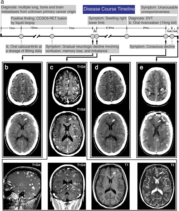
International License.Rivaroxaban triggered multifocal brain metastases hemorrhage 619 Figure 1: Timeline of major disease-related events and representative images of head imaging. (a) A schematic timeline displayed the major events during disease progression. (b) The initial head CT revealed extensive iso-dense and several hyperdense ill-defined masses. (c) CE- MRI identified diffuse T1 gadolinium-positive (T1 Gd+) lesions. Several high signals on T1 image indicated ITH, and typically hyperintense metastases were identified without significant mass effects. (d) There was negative finding on head CT imaging when the patient was diagnosed with DVT. (e) The last head CT showed diffuse open-ring or hollow circles of hyperintensities with sulci effacement, compatible with diffuse hemorrhagic metastases. The white arrow pointed to a same hemorrhagic metastasis.
620 Luyue Chen et al. with cabozantinib, an oral inhibitor against the tyrosine and international normalized ratio were slightly elevated, kinase domain of CCDC6-RET fusion, at a dosage of 60 mg and platelet count was normal. The head CT scan con- daily. The chief complaints of this patient were frequent firmed the diagnosis and revealed the extensive but sepa- fatigue, multiple bone pain, and occasional dizziness. No rate foci of intraparenchymal hemorrhage and general neurological deficits were found on the initial physical brain swelling (Figure 1e). Based on her previous history examination. After 17 months of medication, the patient of extensive BMs, the open-ring or hollow circles of hyper- visited our emergency room (ER) for the first time and intensities which occupied the majority of hemorrhagic presented with a 1-month history of gradual neurologic sites were suspected to be hemorrhagic metastases. The decline involving confusion, memory loss, and imbal- patient developed dyspnea right after the CT scan. How- ance. A head computed tomography (CT) performed ever, after discussions with the patient’s family, the goals without the administration of contrast material revealed of care were shifted toward comfort measures and no intu- extensive iso-dense and several hyperdense ill-defined bation was performed. mass located supratentorially at the junction of gray and white matters. Only sporadic minor peritumoral Consent for publication: Written informed consent for edema and ITH or calcification were observed at the publication of their clinical details and/or clinical images initial CT scan (Figure 1b). The patient was assigned to was obtained from the daughter of the patient. A copy of an observation room for subsequent contrast-enhanced the consent form is available for review by the Editor of magnetic resonance imaging (CE-MRI). After 3 days, a this journal. gadolinium-based CE-MRI was performed, and during this period, no further deterioration in clinical status Ethics approval and consent to participate: The CARE was encountered. Contrasted MRI showed innumerable guidelines were thoroughly followed to present this case. avid enhancements of the parenchymal metastases, which distribute across brain lobes, cerebrum, cere- bellum, and brain stem. Sporadic hemorrhagic other than calcified metastases were confirmed on T1, which 3 Discussion was indicated by intrinsic high signal (Figure 1c). Only mild peritumoral edema was identified on T2, and the use BMs are the most common intracranial tumors, and the of mannitol or corticosteroids was suspended. As a con- incidence is estimated at least three times the number of cern of disease progression at the primary and metastatic newly diagnosed primary malignant brain tumors [9]. sites, a tissue biopsy and WBT were recommended and The most frequent primary sites for BMs are lung cancer, again denied. The patient was then discharged in a week. breast cancer, and melanoma, accounting for 67–80% of After taking cabozantinib for 26 months, this patient was all cancers [10,11]. In patients with four or more BMs, transferred to our ER in a coma with a Glasgow Coma incidence in patients was negatively correlated with the Scale score of 3 (E1 V1 M1). This patient had a 7-day number of metastases, and only less than 5% of patients history of conscious decline and was found to be unre- were diagnosed with 10 or more BMs [12]. With regard sponsive for 2 h. Before 2 weeks, because of a swelling to this patient, countless number of BMs is a very rare right lower limb for over 2 months, DVT was diagnosed performance and passive attitude to receive standard at our department of vascular surgery by compression treatment could exacerbate the disease progression. CCDC6- ultrasonography (CUS). The laboratory test revealed no RET rearrangement identified in circulating tumor DNA coagulopathy, thrombocytopenia, or severe liver/renal provided an acceptable option of cancer management, dysfunction. The D-dimer level was 6,950 ng/mL (normal and the patient settled for targeted treatment. Cabozan- level:
Rivaroxaban triggered multifocal brain metastases hemorrhage 621
intracranial activity of cabozantinib in MET-positive lung incidence of VTE or death because of VTE in high-risk
cancer with BMs [15]. Another report indicated that cabo- ambulatory patients with cancer. However, during the
zantinib was able to reach brain tumors and induce signifi- 180-day trial period, rivaroxaban led to a substantially
cant regression in two patients with radioresistant BMs lower incidence of such events, with a low incidence of
from renal cell carcinoma [16]. Therefore, it is likely that major bleeding [20]. As the aforementioned concern of
this patient could benefit from cabozantinib treatment at anticoagulant-related intracranial bleeding, both trials
the primary and metastatic tumors, though histopathology exclude patients with BMs and whether these population
remains unknown. with VTE could safely benefit from rivaroxaban treatment
Recent advances in chemotherapy and targeted remains to be further investigated.
therapies have significantly prolonged the life expec- In our case, the patient received unexpected clinical
tancy in patients with systemic malignancy, but in the benefit from cabozantinib treatment. The patient survived
meantime, the lifetime risks to develop BMs and cancer- over 2 years until the presence of catastrophic ITH or ICH.
associated comorbidities increase [17]. Patients with Prior minor bleeding of BMs, therapeutic anticoagula-
intracranial tumors are at greater risk for thrombotic tion, and cabozantinib treatment were possible causes
complications and ICH, which accelerates the natural responsible for the eventual multifocal ICH of this
course to the end-of-life period. Significant ICH is reported patient. Up to 50% of patients with BMs suffered from
to occur in 20–50% of patients with BMs [2]. Therefore spontaneous ICH [2], and the initial head CT/MRI ima-
BMs are considered to be a bleeding risk factor and ging of this patient had revealed several minor intra-
excluded from most of the selected anticoagulant trials. tumoral bleedings, indicating the high-risk hemorrhagic
Another leading cause of morbidity and mortality in potential (Figure 1b and c). Prior hemorrhagic BMs is a
this population is the thrombotic complications. Active relative contraindication in therapeutic anticoagulation,
cancer is a well-established risk factor for DVT and pul- while the active major bleeding is a situation in which
monary embolism, collectively referred to as VTE. Throm- anticoagulation should not be given. Therefore after
boprophylaxis and remedy with anticoagulants may be confirmation of DVT by CUS and significantly elevated
offered by clinician to selected high-risk cancer patients D-dimer level, the patient received the second head
who will inevitably suffer from a high rate of VTE recur- CT and no trace of hemorrhage diminished the concern
rence and bleeding complications [4]. A meta-analysis of hemorrhagic complication of therapeutic rivaroxaban
focused on ICH in patients with brain tumors receiving (Figure 1d). Anticoagulant therapy with NOACs is a
therapeutic anticoagulation, and the conclusion is that simple regimen for treating acute DVT without the need
no significantly increased risk of ICH in patients with for repeated subcutaneous injection and laboratory mon-
BMs [7]. The safety of long-term anticoagulation in 125 itoring. Increasing evidence supports the safety of NOACs
patients with BMs has been reviewed by a retrospective in patients with BMs, without significant increase in the
study, and the result demonstrated that the incidence risk of ICH, but the risk of spontaneous ICH can reach as
of ICH did not increase with the use of anticoagulant high as 50% in this population [2,7,8]. The exact inci-
therapy [8]. A matched cohort study on ICH in patients dence of rivaroxaban-associated ICH in patients with
with BMs treated with therapeutic enoxaparin concluded BMs is still unknown, and further clinical studies are
that therapeutic anticoagulation in such population did required to ascertain whether the risk of harm associated
not increase the risk of ICH, but there was four-fold with hemorrhagic complications exceeds the potential
higher about the risk of ICH in patients with melanoma benefit from rivaroxaban. Compared with warfarin-asso-
or renal cell carcinoma [2]. The American Society of Clini- ciated ICH, relatively smaller volume of hemorrhage, less
cal Oncology recommends that intracranial malignancy chance of hematoma expansion, and more favorable out-
or BMs should not be regarded as an absolute contrain- comes were found in rivaroxaban-associated ICH [21].
dication for therapeutic anticoagulation [6]. Rivaroxaban The hemorrhagic features of this patient, small ICH and
is a potent NOAC with the high selectivity of direct factor no expansion of hematoma, shared the similar features of
Xa inhibition, and it is recommended in cancer patients rivaroxaban-associated ICH, but the major difference was
for prevention and treatment of VTE [18]. The EINSTEIN the unfavorable outcome. We suspected that the onset of
program demonstrated that a single drug, rivaroxaban, ICH occurred within the first week of rivaroxaban treat-
offered the benefit-to-risk profile of anticoagulation in ment, and no special attention was paid to the neuro-
the short-term and continued treatment of symptomatic logical decline of the patient until the sudden loss of
venous thrombosis [19]. The CASSINI trial provided infor- consciousness, which had been 2 weeks after the possible
mation regarding the non-superiority in lowering the initial symptomatic ICH. This patient was transferred to622 Luyue Chen et al.
our ER at a severe condition and received comfort mea- Funding information: Language editing, data analysis,
sures after rapid disease exacerbation. Apart from the literature collection, and figure organization were funded
two major risk factors of hemorrhage, complication of by Training Program for The Young Talents of The Fujian
cabozantinib regimen should not be ignored. The safety Health System (grant number 2017-ZQN-90) and The
of cabozantinib has been evaluated by various clinical Natural Science Foundation of Fujian Province (grant
trials. However, 3–5% of patients receiving cabozantinib number 2018J01399).
treatment are at risk of hemorrhagic events [13,14]. A
phase 1 trial of cabozantinib in the newly diagnosed Authors contribution: L. Y. C. and E. C. retrieved and
patients with high-grade gliomas reported that 31% of analyzed patient data and imaging. L. Y. C. and Y. L. H.
patients experienced the grade 3/4 adverse event of searched literatures and wrote the original draft. X. H. T.
thrombocytopenia. One of the 26 recruited patients and L. Y. C. reviewed and edited the manuscript. Y. L. H.
encountered with an adverse event of ICH but do not and X. H. T. made substantial contributions in data
require surgical intervention [22]. In addition, cabozan- retrieval and data interpretation. All authors read and
tinib and rivaroxaban are both substrates of CYP3A4 and approved the final manuscript.
P-gp, two important factors in drug metabolism [13,14,23].
It is unclear whether the coexistence of both drugs posts Conflict of interest: The authors declare that the manu-
any impact on a single drug exposure, increasing the script was written in the absence of any commercial
possibility of hemorrhagic complications. It is believed or financial relationships that could be construed as a
that the oral rivaroxaban may trigger the diffuse hemor- potential conflict of interest.
rhagic transformation of BMs in this patient, and, to
some extent, diffuse hemorrhage-prone BMs and cabo- Availability of data and material: All data and images are
zantinib treatment may also contribute to this malignant available on demand to the corresponding author.
transformation.
Emerging evidence supports the use of therapeutic
anticoagulants in patients with BMs. Because of the mis-
erable prognosis of anticoagulant-associated ICH, more References
studies with a greater sample size should be performed to
address concerns and guide decisions in clinical practice. [1] Blom JW, Vanderschoot JPM, Oostindiër MJ, Osanto S, Van Der
Besides, sporadic case reports will also provide novel Meer FJM, Rosendaal FR. Incidence of venous thrombosis in a
insights for patient stratification. Clinical characteristics large cohort of 66,329 cancer patients: results of a record
linkage study. J Thromb Haemost. 2006;4:529–35.
on those patients with severe adverse events will help to
doi: 10.1111/j.1538-7836.2006.01804.x.
identify stratification factors for absolute contraindica-
[2] Donato J, Campigotto F, Uhlmann EJ, Coletti E, Neuberg D,
tions for anticoagulation. Therefore, it is recommended Weber GM, et al. Intracranial hemorrhage in patients with
that clinicians should still be vigilant to balance the brain metastases treated with therapeutic enoxaparin:
desirable and undesirable effects in this setting. a matched cohort study. Blood. 2015;126:494–9.
doi: 10.1182/blood-2015-02-626788.
[3] Navi BB, Reichman JS, Berlin D, Reiner AS, Panageas KS,
Segal AZ, et al. Intracerebral and subarachnoid hemorrhage
Abbreviations list in patients with cancer. Neurology. 2010;74:494–501.
doi: 10.1212/WNL.0b013e3181cef837.
BMs brain metastases [4] Timp JF, Braekkan SK, Versteeg HH, Cannegieter SC.
CE-MRI contrast-enhanced magnetic resonance Epidemiology of cancer-associated venous thrombosis. Blood.
2013;122:1712–23. doi: 10.1182/blood-2013-04-460121.
imaging
[5] Di Nisio M, van Es N, Büller HR. Deep vein thrombosis and
CT computed tomography
pulmonary embolism. Lancet. 2016;388:3060–73.
CUS compression ultrasonography doi: 10.1016/S0140-6736(16)30514-1.
DVT deep vein thrombosis [6] Key NS, Khorana AA, Kuderer NM, Bohlke K, Lee AYY, Arcelus JI,
ER emergency room et al. Venous thromboembolism prophylaxis and treatment in
ICH intracranial hemorrhage patients with cancer: ASCO clinical practice guideline update.
J Clin Oncol. 2019;38(5):496–520. doi: 10.1200/jco.19.01461.
ITH intratumoral hemorrhage
[7] Zwicker JI, Karp Leaf R, Carrier M. A meta-analysis of intra-
NOACs non-vitamin K oral anticoagulants cranial hemorrhage in patients with brain tumors receiving
VTE venous thromboembolism therapeutic anticoagulation. J Thromb Haemost.
WBT whole brain radiotherapy 2016;14:1736–40. doi: 10.1111/jth.13387.Rivaroxaban triggered multifocal brain metastases hemorrhage 623
[8] Horstman H, Gruhl J, Smith L, Ganti AK, Shonka NA. Safety of [17] Johung KL, Yeh N, Desai NB, Williams TM, Lautenschlaeger T,
long-term anticoagulation in patients with brain metastases. Arvold ND, et al. Extended survival and prognostic factors for
Med Oncol. 2018;35:43. doi: 10.1007/s12032-018-1101-z. patients with ALK-rearranged non-small-cell lung cancer and
[9] Davis FG, Dolecek TA, McCarthy BJ, Villano JL. Toward deter- brain metastasis. J Clin Oncol. 2016;34:123–9. doi: 10.1200/
mining the lifetime occurrence of metastatic brain tumors JCO.2015.62.0138.
estimated from 2007 United States cancer incidence data. [18] Prins MH, Lensing AWA, Brighton TA, Lyons RM, Rehm J,
Neuro Oncol. 2012;14:1171–7. doi: 10.1093/neuonc/nos152. Trajanovic M, et al. Oral rivaroxaban versus enoxaparin with
[10] Nayak L, Lee EQ, Wen PY. Epidemiology of brain metastases. Curr vitamin K antagonist for the treatment of symptomatic venous
Oncol Rep. 2012;14:48–54. doi: 10.1007/s11912-011-0203-y. thromboembolism in patients with cancer (EINSTEIN-DVT and
[11] Cagney DN, Martin AM, Catalano PJ, Redig AJ, Lin NU, Lee EQ, EINSTEIN-PE): A pooled subgroup analysis of two randomised
et al. Incidence and prognosis of patients with brain metas- controlled trials. Lancet Haematol. 2014;1:e37–46.
tases at diagnosis of systemic malignancy: a population- doi: 10.1016/S2352-3026(14)70018-3.
based study. Neuro Oncol. 2017;19:1511–21. doi: 10.1093/ [19] Bertoletti L, Mismetti P. Oral rivaroxaban for symptomatic
neuonc/nox077. venous thromboembolism. N Engl J Med. 2010;363:2499–5.
[12] Bhatnagar AK, Flickinger JC, Kondziolka D, Lunsford LD. doi: 10. 10.1056/NEJMoa1007903.
Stereotactic radiosurgery for four or more intracranial metas- [20] Khorana AA, Soff GA, Kakkar AK, Vadhan-Raj S, Riess H, Wun T,
tases. Int J Radiat Oncol. 2006;64:898–903. doi: 10.1016/ et al. Rivaroxaban for thromboprophylaxis in high-risk ambu-
j.ijrobp.2005.08.035. latory patients with cancer. N Engl J Med. 2019;380:720–8.
[13] Exelixis Inc. Cometriq (Cabozantinib) [Labeling-Package doi: 10.1056/NEJMoa1814630.
Insert]. U.S. Food and Drug Administration Website; 2020. [21] Hagii J, Tomita H, Metoki N, Saito S, Shiroto H, Hitomi H, et al.
https://www.accessdata.fda.gov/drugsatfda_docs/label/ Characteristics of intracerebral hemorrhage during rivarox-
2020/203756s009lbl.pdf aban treatment: comparison with those during warfarin.
[14] Exelixis Inc. Cabometyx (Cabozantinib) [Efficacy-New Stroke. 2014;45:2805–7. doi: 10.1161/
Indication]. U.S. Food and Drug Administration Website; 2021. STROKEAHA.114.006661.
https://www.accessdata.fda.gov/drugsatfda_docs/label/ [22] Schiff D, Desjardins A, Cloughesy T, Mikkelsen T, Glantz M,
2021/208692s010lbl.pdf Chamberlain MC, et al. Phase 1 dose escalation trial of the
[15] Klempner SJ, Borghei A, Hakimian B, Ali SM, Ou SHI. safety and pharmacokinetics of cabozantinib concurrent with
Intracranial activity of cabozantinib in MET exon 14 – positive temozolomide and radiotherapy or temozolomide after radio-
NSCLC with brain metastases. J Thorac Oncol. 2017;12:152–6. therapy in newly diagnosed patients with high-grade gliomas.
doi: 10.1016/j.jtho.2016.09.127. Cancer. 2016;122:582–7. doi: 10.1002/cncr.29798.
[16] Négrier S, Moriceau G, Attignon V, Haddad V, Pissaloux D, [23] Janssen Pharms. Xarelto (Rivaroxaban) [Labeling-Package
Guerin N, et al. Activity of cabozantinib in radioresistant brain Insert]. U.S. Food and Drug Administration Website; 2020.
metastases from renal cell carcinoma: Two case reports. J Med https://www.accessdata.fda.gov/drugsatfda_docs/label/
Case Rep. 2018;12:1–6. doi: 10.1186/s13256-018-1875-9. 2020/202439s031,022406s035lbl.pdfYou can also read