Clinical Analysis of Classication for Tibial Plateau Fractures and Popliteal Artery Injury
←
→
Page content transcription
If your browser does not render page correctly, please read the page content below
Clinical Analysis of Classi cation for Tibial Plateau
Fractures and Popliteal Artery Injury
Yilun Yao
Nanjing First Hospital
Xiaoshu Wu
Nanjing First Hospital
Lei Wu
Nanjing First Hospital
Lei Yang
Nanjing First Hospital
Chunzhi Jiang
Nanjing First Hospital
Wengbo Yang ( shock_melon1116@163.com )
Research article
Keywords: tibial plateau fractures, popliteal artery, amputation, clinical diagnosis
DOI: https://doi.org/10.21203/rs.3.rs-33674/v1
License: This work is licensed under a Creative Commons Attribution 4.0 International License.
Read Full License
Page 1/17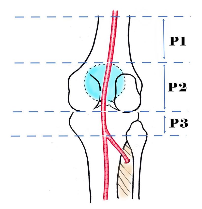
Abstract
Objective To explore the association between the classi cation for tibial plateau fractures (TPF) and the
popliteal artery injury (PAI).

Methods 22 TPF patients accompanied by PAI who were treated from May 2012 to July 2019 were
retrospectively analyzed. There were 19 males and 3 females with an average age of 49.43 years. The
Schatzker classi cation and three-column classi cation were performed for TPF. The severity of
extremity injury was evaluated using the mangled extremity severity score (MESS). Except 3 patients
treated with amputation, the remaining patients underwent surgical repair of popliteal artery and fracture
external xation. The e cacy was evaluated using the Rasmussen score for tibial head fractures.

Results There were 10 cases of Schatzker type IV fractures, 1 case of type V fractures and 11 cases of
type VI fractures. Based on the three-column classi cation, the posterior column was involved in 22
cases, 2 columns in 15 cases and 3 columns in 6 cases. The MESS was 6-10 points, with an average of
7.59 points. Except 1 case directly receiving amputation, 3 cases of segment P1 injury was observed via
preoperative DSA + intraoperative exploration, while segment P2 in 6 cases and segment P3 in 12 cases.
Popliteal artery was found completely ruptured in 11 cases, partially ruptured in 1 case, and severely
contused with thrombosis in 10 cases. The Rasmussen score was given to 19 patients at the last follow-
up,except for the cases undergoing amputation. The e cacy was excellent in 8 cases, good in 7 cases,
moderate in 3 cases and poor in 1 case.

Conclusion: In patients with complex TPF, the risk of PAI becomes higher with the increase of Schatzker
classi cation level. Knee CT scan is helpful in determining the severity of fractures and evaluating PAI.
Based on the three-column classi cation, PAI should be suspected when the fractures involve the medial
and posterior column.PAI is mainly in the segment P3, and artery rupture or severe contusion with
extensive thrombosis may occur.The postoperative knee function of patients with complex TPF can be
well restored by early active surgical repair of popliteal artery.

Introduction
Popliteal artery injury (PAI) is the most dangerous among concomitant injuries of complex tibial plateau
fractures (TPF), and the amputation rate can be up to 51.8% [1]. Liu et al [2] argued that PAI may be
caused by even mild TPF, and Magnotti [3] et al believed that early effective improvement of popliteal
artery blood ow is the only effective factor ameliorating the postoperative knee function. However, the
speci c relation between the classi cation for TPF and PAI has been rarely reported. Therefore, a
retrospective study was conducted about the association between the two.

In this paper, the data of 22 TPF patients accompanied by PAI who were treated from May 2012 to July
2019 and followed up were retrospectively analyzed, and the Schatzker classi cation, CT-based three-
column classi cation and force mechanism were detected in patients, so as to explore the association
between the classi cation for TPF and PAI. It is now reported as follows:
                                                    Page 2/17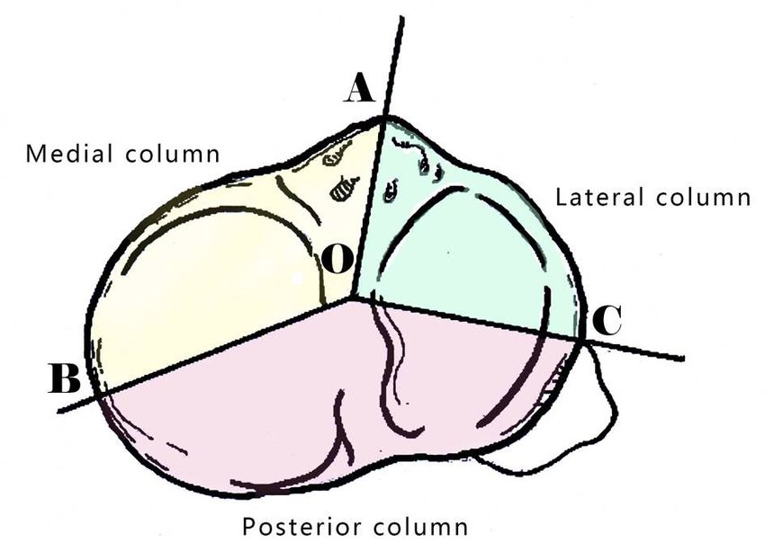
Patients And Methods
General data

A total of 22 patients were enrolled in this study, including 19 males and 3 females aged 11-71 years old,
with an average of 50 years old. The fractures were on the left side in 12 cases and on the right side in 10
cases. The duration from injury to surgical exploration and repair of popliteal artery was 1 week in 2 cases, as late as 12 d after injury. In terms of
the cause of injury, there were 16 cases of tra c injury, 2 cases of falling injury and 4 cases of crushing
injury. In terms of the concomitant injuries, ipsilateral knee dislocation occurred in 2 cases,
fractures at other sites in 14 cases, popliteal vein injury in 7 cases, common peroneal nerve injury and
tibial nerve injury in 6 cases, craniocerebral trauma in 5 cases, chest trauma in 2 cases and traumatic
shock in 2 cases. The mangled extremity severity score (MESS) [4] was 6-10 points, with an average of
7.59 points. According to the Schatzker classi cation criteria [5], there were 10 cases of type IV fractures,
1 case of type V fractures and 11 cases of type VI fractures. Based on the CT-based three-column
classi cation [6], the posterior column was involved in 22 cases, 2 columns in 15 cases and 3 columns in
6 cases. 19 patients were diagnosed with TPF accompanied by PAI in our hospital or other hospitals. In 2
patients, PAI was missed in diagnosis in other hospitals, but found after fracture internal xation was
performed, and then they were transferred to our hospital. The remaining 1 patient was diagnosed with
PAI and treated with failed fracture xation and repair of artery injury, and then he was transferred to our
hospital. All patients underwent anteroposterior and lateral X-ray and CT scan of the knee joint. Whether
there was PAI was determined using physical examination, and digital subtraction angiography (DSA) or
CT angiography (CTA) was used promptly if failed.

Classi cation methods

In addition to the traditional Schatzker classi cation, namely the X-ray-based TPF classi cation, the CT-
based three-column classi cation for TPF was also adopted in this paper. In the classi cation (Figure 1),
from an aerial perspective of the tibial plateau, the point A, O, C and B indicates the tibial tubercle, the
midpoint of the tibial spine line, the anterior border of the bular head, and the crista medialis tibialis,
respectively. The tibial plateau is divided into three parts (lateral column, medial column and posterior
column) by the line OA, OC and OB, and column fractures are de ned as cortical rupture involved.
According to the bone landmarks, the popliteal artery is divided into three segments through the
anatomical landmarks: segment P1: from hiatus of adductor to superior border of patella, segment P2:
from superior border of patella to tibial plateau, and segment P3: from tibial plateau to the beginning of
arteriae tibialis anterior (Figure 2). The classi cation results are shown in Table 1 and 2.

Table 1: Schatzker classification of 22 cases of TPF accompanied by PAI

                                                   Page 3/17
Schatzker classification
Classificatio I II III IV V VI
n
n - - - 10 1 11
Table 2: Three-column classification of 22 cases of TPF accompanied by PAI
Three-column classification
Classifica Simple L M N L+M L+N M+N L+M+N
tion compress
ion
fractures
n - - - 1 - 5 10 6
Note: L: lateral column, M: medial column, N: posterior column. N: only posterior column involved, L+N: lateral
column + posterior column involved, M+N: medial column + posterior column involved, L+M+N: lateral column
+ medial column + posterior column involved
Treatment methods
TPF patients accompanied by PAI initially diagnosed in our hospital or con rmed in other hospitals
underwent related emergency examinations, and received operation after the surgical contraindications
were excluded (2 cases of right lower extremity vein thrombosis shown in preoperative DSA were rst
treated with lower extremity vein lter implantation). Except 1 case directly receiving leg amputation, the
remaining 21 cases underwent fracture reduction and external xation + surgical repair of popliteal
artery.
Operation methods
After anesthesia, routine disinfection and draping in a supine position (if
osteofascial compartment syndrome occurred in the leg before operation, incision decompression of
osteofascial compartment was performed rst), 2 Schanz screws were percutaneously drilled into the
distal femur and proximal tibia, the external xation support was placed, and the screws were tightened
at about 30° of knee exion. Then after disinfection and draping again in a prone position, an S-shaped
incision (about 20 cm long) was made at the posterior popliteal fossa, the skin and subcutaneous tissues
were cut open, and the popliteal artery and popliteal vein were explored. If the popliteal artery defect was
less than 2 cm, vascular anastomosis was performed directly. If the defect was larger than 4 mm after
dissociation, the great saphenous vein on the unaffected side was taken for vascular anastomosis. The
Page 4/17posterior incision at the knee joint was sutured properly, the leg was covered with VSD device for
decompression, and the affected limb was wrapped with a semi-permeable membrane.
Postoperative follow-up
All patients were followed up routinely in outpatient clinic at 1, 2 and 3 months after operation, and then
in outpatient clinic once every 6 months. The blood supply of the affected limb, whether TPF was healed,
and the mobility and stability of the knee joint were detected. At the last follow-up, the e cacy was
evaluated using the Rasmussen score for tibial head fractures [7]: 30-27 points (excellent), 26-20 points
(good), 19-10 points (moderate), and 9-6 points (poor).
A typical case
A 42-year-old man suffered from right TPF accompanied by PAI due to a tra c accident. After treatment
in another hospital for 10 d, disturbance of blood circulation of the right foot was found, and the pulse of
dorsal pedal artery disappeared. It was shown in CTA that the right popliteal artery had no image. The
MESS at admission was 8 points, and the patients had type IV fractures according to the Schatzker
classi cation. In the three-column classi cation, the medial and posterior columns were involved. Then
external xation of right TPF and surgical repair of PAI were performed. Popliteal artery rupture and
popliteal vein contusion with thrombosis was observed during operation, and the popliteal artery and
popliteal vein were repaired. The blood supply of the right foot was restored after operation, and the
patient was discharged at 2 weeks after operation. At the last follow-up, the Rasmussen score indicated
the excellent e cacy (Figure 3A: Preoperative photos, Figure 3B: Preoperative anteroposterior and lateral
X-rays of knee, Figure 3C: Preoperative CT cross section, Figure 3D: Preoperative CTA, Figure 3E:
Intraoperative fracture external xation, Figure 3F: Intraoperative exploration of popliteal artery rupture,
Figure 3G: Intraoperative anastomosis of popliteal artery, Figure 3H: Postoperative review).
Results
According to the Schatzker classi cation criteria, there were 10 cases of type IV fractures, 1 case of type
V fractures and 11 cases of type VI fractures. Based on the three-column classi cation, the posterior
column was involved in 22 cases, 2 columns in 15 cases and 3 columns in 6 cases. The MESS at
admission was 6–10 points, with an average of 7.59 points. PAI was con rmed in 10 cases via physical
examination + DSA or CTA, 1 case via physical examination + vascular B ultrasound, and 11 cases via
physical examination. 1 case directly underwent leg amputation, and the remaining 21 cases underwent
surgical repair of popliteal artery + fracture reduction and external xation. After operation, 2 cases had
severe infection and muscle necrosis, and the leg was amputated. Finally, the limb was saved
successfully in 19 cases. The rst amputated patient was a 68-year-old elderly woman with a poor
cardiopulmonary function, the MESS at admission was 9 points, and the duration from injury to
admission was 9 h. Finally, the left leg amputation was conducted. The second amputated patient was
diagnosed with right TPF accompanied by PAI after trauma in another hospital, with the MESS at
Page 5/17admission of 9 points. Then the patient underwent surgical repair of popliteal artery + fracture internal
xation. After operation, there was no signi cant improvement in the blood supply of the affected limb,
and the pulse of dorsal pedal artery still failed to be felt. On the 5th day after injury, he was transferred to
our hospital for further treatment. After emergency debridement of the right leg and surgical repair of
popliteal artery, the blood supply of the affected limb was improved, but leg amputation was conducted
ultimately due to severe infection with extensive muscle necrosis. The third amputated patient was
transferred from another hospital to our hospital at 11 d after injury. At admission, the MESS was 10
points, and the right lower extremity was black and had skin vesicles. The right popliteal artery rupture
was found in the emergency DSA in our hospital, so emergency surgical repair of right popliteal artery
was performed. However, due to extensive necrosis of muscles and soft tissues, the right leg was
amputated. Except 1 case directly receiving amputation, the remaining patients underwent surgical repair
of popliteal artery.
According to the location of popliteal artery injury, there were 3 cases of P1 segment injury, 7 cases of P2
segment injury and 12 cases of P3 segment injury. Table 3 and Table 4 showed the details of location of
PAI based on Schatzker and three-column classi cation.
Table 3
Location of PAI based on Schatzker classi cation
Location of PAI Schatzker type IV Schatzker type V Schatzker type VI
P1 2 0 1
P2 3 1 3
P3 5 0 7
Table 4
Location of PAI based on three-column classi cation
Location of PAI N L+N M+N L+M+N
P1 0 1 2 0
P2 0 1 4 2
P3 1 3 5 3
According to the type of popliteal artery injury, 10 cases were contusion, 1 case was partial rupture, and
11 cases were complete rupture. Table 5 and Table 6 showed the details of type of PAI based on
Schatzker and three-column classi cation.
Page 6/17Table 5
Type of PAI based on Schatzker classi cation
Type of PAI Schatzker type IV Schatzker type V Schatzker type VI
Popliteal artery 4 1 5
contusion
Partial rupture of 1 0 0
popliteal artery
Complete rupture of 5 0 6
popliteal artery
Table 6
Type of PAI based on three-column classi cation
Type of PAI N L+N M+N L+M+N
Popliteal artery 0 4 4 2
contusion
Partial rupture of 0 0 1 0
popliteal artery
Complete rupture 1 1 6 3
of popliteal
artery
According to the types of popliteal vein injury, 15 cases without obvious popliteal vein injury, 3 cases with
popliteal vein contusion and 4 cases with popliteal vein rupture. Table 7 and Table 8 showed the details
of type of popliteal vein injury based on Schatzker and three-column classi cation.
Table 7
Type of popliteal vein injury based on Schatzker classi cation
Type of popliteal vein Schatzker type IV Schatzker type V Schatzker type VI
injury
No obvious popliteal 6 1 8
vein injury
Popliteal vein 1 0 2
contusion
Popliteal vein rupture 3 0 1
Page 7/17Table 8
Type of popliteal vein injury based on three-column classi cation
Type of popliteal N L+N M+N L+M+N
vein injury
No obvious 1 4 6 4
popliteal vein
injury
Popliteal vein 0 1 1 1
contusion
Popliteal vein 0 0 4 0
rupture
In the case of both popliteal artery and vein injuries, the patients received popliteal artery repair rst and
then popliteal vein repair. 20 patients underwent fracture reduction and external xation, and then
surgical repair of popliteal artery. 6 patients with osteofascial compartment syndrome underwent incision
decompression. The Rasmussen score was given to 19 limb-saved patients at the last follow-up. It was
con rmed that the e cacy was excellent in 8 cases, good in 7 cases, moderate in 3 cases and poor in 1
case, with an excellent/good rate of 78.95%.
Discussion
TPF is usually caused by lateral violence, axial violence and combination of lateral and axial violence.
The shape of fractures is closely related to the injury mechanism. The tibial plateau is subject to
compression and shear stress of the femoral condyle, so it may suffer from cleavage, collapse or both.
The degree and direction of violence can be roughly determined based on the size and displacement
direction and degree of fracture blocks [8]. The knee joint is subject to axial stress under exion or semi-
exion, often leading to fractures of the posterior coronal plane. Compression fractures often occur under
90° of exion, while cleavage fractures often occur under 30° and 60° of exion [9]. Under knee exion,
vertical violence combined with valgus stress can lead to lateral and posterior TPF, while vertical violence
combined with varus stress can cause medial and posterior TPF. Furthermore, the increased stress can
cause bicondylar fractures [10, 11].
X-ray-based Schatzker classi cation (Schatzker - ) is a traditional classi cation for TPF. Generally, the
higher fracture classi cation corresponds to the greater injury, severer fracture displacement and more
serious soft tissue injury. However, there are few studies on the relation between Schatzker classi cation
and PAI, and its mechanism analysis. Wicky [12] and Macarini [13] et al found that using X-ray-based
Schatzker classi cation alone may underestimate the severity of fractures, thus affecting the diagnosis
and correct treatment of TPF, and also in uencing the correct determination of degree of soft tissue injury
and PAI. Therefore, CT-based three-column classi cation was introduced to evaluate the severity of TPF
more comprehensively, so as to discover the association between classi cation for TPF and PAI.
Page 8/17In this study, in terms of the cause of TPF, there were 16 cases of tra c injury, 2 cases of falling injury
and 4 cases of crushing injury, and all patients had high-violence injury. According to the analysis of
injury mechanism, such patients are injured mostly under exion or semi- exion of knee joints, and
fractures of the posterior coronal plane, namely posterior column fractures, can be caused when
subjected to axial stress. At this moment, Schatzker type fractures can be caused when subjected to
varus stress. In other words, the fracture line leans posteriorly, and the medial and posterior columns are
further involved, so the popliteal artery is squeezed or crushed by posterior bone blocks, resulting in
popliteal artery contusion or rupture. If the knee joint is subjected to axial stress and valgus stress under
exion or semi- exion, the fractures will involve both lateral and posterior columns, and the popliteal
artery may also be injured. In the case of extremely great axial stress, varus stress and valgus stress
under exion of knee joints, the lateral, medial and posterior columns will be affected simultaneously,
causing Schatzker type VI fractures. The reason is that the medial articular surface is larger and its
strength is greater than that of lateral articular surface. Therefore, ligament, vascular and nerve injuries
are likely to occur in the case of medial plateau fractures often caused by great violence. Moore et al [14,
15]
also argued that the risk of vascular and nerve injuries is the highest in Schatzker type IV fractures. In
this study, Schatzker type VI fractures accounted for 50%, type V fractures for 4.54%, and type IV fractures
for 45.45%. Based on the CT-based three-column classi cation, the posterior column was involved in all
22 cases, 2 columns in 15 cases, 3 columns in 6 cases, and only the posterior column in 1 case.
Therefore, it is believed that PAI should be suspected when the posterior column is involved in complex
TPF, and the risk of PAI is the highest when the medial and posterior columns are involved in TPF.
The popliteal artery is located behind the knee joint in a shallow position, and it starts from the femoral
artery upwards to downwards closely clinging to the femoral condyle and lip-shaped protrusion at the
posterior border of tibial plateau, which is divided into arteriae tibialis anterior and arteria tibialis posterior
at the inferior border of popliteus. At the popliteal fossa, there are 5 branches from the knee arterial
network, and they act as ropes to x the popliteal artery behind the knee joint, without displacement. The
results in this study indicate that when the posterior column or tibial shaft is involved in complex TPF, the
popliteus can pull the entire tibial plateau or fracture blocks of posterior column move backwards due to
great violent injury. As a result, the popliteal artery is directly impacted, and stabbed, squeezed or crushed
by fracture blocks, thus resulting in popliteal artery rupture or severe contusion and then extensive
thrombosis.
In this study, in terms of the type and location of PAI, PAI was in the segment P1 in 3 cases, segment P2
in 6 cases and segment P3 in 12 cases. It can be seen that the possibility of PAI was the highest in
segment P3 (57.14%), and the same conclusion was made regardless of Schatzker classi cation or CT-
based three-column classi cation. The possible reason is that the popliteal artery at the same plane
(segment P2 and P3 of popliteal artery) is directly squeezed or crushed by the posterior border of tibial
plateau and fracture blocks of posterior column. However, during surgical exploration of PAI, it is
recommended that an S-shaped incision be made at the posterior popliteal fossa, and the popliteal artery
Page 9/17be explored from the distal end. This is because the residual end may retract to the distal and proximal
ends after PAI, so exploring from far to near can effectively avoid omissions.
It was found via intraoperative exploration that the popliteal artery was completely ruptured in 11 cases,
partially ruptured in 1 case, and severely contused with thrombosis in 10 cases. At the same time,
popliteal vein rupture and popliteal vein contusion with thrombosis were found in 3 cases and 3 cases,
respectively, so the popliteal vein was also repaired during operation. It is believed that in TPF
accompanied by PAI, both popliteal artery rupture and severe popliteal artery contusion with extensive
thrombosis may occur regardless of Schatzker type fractures or Schatzker type VI fractures, consistent
with the research results of Drapanas et al [16]. Moreover, the popliteal vein injury is more likely to occur in
Schatzker type TPF accompanied by PAI, which was also con rmed by the CT-based three-column
classi cation that the risk of popliteal vein injury was the highest when the medial and posterior columns
were involved in fractures. In clinic, the pulse of dorsal pedal artery can still be felt in some TPF patients
with PAI in the early stage. Meek [17] et al found that the weaker pulse than normal can be felt at the distal
end in about 22% of patients with injury of major limb arteries, also consistent with the ndings in this
study that the weaker pulse of dorsal pedal artery than that at the contralateral side could be felt in 5
cases in the early stage. The possible reason is that the popliteal artery has not been completely ruptured
but severely contused in some patients with PAI, causing damage to the artery intima and thrombosis, so
some blood supply remains in the initial stage. Later, due to the worsening of thrombosis, the artery
becomes completely occluded, the blood supply of popliteal artery completely disappears, and lower limb
ischemia is gradually enhanced, leading to limb necrosis. Therefore, it is necessary to determine whether
PAI is accompanied in TPF in the clinical examination, rather than only based on the presence or absence
of pulse of dorsal pedal artery. Besides, bilateral comparison is needed, and CTA or DSA should be
performed promptly for de nite diagnosis if it is unable to determine accurately. If there are no conditions
for CTA or DSA in a hospital, the patients should be transferred to the conditional hospital in time to avoid
missed diagnosis.
In this study, the MESS of the 22 patients at admission was 6–10 points, with an average of 7.59 points.
In the case of MESS ≥ 9 points, the possibility of amputation was greatly raised. There were 20 cases
with the MESS ≥ 7 points, and the amputation rate was 15%, lower than the traditional rate in PAI (30–
50%) [6]. The amputation rate was 60% when the MESS ≥ 9 points. Therefore, PAI patients with the MESS
at admission ≥ 9 points should be highly alerted, in which case the amputation rate is signi cantly
increased. In addition, Mullenix et al [18] argued that incision decompression is needed for patients with
the increased pressure of osteofascial compartment. Similarly, the results in this study demonstrate that
therapeutic or prophylactic incision decompression is necessary when the pressure of osteofascial
compartment rises in the leg. At the same time, the deep fascia and other osteofascial compartments
should be completely separated during incision decompression. Otherwise, the decompression effect will
be unsatisfactory, and the treatment of PAI will not be bene ted.
Page 10/17It is reported by Wani et al [19] that the amputation rate is 42.1% in patients undergoing operation 6 h after
PAI, the success rate of popliteal artery repair is 89% within 8 h, and the amputation rate is up to 86%
after 8 h. Wager et al [20] also thought that the success rate of blood circulation reconstruction will
dramatically decline with the prolongation of duration of limb ischemia. In this study, the amputation rate
was 20% in patients undergoing operation after 6 h, while it was 27.27% after 8 h. It is believed that the
duration of injury is not the absolute criterion for deciding whether amputation is needed. Limb salvage
can still be considered if the patients have no severe infection or extensive leg muscle necrosis. This is
because PAI patients are mostly adult men, and they have higher expectations and shoulder greater
social pressure [21]. Therefore, retaining the good function of knee is of great importance for the quality of
life after operation.
Glass [22] and Percival [23] et al showed that whether fractures or vascular injury should be treated rst
during operation depends on the duration of ischemia. In the case of duration of injury < 6 h, the fractured
bones should be xed rst, followed by vascular repair. On the contrary, in the case of duration of injury >
6 h, the blood vessels should be repaired rst, followed by xation of the fractured bones, so as to reduce
the time of tissue ischemia and hypoxia. However, it is believed that except acute open PAI in which
surgical repair is needed rst, fracture reduction and external xation should be considered rst in other
cases, so that the re-rupture of damaged artery can be avoided when the patient's posture is changed
after artery repair.
In this study, the oldest patient was aged 71 years old, and the duration from admission to surgical repair
was 6 h. With the timely diagnosis of PAI, and early fracture external xation + surgical repair of popliteal
artery, the limb was saved successfully. At the last follow-up, it was con rmed using the Rasmussen
score that the e cacy was good, suggesting that age is not a contraindication for limb salvage of
patients.
Conclusion
It is recommended that in addition to routine anterioposterior and lateral X-ray examinations of knee, TPF
patients undergo CT examination, as well as three-dimensional reconstruction if necessary. Such a
measure can help determine the severity of TPF, assist clinicians in developing a reasonable plan of
fracture operation and determining whether there is PAI, so as to avoid missed diagnosis. In the case of a
higher Schatzker classi cation level, and involvement of both medial and posterior columns according to
CT-based three-column classi cation, the risk of PAI signi cantly rises. PAI is mainly in the segment P3,
and artery rupture or severe contusion with extensive thrombosis may occur. In the case of Schatzker type
IV fractures, and involvement of both medial and posterior columns, the possibility of popliteal vein injury
also signi cantly rises, so it is also important to repair the popliteal vein injury. If the MESS is ≥ 9 points,
the possibility of amputation is greatly raised. In conclusion, age is not the absolute criterion for deciding
whether amputation is needed, and the postoperative knee function can be well restored by early active
surgical repair of popliteal artery.
Page 11/17Abbreviations
TPF: tibial plateau fractures; PAI: popliteal artery injury; MESS: mangled extremity severity score
Declarations
Acknowledgements
No
Authors’ contributions
All authors were involved in the conception and design. Study design: YLY, WBY, LY, XSW, LW and CZJ.
Study conduct: YLY, LW and WBY. Data collection and analysis: YLY, WBY and LY. Data interpretation:
WBY, XSW, CZJ and LY. Drafting of the manuscript: YLY, WBY, and LY. All authors take responsibility for
the integrity of the data analysis. The author(s) read and approved the nal manuscript.
Funding
No funding
Availability of data and materials
The datasets used and analyzed during the current study are available from the corresponding author on
reasonable request.
Ethics approval and consent to participate
The study was reviewed and approved by the institutional review board and the ethics committee of our
institution. Patients or their family members agreed to our study and signed the informed consents.
Consent for publication
Written informed consent for publication was obtained from all participants.
Competing interests
All authors state that they have no competing interests.
References
1.W Schlickewei, E H Kuner, AB Mullaji,et al. Upper and lower limb fractures with concomitant arterial
injury[J]. Journal of Bone & Joint Surgery British Volume, 1992, 74(2):181-188.
Page 12/172.Liu Yan-Wei,Li Yan-Hui,Yu Tiecheng et al. Popliteal artery transection associated with a minimally
displaced tibial plateau fracture: a case report and review of the literature.[J] .BMC Musculoskelet Disord,
2020, 21: 59.
3.Magnotti Louis J,Sharpe John P,Tolley Betsy et al. Long-term functional outcomes after traumatic
popliteal artery injury: A 20-year experience.[J] .J Trauma Acute Care Surg, 2020, 88: 197-206.
4.Helfet D L , Howey T , Sanders R , et al. Limb Salvage Versus Amputation[J]. Clinical Orthopaedics and
Related Research, 1990, &NA;(256):80-86.
5.Schatzker J, Mcbroom R, Bruce D. The tibial plateau fracture. The Toronto experience 1968-1975[J].
2015, 138(138):94.
6.LUO Cong-feng,HU Cheng-fang,GAO Hong, et al. Three Column Classi cation for tibial plateau
fractures[J]. Chinese Journal of Orthopaedic Trauma , 2009, 11(3):201-205.
7.RASMUSSEN,P. S. Tibial Condylar Fracture. Impairment of knee stability as an indication for surgical
treatment[J]. Journal of Bone & Joint Surgery American Volume, 1973, 55.
8.Thomas T P, Anderson D D, Mosqueda T V, et al. OBJECTIVE CT-BASED METRICS OF ARTICULAR
FRACTURE SEVERITY TO ASSESS RISK FOR POST-TRAUMATIC OSTEOARTHRITIS[J]. 2010, 24(12):764.
9.Yi Zhu, Severin Meili, Ming-Jie Dong,et al. Pathoanatomy and incidence of the posterolateral fractures
in bicondylar tibial plateau fractures: a clinical computed tomography-based measurement and the
associated biomechanical model simulation[J]. Archives of Orthopaedic & Trauma Surgery, 2014,
134(10):1369-1380.
10.Yang G , Zhai Q , Zhu Y , et al. The incidence of posterior tibial plateau fracture: an investigation of 525
fractures by using a CT-based classi cation system[J]. Archives of Orthopaedic and Trauma Surgery,
2013, 133(7):929-934.
11.J C Kennedy, W H Bailey. Experimental tibial-plateau fractures. Studies of the mechanism and a
classi cation[J]. Journal of Bone & Joint Surgery, 1969, 50(8):1522-1534.
12.Wicky S , Blaser P F , Blanc C H , et al. Comparison between standard radiography and spiral CT with
3D reconstruction in the evaluation, classi cation and management of tibial plateau fractures[J].
European Radiology, 2000, 10(8):1227-1232.
13.Macarini L, Murrone M, Marini S, et al. Tibial plateau fractures: evaluation with multidetector-CT.[J].
2004, 108(5-6):503.
14.Moore T M . Fracture--dislocation of the knee[J]. Prov Med J Retrosp Med Sci, 1981, 88(1):66.
Page 13/1715.Moore T . Tibial plateau fractures : de nition, demographics, treatment rationale, and long-term results
of closed traction management or operative reduction[J]. J Orthop Trauma, 1987, 2(2):97.
16.DRAPANAS, THEODORE, HEWITT, ROBERT L, WEICHERT, RUDOLPH F,et al. A Critical Appraisal of
Three Decades of Management[J]. Annals of Surgery, 172(3):351-360.
17.Meek A C , Robbs J V . Vascular injury with associated bone and joint trauma[J]. British Journal of
Surgery, 1984, 71(5):341-344.
18.Mullenix, Philip S, Steele, Scott R, Andersen, Charles A,et al. Limb salvage and outcomes among
patients with traumatic popliteal vascular injury: An analysis of the National Trauma Data Bank[J].
Journal of Vascular Surgery, 44(1):94-100.
19.Wani, Mohd Lateef, Ahangar, Ab Gani, Wani, Shadab Nabi,et al. Peripheral vascular injuries due to
blunt trauma (road tra c accident): Management and outcome[J]. International Journal of Surgery,
10(9):560-562.
20.Willis H. Wagner, Albert E. Yellin, Fred A. Weaver,et al. Acute Treatment of Penetrating Popliteal Artery
Trauma: The Importance of Soft Tissue Injury[J]. Annals of Vascular Surgery, 1994, 8(6):557-565.
21.Willis H. Wagner, Albert E. Yellin, Fred A. Weaver,et al. Acute Treatment of Penetrating Popliteal Artery
Trauma: The Importance of Soft Tissue Injury[J]. Annals of Vascular Surgery, 1994, 8(6):557-565.
22.G.E. Glass, M.F. Pearse, J. Nanchahal. Improving lower limb salvage following fractures with vascular
injury: a systematic review and new management algorithm[J]. 62(5):571-579.
23.T J Percival, T E Rasmussen. Reperfusion strategies in the management of extremity vascular injury
with ischaemia[J]. Br J Surg, 2012, 99 Suppl 1(S1):66-74.
Figures
Page 14/17Figure 1
Schematic diagram for three-column classi cation
Page 15/17Figure 2
The popliteal artery is divided into three segments through the anatomical landmarks: Segment P1: from
hiatus of adductor to superior border of patella, segment P2: from superior border of patella to tibial
plateau, and segment P3: from tibial plateau to the beginning of arteriae tibialis anterior.
Page 16/17Figure 3
A typical case. A 42-year-old man suffered from right TPF accompanied by PAI due to a tra c accident.
(A) Preoperative photos; (B) Preoperative anteroposterior and lateral X-rays of knee; (C) Preoperative CT
cross section; (D) Preoperative CTA; (E) Intraoperative fracture external xation; (F) Intraoperative
exploration of popliteal artery rupture; (G) Intraoperative anastomosis of popliteal artery; (H)
Postoperative review.
Page 17/17You can also read