Long-Term Clinically Significant Posterior Capsular Opacification Development Pattern in Eyes Implanted with an Aspheric Monofocal Intraocular ...
←
→
Page content transcription
If your browser does not render page correctly, please read the page content below
Hindawi
Journal of Ophthalmology
Volume 2021, Article ID 4566436, 7 pages
https://doi.org/10.1155/2021/4566436

Research Article
Long-Term Clinically Significant Posterior Capsular
Opacification Development Pattern in Eyes Implanted with an
Aspheric Monofocal Intraocular Lens with a Square Optic Edge

          Javier Placeres Dabán,1 Juan Carlos Elvira,2 César Azrak,2 Lucı́a Rial,1 David P. Piñero ,3
          and José I. Belda 1
          1
            Department of Ophthalmology, University Hospital of Torrevieja, Alicante, Spain
          2
            Department of Ophthalmology, University Hospital of Vinalopó, Elche, Spain
          3
            Department of Optics,Pharmacology and Anatomy, University of Alicante, Alicante, Spain

          Correspondence should be addressed to David P. Piñero; david.pinyero@ua.es and José I. Belda; drbelda@gmail.com

          Received 28 July 2021; Revised 22 September 2021; Accepted 23 September 2021; Published 30 September 2021

          Academic Editor: Alessandro Meduri

          Copyright © 2021 Javier Placeres Dabán et al. This is an open access article distributed under the Creative Commons Attribution
          License, which permits unrestricted use, distribution, and reproduction in any medium, provided the original work is
          properly cited.
          Purpose. To analyse the posterior capsular opacification (PCO) development pattern in the long term in eyes implanted with a
          monofocal intraocular lens (IOL) with a square edge all around the optic. Methods. Longitudinal retrospective study is data
          analyzed from a total of 7059 eyes from 4764 patients (mean age: 75.8 years) undergoing cataract surgery with implantation of an
          aspheric monofocal IOL (Bi-Flex HL 677AB/677P, Medicontur, Budapest, Hungary). These data were retrospectively collected
          using the electronic medical record of the hospitals involved. Nd : YAG capsulotomy rates were calculated per year during a
          follow-up of more than 10 years. The Kaplan–Meier analysis was used to establish the transparent capsule survival rate. Results.
          The Nd : YAG capsulotomy rate increased from 1.1% at 1 year postoperatively to 17.2% at 5 years after surgery. No significant
          differences were found between eyes with and without capsulotomy in terms of age (p � 0.202), gender (p � 0.061), type of
          anaesthesia used (p � 0.128), and presence of conditions such as hard cataract (p � 0.111) or pseudoexfoliation (p � 0.137). IOL
          power was significantly lower in those eyes of patients requiring Nd : YAG capsulotomy during the follow-up (p < 0.001).
          Significantly more eyes implanted with the preloaded model of the IOL required capsulotomy (p < 0.001). Mean survival time and
          rate were 9.38 years and 85.9%, respectively. Conclusions. Most eyes undergoing cataract with implantation of the Bi-Flex IOL do
          not develop a clinically significant PCO requiring Nd : YAG capsulotomy in the long term. IOL material and design may be the
          main factors accounting for this finding.

1. Introduction                                                        when using IOLs made of hydrophilic material instead of
                                                                       hydrophobic material [3–6]. Some experimental data pre-
Posterior capsular opacification (PCO) is a relatively fre-             viously reported suggested that that interleukin-6 (IL-6)
quent complication after cataract surgery, which is the result         contributes to the development of PCO by promoting the
of the proliferation and migration of residual crystalline             transformation of the growth factor β2 (TGF-β2) activation
epithelial cells from the posterior periphery of the capsular          and extracellular matrix (ECM) synthesis through a JAK/
bag towards the space between the capsule and the optics of            STAT3 signalling-dependent mechanism [7].
the intraocular lens (IOL) [1]. Its prevention is crucial since            Besides material, other factors, such as the design of the
it induces a significant decrease in visual acuity and quality          IOL, the configuration of the IOL optics, IOL power, and the
deterioration [2]. The material of the IOL is one relevant             positioning of the IOL into the capsular bag, have also great
factor for the development of both anterior and posterior              relevance [8–14]. A meta-analysis of the studies evaluating
capsular opacification, with a trend to higher rates of PCO             the impact of IOL design on PCO concluded that IOLs made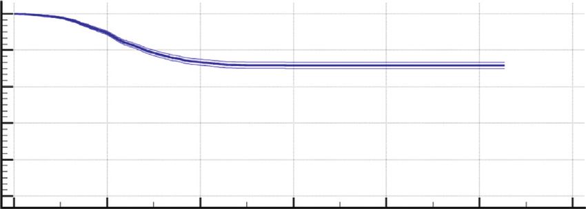
2                                                                                                     Journal of Ophthalmology

of acrylic material and silicone, as well as those with sharp      involved in the study. The surgical procedure began with
optic edges, were superior in terms of a minor incidence of        disinfection of the operative area using povidone iodine or
PCO. In addition, some studies have confirmed the benefit            chlorhexidine. After this, the surgical field was prepared, and
of a square edge all around the optic to control cell migration    the anaesthesia was applied through the topical use of drops
[15, 16].                                                          or by peribulbar injection of anaesthetic depending on the
    The prevention of PCO is crucial, and its solution is the      potential level of collaboration of the patient. Once the
creation of a hole in the posterior capsule (capsulotomy)          surgical field was prepared, a 2.2 mm peripheral corneal
using YAG laser. This hole in the posterior capsule promotes       incision was made manually with a calibrated knife. A
the migration of epithelial cells to the periphery and the         viscoelastic substance was then introduced into the anterior
transparency of the central area of the optic [17]. Despite        chamber to maintain its volume, allowing the surgeon to
YAG capsulotomy is a procedure easy to perform, it should          manoeuvre with sufficient safety. At this moment, the
be considered that it has some risks [18–20] and economic          capsulorhexis was performed using a manual technique
costs associated [21], being preferrable to delay it as much as    followed by cataract partition and aspiration using different
possible. It should be considered that relevant complica-          extraction techniques by microinfiltrated ultrasound
tions, such as an accidental macular hole [19], retinal de-        through the phacoemulsifier. Afterwards, the capsule was
tachment, or cystoid macular oedema [20], have been                cleaned of possible remains of cataract adhered by means of
described after YAG laser capsulotomy. Likewise, Nd : YAG          a specific irrigation-aspiration device. More viscoelastic
laser can induce evident changes in PMMA IOL morphology            product was injected again into the anterior chamber to
and organic alterations in their chemistry that should be          avoid damaging the capsular bag with the introduction of the
considered and controlled [18]. The objective of this study        IOL. Finally, the aspheric monofocal IOL was introduced
was to evaluate the long-term incidence of PCO requiring           into the capsular bag using the MEDJET PIL-MA injector
YAG capsulotomy in a large hospital population of eyes             (Medicontur, Budapest, Hungary). The surgery was finished
implanted with a monofocal IOL with a square edge all              after cleaning the anterior chamber by means of an irri-
around the optic.                                                  gation-aspiration cannula connected to the phacoemulsifier,
                                                                   eliminating all possible remains, with additional prophy-
                                                                   lactic intraocular instillation of antibiotics (cefuroxime),
2. Methods
                                                                   except in case of allergy (use of vancomycin instead), and
2.1. Patient Selection and Data Collection. Longitudinal ret-      topical ocular instillation of antibiotic and anti-inflamma-
rospective study enrolled a total of 7059 eyes undergoing cat-     tory drops.
aract surgery with implantation of a specific model of aspheric
monofocal IOL (Bi-Flex HL 677AB/677P, Medicontur, Geneva,          2.3. Intraocular Lens. The Bi-Flex HL IOL (Medicontur,
Switzerland) at the Department of Ophthalmology of the             Budapest, Hungary) is a single-piece aspherical lens (25%
University Hospitals of Torrevieja and Elche-Vinalopó (Ali-       water content), with a square optic edge at 360°. It is made of
cante, Spain). The primary objective of this retrospective         a copolymer of hydrophobic and hydrophilic monomers,
analysis was to evaluate the incidence of PCO requiring YAG        with 25% water content, and ultraviolet (UV) absorber. The
capsulotomy with this model of IOL. Clinical data were col-        refractive index of the IOL material is 1.46 and the Abbe
lected retrospectively using the electronic medical record         number is 58. Concerning its design and geometry, this IOL
(Florence) and with the help of the IT Department of the           is biconvex, with a total diameter of 13 mm and a diameter of
Hospitals of Torrevieja and Vinalopó. Specifically, this De-       6 mm in the optic zone. The haptic angle is 0°, with an
partment provided an anonymized database in Excel format of        asymmetric design with posterior vaulting. The IOL is
patients who met the study criteria during the period from         available in optic powers from −10.0 to −1.0 D in 1.0 D steps,
January 2007 to October 2020. The study was conducted fol-         from 0.00 to 30.00 D in 0.5 D steps, and from 31 to 35 D in
lowing the tenets of the Declaration of Helsinki and was ap-       1.0 D increments. Two different models of this IOL were
proved by the ethics committee of the University Hospitals         used in the current study: 677AB model, which is the
Torrevieja and Elche-Vinalopó (Alicante, Spain) (MEDI-            conventional model, and 677P model, which is its preloaded
CONTUR-1, data approval 25/09/2020).                               model.
    Inclusion criteria were patients undergoing cataract surgery
without intraoperative complications, including posterior
capsular rupture, vitreous loss, retrobulbar hemorrhage,           2.4. Statistical Analysis. Most data analysis was performed
suprachoroidal effusion/hemorrhage, IOL drop or nucleus             with the commercially available software package SPSS
drop, and implanted with the monofocal IOL Bi-Flex HL.             Version 22.0 (IBM Corporation, Armonk, NY, USA). The
Exclusion criteria for the study were patients implanted with      normality of data distributions was confirmed using the
other different types of monofocal IOL, chronic or recurrent        Kolmogorov–Smirnov test. Mean, standard deviation, and
uveitis, diabetes with retinal changes, keratoconus, and endo-     range were used to characterize the distribution of each
thelial corneal dystrophy.                                         variable evaluated in the sample. The Student t-test for
                                                                   unpaired data was used to compare quantitative variables
                                                                   among the groups of eyes requiring Nd : YAG capsulotomy
2.2. Surgical Procedure. The same protocol for phaco-              during the follow-up and those not requiring it. The com-
emulsification cataract surgery was used in both hospitals          parison of percentages for binary data (male/female, 677AB/
Journal of Ophthalmology 3
677P, or peribulbar/topic) between groups was performed 20
18 17.2%
YAG capsulotomy rate (%)
using the chi-square test. The Kaplan–Meier analysis was
16
used to establish the transparent capsule survival time after 14 13.3%
cataract surgery and YAG capsulotomy-free interval. Sta- 12 10.7%
tistical significance was determined using the log-rank test. 10
This analysis was performed with the MedCalc software 8
6 5.3%
version 19.8 (MedCalc Software Ltd, Ostend, Belgium). All
4
statistical tests were 2 tailed, and p values below 0.05 were 2 1.1%
considered statistically significant. 0
1 years 2 years 3 years 4 years 5 years
3. Results Follow-up
Figure 1: Changes in YAG capsulotomy rate during the follow-up
This retrospective analysis included data from 7059 eyes
in the sample of eyes evaluated.
from 4764 patients ranging in age from 33 to 100 years old
(mean: 75.8; standard deviation, SD: 8.7 years). The distri-
bution of the sample in terms of gender was as follows: 2413 Kaplan–Meier analysis was only repeated considering a
males (50.7%) and 2351 females (49.3%). A total of 3409 follow-up of 7 years as maximum (Figure 4), obtaining a
(48.3%) and 3650 (51.7%) right and left eyes were included, mean survival time of 6.22 years (standard error, 0.020; 95%
respectively. Concerning the IOL model, a total of 2139 eyes confidence interval, 6.19–6.26) and a mean survival rate of
(30.3%) and 4920 eyes (69.7%) were implanted with the 85.9% (standard error, 0.0043).
677AB and 677P models, respectively. The IOL power
implanted ranged from −6.0 to 36.0 D (mean: 20.8 D; SD: 3.6 4. Discussion
D), with a mean target postoperative refraction of −0.14 D
(SD: 0.20; range: −2.00 to 0.50 D). Peribulbar anaesthesia In the current retrospective analysis, an analysis of the
was used in 3776 eyes (53.5%), whereas topic anaesthesia was percentage of eyes needing Nd : YAG capsulotomy was
used in the rest of the sample (3283 eyes, 46.5%). Mean performed in a large sample of eyes (7059 eyes) undergoing
follow-up for the patients included in the study was 4.5 years cataract surgery in a public hospital with implantation of a
(SD: 1.3), ranging from 0.1 to 10.5 years. monofocal IOL with square optic edge at 360°. This IOL is
made of a material combining hydrophobic and hydrophilic
3.1. YAG Capsulotomy Rate. Nd : YAG capsulotomy was monomers. As in other retrospective studies analysing large
performed in a total of 956 eyes (13.5%) of the sample populations [8], Nd : YAG capsulotomies were used as an
retrospectively analysed. Figure 1 shows changes in Nd : estimate of clinically significant PCO. Indeed, different
YAG capsulotomy rate during the follow-up. The YAG studies have shown similar percentage of eyes with clinically
capsulotomy rate increased from a value of 1.1% at 1 year significant PCO and laser capsulotomy, with only a slight
after the implantation of the IOL to 17.2% at 5 years trend to obtain lower values for the Nd : YAG capsulotomy
postoperatively. No significant differences were found be- rate. Maxwell and Suryakumar [22] reported, for a hydro-
tween eyes requiring Nd : YAG capsulotomy or not in terms phobic IOL, rates of clinically significant PCO and laser
of age (p � 0.202), gender distribution (p � 0.061), type of capsulotomy at 3 years after surgery of 2.2% and 1.4%,
anaesthesia used (p � 0.128), presence of hard cataract respectively [4, 21, 23]. In our sample, the YAG capsulotomy
(p � 0.111), pseudoexfoliation (p � 0.137) or intra- rate increased from a value of 1.1% during the first year after
operative floppy iris syndrome (IFIS) (p � 0.382), and the implantation of the IOL to 17.2% at 5 years after surgery.
combined surgery of implantation of an iStent (p � 0.352) It should be considered that the percentage of clinically
or pars plana vitrectomy (PPV) (p � 0.398) (Table 1). In significant PCO might be slightly superior according to what
contrast, patients requiring Nd : YAG capsulotomy were was previously mentioned.
implanted with an IOL of significantly lower power The Nd : YAG rate found in the current study was lower
(p < 0.001). Furthermore, significantly more eyes implanted than that reported for different hydrophilic IOLs [3–6, 24].
with the 677P IOL model required Nd : YAG capsulotomy Vasavada et al. [6] found Nd : YAG capsulotomy rates at 3
(p < 0.001) (Table 1). years after surgery of 12.9% and 16% for two different types
of hydrophilic IOLs. Auffarth et al. [24] reported in a
multicenter study a 3-year laser capsulotomy rate of 31.1%
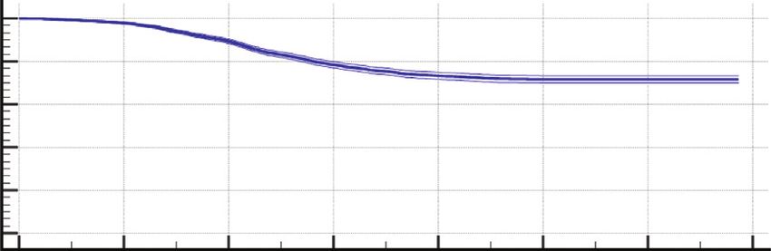
3.2. Survival Analysis. The Kaplan–Meier plot illustrating for a specific model of hydrophilic acrylic IOL. Furthermore,
the transparent posterior capsule survival profile is shown in the laser capsulotomy rates of the sample evaluated were
Figure 2. Most of Nd : YAG capsulotomies were performed similar to those reported for some models of hydrophobic
from the first to the fourth year of follow-up after IOL IOLs [5, 8, 25] but higher than those reported for some other
implantation (843 eyes). The mean survival time was 9.38 models of hydrophobic acrylic IOLs [6, 10, 21, 24, 26, 27].
years (standard error, 0.036; 95% confidence interval, Hecht et al. [8] found in a large population study (14,264
9.31–9.45) and the mean survival rate was 85.9% (standard cases) a Nd : YAG capsulotomy rate for a square edge hy-
error, 0.0043). As most of the patients enrolled in the study drophobic IOL increasing from 1.1% at 1 year after surgery
had a follow-up of 7 years or below (Figure 3), the to 10.2% at 4 years postoperatively. Ling et al. [25] reported4 Journal of Ophthalmology
Table 1: Differences between eyes requiring YAG capsulotomy and those not needing it in different preoperative and intraoperative
variables.
Mean (SD) No YAG capsulotomy
YAG capsulotomy (956 eyes/849 patients) p value
Range (6102 eyes/3914 patients)
75.9 (8.7) 75.5 (9.0)
Age (years) 0.202
33.0 to 100.0 38.0 to 98.0
20.86 (3.48) 20.40 (4.51)
IOL power (D)Journal of Ophthalmology 5
YAG time and rate for the whole follow-up were 9.38 years and
100 85.9%, respectively. Chang and Kugelberg [36] found in a
probability (%)
90 comparative study that the median survival time exceeded 9
Survival
80 years for a hydrophobic IOL and was 2.6 years for a specific type
70
of hydrophilic IOL. As most of the patients from the current
60
50
sample had a follow-up of 7 years or less, this survival analysis
0 1 2 3 4 5 6 7 was repeated considering a period of 7-year follow-up. A mean
Time survival time of 6.22 years was obtained with this new analysis.
Likewise, the survival rate was 85.9%, which was the same rate
Figure 4: Kaplan–Meier survival curve with its confidence interval obtained considering the whole follow-up.
concerning transparent posterior capsule survival after cataract
This study has limitations that should be acknowledged.
surgery (log-rank test: p < 0.01) for the sample of eyes evaluated
with a follow-up of 7 years or below. Mean survival time was 6.22 The most important limitation is the retrospective nature of
years for a 7-year follow-up, and mean survival rate was 85.9%. the study, limiting the type of variables that could be ana-
lysed (only those reported in the clinical histories were
evaluated). Likewise, a comparative study with other types of
normally need a low-power IOL which are those with long IOLs would have been adequate to know exactly the su-
axial lengths. It should be considered that axial length has periority or not of the IOL evaluated in terms of PCO
been shown to be a valuable clue to expected size of capsular formation in comparison with other IOLs.
bag [29], being positively correlated with capsule shrinkage In conclusion, most of eyes undergoing cataract with im-
[30] and capsular bag diameter [31]. Possibly, the optic edge plantation of the monofocal IOL evaluated in the current
has a more limited barrier effect in long eyes due to less sample do not develop a clinically significant PCO requiring
stability and level of adhesion to a larger capsule with more Nd : YAG capsulotomy, with a mean transparent capsule sur-
level of shrinkage. Wang et al. [32] demonstrated that 360° vival rate of 85.9%. The capsulotomy rate of this IOL increases
anterior capsule polishing in high myopes can effectively over time during the four first years after surgery, with a
reduce the extent of the anterior capsule contraction and minimal increase in the long term and a PCO rate maintained
increase the stability of the IOL implanted. below 20%. Eyes implanted with low IOL powers using the
In the sample evaluated, no significant differences were preloaded design seem to be more predisposed to develop PCO
found in age and gender between eyes requiring or not Nd : for the specific IOL type evaluated in the current series. Future
YAG capsulotomy. In addition, the type of anaesthesia used prospective comparative studies should be conducted corrob-
and the presence of IFIS or hard cataract were not differential orating these findings as well as comparing them with those
factors among eyes with and without capsulotomy. Likewise, obtained with other types of IOLs.
pseudoexfoliation was not related to the requirement of Nd :
YAG capsulotomy in medium and long term in eyes implanted Data Availability
with the IOL evaluated. Østern et al. [33] also found that the
development of long-term posterior capsular opacification was The data used to support the findings of this study are
not increased in patients with pseudoexfoliation syndrome after available from the corresponding author upon request.
uncomplicated cataract surgery. No association was found
between the performance of capsulotomy in the medium and Disclosure
long terms and the simultaneous implantation of an iStent for
the management of glaucoma or the combination with pars The authors have no proprietary or commercial interest in
plana vitrectomy (PPV). Previous studies have shown that no the medical devices that are involved in this study.
increased PCO rate was present in eyes undergoing a combined
procedure of PPV and cataract surgery, with rates even lower Conflicts of Interest
than those associated to eyes undergoing sequential surgeries
[34]. Finally, more eyes implanted with the preloaded model of The authors declare that they have no conflicts of interest.
the IOL evaluated in the current sample required Nd : YAG
capsulotomy to treat a clinically significant PCO. It should be Acknowledgments
considered that events such as trapped trailing haptic, problems
This is an investigator-initiated study supported by an
of haptic-optic adhesion, overriding of the plunger over the
unrestricted research grant from Medicontur AVI. David P
optic, and trauma to optic edge have been described when using
Piñero was supported by the Ministry of Economy, Industry
preloaded IOL implantation systems [35]. Possibly, these po-
and Competitiveness of Spain within the program Ramón y
tential events as well as the mode of releasing the lens into the
Cajal (RYC-2016-20471).
capsular bag are related to a less adjusted position of the IOL
into the capsular bag. More studies are needed to corroborate if
less optic edge-capsule adhesion is present in eyes implanted References
with the preloaded version of the IOL evaluated. [1] L. M. Nibourg, E. Gelens, R. Kuijer, J. M. M. Hooymans,
Finally, a Kaplan–Meier analysis was performed to estimate T. G. van Kooten, and S. A. Koopmans, “Prevention of
the transparent posterior capsule survival rate for the eyes posterior capsular opacification,” Experimental Eye Research,
implanted with the monofocal IOL evaluated. Mean survival vol. 136, pp. 100–115, 2015.6 Journal of Ophthalmology
[2] C. Lu, S. Yu, H. Song et al., “Posterior capsular opacification [17] N. Awasthi, S. Guo, and B. J. Wagner, “Posterior capsular
comparison between morphology and objective visual func- opacification,” Archives of Ophthalmology, vol. 127, no. 4,
tion,” BMC Ophthalmology, vol. 19, no. 1, p. 40, 2019. pp. 555–562, 2009.
[3] Y. Zhao, K. Yang, J. Li, Y. Huang, and S. Zhu, “Comparison of [18] A. Meduri, A. A. Severo, A. De Maria et al., “PMMA intra-
hydrophobic and hydrophilic intraocular lens in preventing ocular lenses changes after treatment with Nd:Yag laser: a
posterior capsule opacification after cataract surgery: an scanning electron microscopy and X-ray spectrometry study,”
updated meta-analysis,” Medicine, vol. 96, Article ID e8301, Applied Sciences, vol. 10, Article ID 6321, 2020.
2017. [19] M. Munteanu, Z. Petrovic, H. Stanca, C. Rosca, A. Jianu, and
[4] Y. Li, J. Wang, Z. Chen, and X. Tang, “Effect of hydrophobic O. Boruga, “Accidental macular hole following neodymium:
acrylic versus hydrophilic acrylic intraocular lens on posterior YAG posterior capsulotomy,” Srpski Arhiv Za Celokupno
capsule opacification: meta-analysis,” PLoS One, vol. 8, Article Lekarstvo, vol. 142, no. 7-8, pp. 468–471, 2014.
ID e77864, 2013. [20] M. A. Burq and A. M. Taqui, “Frequency of retinal detach-
[5] M. U. Saeed, A. J. Jafree, M. S. Saeed, R. Zia, I. M. Sheikh, and ment and other complications after neodymium:Yag laser
M. Heravi, “Intraocular lens and capsule opacification with capsulotomy,” JPMA. The Journal of the Pakistan Medical
hydrophilic and hydrophobic acrylic materials,” Seminars in Association, vol. 58, pp. 550–552, 2008.
Ophthalmology, vol. 27, no. 1-2, pp. 15–18, 2012. [21] F. Cullin, T. Busch, and M. Lundström, “Economic consid-
[6] A. R. Vasavada, S. M. Raj, A. Shah, G. Shah, V. Vasavada, and erations related to choice of intraocular lens (IOL) and
V. Vasavada, “Comparison of posterior capsule opacification posterior capsule opacification frequency—a comparison of
with hydrophobic acrylic and hydrophilic acrylic intraocular three different IOLs,” Acta Ophthalmologica, vol. 92, no. 2,
lenses,” Journal of Cataract & Refractive Surgery, vol. 37, no. 6, pp. 179–183, 2014.
pp. 1050–1059, 2011. [22] A. Maxwell and R. Suryakumar, “Long-term effectiveness and
[7] M. H. Shihan, M. Kanwar, Y. Wang, E. E. Jackson, safety of a three-piece acrylic hydrophobic intraocular lens
A. P. Faranda, and M. K. Duncan, “Fibronectin has multi- modified with hydroxyethyl-methacrylate: an open-label, 3-
functional roles in posterior capsular opacification (PCO),” year follow-up study,” Clinical Ophthalmology, vol. 12,
Matrix Biology, vol. 90, pp. 79–108, 2020. pp. 2031–2037, 2018.
[8] I. Hecht, B. Dubinsky-Pertzov, P. Karesvuo, A. Achiron, and [23] B. Johansson, “Glistenings, anterior/posterior capsular opa-
R. Tuuminen, “Association between intraocular lens diopter cification and incidence of Nd:YAG laser treatments with two
and posterior capsular opacification,” Clinical and Experi- aspheric hydrophobic acrylic intraocular lenses—a long-term
mental Ophthalmology, vol. 48, no. 7, pp. 889–894, 2020. intra-individual study,” Acta Ophthalmologica, vol. 95, no. 7,
[9] H. V. Pai, A. Pathan, and Y. S. Kamath, “A comparison of pp. 671–677, 2017.
posterior capsular opacification after implantation of three [24] G. Auffarth, A. Brezin, A. Caporossi et al., “Comparison of Nd
different hydrophobic square edge intraocular lenses,” Indian : YAG capsulotomy rates following phacoemulsification with
Journal of Ophthalmology, vol. 67, pp. 1424–1427, 2019. implantation of PMMA, silicone, or acrylic intra-ocular lenses
[10] T. Kohnen, E. Fabian, R. Gerl et al., “Optic edge design as in four European countries,” Ophthalmic Epidemiology,
long-term factor for posterior capsular opacification rates,” vol. 11, no. 4, pp. 319–329, 2004.
Ophthalmology, vol. 115, no. 8, pp. 1308–1314, 2008. [25] R. Ling, E.-M. Borkenstein, and A. F. Borkenstein, “Evalua-
[11] Y. Nishi, T. M. Rabsilber, I.-J. Limberger, A. J. Reuland, and tion of Nd:YAG laser capsulotomy rates in a real-life pop-
G. U. Auffarth, “Influence of 360-degree enhanced optic edge ulation,” Clinical Ophthalmology, vol. 14, pp. 3249–3257,
design of a hydrophilic acrylic intraocular lens on posterior 2020.
capsule opacification,” Journal of Cataract & Refractive [26] R. Duman, F. Karel, P. Özyol, and C. Ateş, “Effect of four
Surgery, vol. 33, no. 2, pp. 227–231, 2007. different intraocular lenses on posterior capsule opacifica-
[12] J.-W. Cheng, R.-L. Wei, J.-P. Cai et al., “Efficacy of different tion,” International Journal of Ophthalmology, vol. 8,
intraocular lens materials and optic edge designs in pre- pp. 118–121, 2015.
venting posterior capsular opacification: a meta-analysis,” [27] A. Haripriya, D. F. Chang, B. Vijayakumar et al., “Long-term
American Journal of Ophthalmology, vol. 143, no. 3, posterior capsule opacification reduction with square-edge
pp. 428–436, 2007. polymethylmethacrylate intraocular lens,” Ophthalmology,
[13] G. U. Auffarth, A. Golescu, K. A. Becker, and H. E. Völcker, vol. 124, no. 3, pp. 295–302, 2017.
“Quantification of posterior capsule opacification with round [28] M. R. Praveen, G. D. Shah, A. R. Vasavada, and K. H. Dave,
and sharp edge intraocular lenses,” Ophthalmology, vol. 110, “The effect of single-piece hydrophobic acrylic intraocular
no. 4, pp. 772–780, 2003. lenses on the development of posterior capsule opacification,”
[14] R. G. Martin, D. R. Sanders, J. Souchek, M. G. Raanan, and American Journal of Ophthalmology, vol. 160, no. 3,
M. DeLuca, “Effect of posterior chamber intraocular lens pp. 470–478, 2015.
design and surgical placement on postoperative outcome,” [29] E. Y. Dong and C. K. Joo, “Predictability for proper capsular
Journal of Cataract & Refractive Surgery, vol. 18, no. 4, tension ring size and intraocular lens size,” Korean Journal of
pp. 333–341, 1992. Ophthalmology, vol. 15, no. 1, pp. 22–26, 2001.
[15] L. Werner, N. Mamalis, S. K. Pandey et al., “Posterior capsule [30] M. Tehrani, B. H. Dick, F. Krummenauer, G. Pfirrmann,
opacification in rabbit eyes implanted with hydrophilic acrylic T. Boyle, and B. M. Stoffelns, “Capsule measuring ring to
intraocular lenses with enhanced square edge,” Journal of predict capsular bag diameter and follow its course after
Cataract & Refractive Surgery, vol. 30, no. 11, pp. 2403–2409, foldable intraocular lens implantation,” Journal of Cataract &
2004. Refractive Surgery, vol. 29, no. 11, pp. 2127–2134, 2003.
[16] M. Tetz and A. Wildeck, “Evaluating and defining the [31] J.-H. Kim, D. Lee, Y.-D. Cha, S.-H. Oh, K.-C. Mah, and
sharpness of intraocular lenses,” Journal of Cataract & Re- M.-S. Lee, “The analysis of predicted capsular bag diameter
fractive Surgery, vol. 31, no. 11, pp. 2172–2179, 2005. using modified model of capsule measuring ring in Asians,”Journal of Ophthalmology 7
Clinical and Experimental Ophthalmology, vol. 36, no. 3,
pp. 238–244, 2008.
[32] D. Wang, X. Yu, Z. Li et al., “The effect of anterior capsule
polishing on capsular contraction and lens stability in cataract
patients with high myopia,” Journal of Ophthalmology,
vol. 2018, Article ID 8676451, 7 pages, 2018.
[33] A. E. Østern, M. Saethre, G. Sandvik, M. Råen, and
L. Drolsum, “Posterior capsular opacification in patients with
pseudoexfoliation syndrome: a long-term perspective,” Acta
Ophthalmologica, vol. 91, no. 3, pp. 231–235, 2013.
[34] J. H. Roh, H. J. Sohn, D. Y. Lee, K. H. Shyn, and D. H. Nam,
“Comparison of posterior capsular opacification between a
combined procedure and a sequential procedure of pars plana
vitrectomy and cataract surgery,” Ophthalmologica, vol. 224,
no. 1, pp. 42–46, 2010.
[35] H. S. Ong, M. Subash, A. Sandhu, and M. R. Wilkins, “In-
traocular lens delivery characteristics of the preloaded
AcrySof IQ SN60WS/AcrySert injectable lens system,”
American Journal of Ophthalmology, vol. 156, no. 1, pp. 77–81,
2013.
[36] A. Chang and M. Kugelberg, “Posterior capsule opacification
9 years after phacoemulsification with a hydrophobic and a
hydrophilic intraocular lens,” European Journal of Ophthal-
mology, vol. 27, no. 2, pp. 164–168, 2017.You can also read