Indications of sole lift and foot orthoses in the management of mild idiopathic scoliosis -a review - J-Stage
←
→
Page content transcription
If your browser does not render page correctly, please read the page content below
J. Phys. Ther. Sci. 32: 251–256, 2020

 The Journal of Physical Therapy Science

Review Article

Indications of sole lift and foot orthoses
in the management of mild idiopathic scoliosis
—a review

Diego Rothschild, DC1)*, Shu Yan Ng, DC, MS, CPed1),
Yin Ling Elaine Ng, BS, MSc, MBA, CPed2)
1) Hong-Kong Chiropractic College Foundation: 11&12/F China Hong Kong Tower,
   8-12 Hennessy Road, Wan chai, Hong-Kong SAR
2) Wanchai Pedorthic Centre, Hong-Kong SAR

        Abstract. [Purpose] Leg length discrepancy is common among patients with scoliosis. Some studies reported
        reduced functional scoliosis curves with correction of leg length discrepancy. Others, however, have shown that
        induced leg length discrepancy has little effects on spinal deformities. Also, small number of studies assessed the
        use of foot orthoses in patients with faulty foot biomechanics and their impact on idiopathic scoliosis. In this con-
        text, a review of the literature is needed to determine the current evidence for the appropriate use of sole lift and
        foot orthoses in a context of scoliosis. [Methods] A literature review was performed. [Results] It appeared that sole
        lifts are indicated for functional lumbar scoliosis when the level of the sacrum is parallel to that of the hips. Sole
        lifts may not be indicated for patients with structural scoliosis, seemingly inducing a compensatory curve. Custom
        foot orthoses were found to reduce spinal curves in juvenile patients with mild idiopathic scoliosis and concomitant
        abnormal foot biomechanics. [Conclusion] Sole lift appeared to be indicated in the presence of certain types of func-
        tional scoliosis. Custom foot orthoses can be considered in the management of mild idiopathic scoliosis in juvenile
        patients. Evidence, however, is low and quality studies are needed to validate these findings.
        Key words: Adolescent idiopathic scoliosis, Leg length discrepancy, Custom foot orthoses
                                                      (This article was submitted Aug. 2, 2019, and was accepted Dec. 24, 2019)

                                                          INTRODUCTION
   Leg length discrepancy (LLD) is a common condition. The threshold of LLD which is of clinical significance varies with
studies and there is at present no universal consensus. LLD of 0.5 cm is thought to affect approximately half of the population
and is associated with an increased prevalence of low back pain1). Epidemiological studies have shown that LLD of less than
2 cm, and more than 0 cm by definition, affects 40–70% of the adult population2, 3). Moreover, several studies have shown
that adolescent idiopathic scoliosis (AIS) is associated with a high prevalence of lower extremities biomechanics abnormali-
ties4–6), including pelvic unlevelness and LLD.
   In clinical practice it is not uncommon to see patients with scoliosis and with leg length discrepancy, when it has to be
decided whether sole lift or foot orthoses are indicated in the management.
   Many studies have attempted to elucidate the relationship between LLDs and scoliosis, but results have not been conclu-
sive. Some studies have shown that correction of LLDs levels pelvic imbalances and reduces the magnitude of functional
scoliosis angles1, 7–10). Other studies, however, have shown that artificially induced LLDs have little effects on the lumbar
curve deformity11, 12).
   We attempt to review the literature to determine the indications of sole lift and foot orthoses in the management of
idiopathic scoliosis.

*Corresponding author. Diego Rothschild (E-mail: rothschild.dc@gmail.com)
©2020 The Society of Physical Therapy Science. Published by IPEC Inc.
                    This is an open-access article distributed under the terms of the Creative Commons Attribution Non-Commercial No Derivatives
                    (by-nc-nd) License. (CC-BY-NC-ND 4.0: https://creativecommons.org/licenses/by-nc-nd/4.0/)

                                                                                                                                           251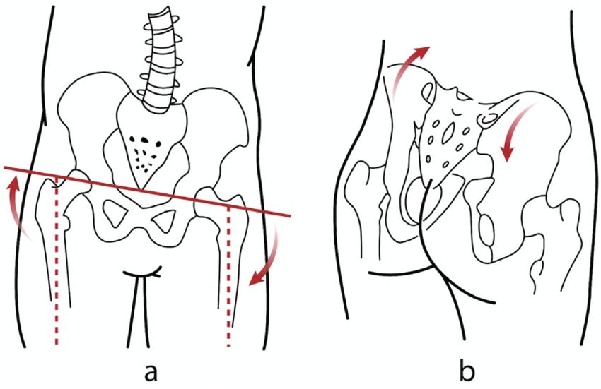
METHODS
   An electronic search was conducted in PUBMED to identify English language papers that examined the relationship
between scoliosis, leg length discrepancy and their management with sole lift and foot orthoses. The databases included were
from 1980 to 2019 using the key words: scoliosis and leg length discrepancy as well as scoliosis and foot. The literature in
foreign language, other than English was excluded. The titles of the remaining papers were then screened. Papers that were
solely related to scoliosis, leg length discrepancy or foot biomechanics were excluded and so were papers on secondary
scoliosis and surgery. For the remaining papers, the abstracts were read and the relevancy to the present review determined.
Only papers that studied or reviewed the relationship between scoliosis and leg length discrepancy or foot biomechanics were
included. Full-text copies of these papers were retrieved, evaluated, and relevant information was included in this review.

                                                            RESULTS
   In total, the search on scoliosis and leg length discrepancy produced 137 papers, 32 of which were in foreign language.
Screening of the titles showed that 92 papers were not relevant. This left 13 papers for review. The search on scoliosis and
foot for the period between 1980 and 2019 produced 356 papers, 40 of which were in foreign language. Three hundred and
eight papers were not relevant, leaving 8 papers for review.
   The review showed that lumbar scoliosis and leg length discrepancy are related. There are different types of LLD and sco-
liosis. LLD can be subdivided into two groups, structural LLD, when there is structural shortening of lower extremity in one
side, and functional LLD, which is a result of altered biomechanics of the lower extremity including the pelvis13). Likewise,
scoliosis can be divided into structural and functional scoliosis, both of which have different biomechanical characteristics.
Functional scoliosis does not present with a hump in the forward bending test as opposed to structural scoliosis.
   Concerning structural scoliosis and how it relates to pelvis disposition and the leg length:
   Sekiya et al.14) used EOS imaging system to evaluate 82 AIS patients, to determine if they had anatomical LLDs. They
found that structural LLDs were not common in AIS patients as opposed to functional LLDs. Of the cohort, none had
structural LLD in excess of 10 mm, whereas 18 of them had functional LLD in excess of 10 mm14).
   The functional LLDs appeared to be correlated with lumbar Cobb angles but not thoracic Cobb angles14). This sug-
gested that functional LLD is possibly compensatory to lumbar curves. The mechanisms involved, however, have not been
investigated.
   While isolated thoracic scoliosis curve can be compensated before reaching the lumbosacral junction, thoraco-lumbar
(TL) and lumbar scoliosis are generally compensated caudally by a lumbosacral hemi curve.
   Schwender et al.15) studied 50 AIS patients with left lumbar curve in excess of 40°, in an attempt to determine if the
lumbosacral hemicurve predisposed AIS patients to coronal decompensation. They found that in 90% of cases, the sacrum,
representing a component of the hemicurve, was tilted into the lumbar curve and 63% of patients had an iliac tilt as well. For
patients with primary lumbar or TL curves, 100% had sacral and iliac tilt into the lumbar curve15). Sacral tilt could thus be a
compensatory mechanism for lumbar curves, which through its connection to the pelvis would contribute to LLD15).
   Cho et al.16) studied the correlation between sacral slant, pelvic obliquity and lumbar curve angles on radiographs of
303 AIS patients. Their analysis supported the previous findings that pelvic obliquity is linked to sacral tilt, compensating
for large lumbar curves16). However, the exact cause and effect relationship between the different parameters could not be
determined. Also, it is to be noted that partial correlation analysis in this study concluded that LLD was not directly linked
with sacral tilt16).
   Concerning structural LLD and how it relates to the pelvis disposition and the spinal curvatures:
   Multiple studies have attempted to understand the effects of structural LLDs on the pelvic disposition using different tools
including motion analysis system, rasterstereography and x-rays. These have shown that LLD affects the pelvis in different
planes, including the coronal, sagittal and transverse planes.
   Several studies found that LLD is correlated with pelvic tilt in the coronal plane, also called pelvic obliquity1, 2, 13, 14, 16–18).
   Some have studied the immediate effects of artificially induced LLDs, using different measuring techniques and showed
that LLD affected the pelvic disposition creating pelvic obliquity. Different sizes of sole lifts or elevating platforms were
used to induce LLD in healthy participants, including adolescents in one study13) and a group of healthy soldiers in another1)
to determine their impact on the pelvic disposition. Results were all consistent, increase in sole height elevating ipsilateral
pelvis resulted in pelvic obliquity as illustrated in Fig. 1.
   Studies have also shown that LLD affects sagittal pelvic tilt. Betsch et al.11) artificially induced LLDs, varying from 5 to
15 mm, in 115 healthy participants. They found that sole lift increased the sagittal pelvic tilt. It was noted that only half of
the increase in sole height was passed onto the sagittal pelvic tilt11). With unclear mechanism, they however thought that this
was possibly due to the mobility of the sacroiliac joints, the bony asymmetry of the pelvis and the hypertonic supra-pelvic
muscles compensating for some of the sole height difference11).
   Many studies have shown that the two innominate bones do not move in tandem in the compensatory pelvic tilt. Beaudoin
et al.19) used motion analysis system in 20 female participants to quantify the 3D postural changes to the pelvis, trunk,

J. Phys. Ther. Sci. Vol. 32, No. 3, 2020                                                                                           252
Fig. 1. Artificial elevation of sole lift on the right would raise the ipsilateral pelvis and induce a func-
tional left lumbar scoliosis (a). In the sagittal plane, the right sole lift would cause posterior iliac
rotation ipsilaterally and anterior iliac rotation contralaterally in the majority of cases (b).
scapular belt and head induced by shoe lift. They showed asymmetric version of the right and left iliac bones in the presence
of sole lift, with a posterior rotation of the innominate bone on the side of the sole lift, or long leg, and an anterior rotation
on the side of the short leg19) (Fig. 1). These results confirmed the findings by other studies2, 20–22). The average total range of
version angle was estimated to be 11 degrees22). It is of note that some studies23, 24), however, showed contradicting results.
The iliac bone tended to rotate anteriorly on the side of long leg, and posteriorly on the side of short leg23, 24).
Few studies have intended to show the effects of LLD on pelvic disposition in the transverse plane, or pelvic rotation13, 19).
Grivas et al.13) using Formetric 4D, found that artificially induced LLD was significantly correlated to pelvic rotation,
however the compensatory mechanism was not described.
Concerning structural scoliosis and how it relates to lower extremity biomechanics:
Apart from apparent leg length discrepancy, AIS has been found to be associated with several lower extremity biomechan-
ics abnormalities4–6) including pes valgus4), pes cavus25), internal leg rotation4), increased femoral shaft angle26), increase
femoral anteversion/tibial torsion ratio5) and contractures of hip abductors27).
Concerning the prevalence, Lehnert-Schroth4), reported a higher incidence of pes valgus in patients with double major
scoliosis, thoracolumbar and lumbar curves4). Of 115 patients with right thoracic and left lumbar curve, 70.3% of them
presented with pes valgus on the side of thoracic concavity and 20.8% of them bilaterally. On the side of heel valgus, 79.1%
of the patients had concomitant internal rotation of the leg. On the contrary, 5.2% of the patients had internal rotation of the
leg on the convex side4).
Carpintero et al.25) on the other hand, showed that majority of AIS patients had pes cavus with a prevalence of 65.38% of
the 138 AIS patients examined. Interestingly, the incidence of the pes cavus did not relate to the severity and location of the
scoliotic curves25).
Concerning the use of sole lift and foot orthoses in a context of LLD and scoliosis:
In the presence of sacral unlevelness and leg length discrepancy, it is intuitive to add a sole lift on the side of the short leg
to level the sacrum and thus reduce the lumbar curve.
Papaionnou et al.7) showed that LLD induced scoliosis is non-structural and non-progressive, so called functional scolio-
sis. Leveling the pelvis by correcting the LLD reduced the lumbar curve. Similarly, Sheha et al.9) and Friberg1) showed that
correction of the LLD reduced the pelvic tilt and the functional lumbar scoliosis in a young patient with 20 mm leg length
inequality and in a group of soldiers respectively.
Similar results were seen in a study on 369 adolescents aged 5–17 with functional scoliosis10). Raczkowski et al.10) found
that correction of LLD helped reduce the magnitude of scoliosis curves in 316 participants (83.7%). Interestingly, 80% of
the participants had legs equalization between 8–16 months of treatment; the mean time required to equalize 1 cm LLD was
about 11.3 months10).
Juhl et al.28) described 6 types of pelvic asymmetries, where the sacral tilt can be parallel to the hip height difference or
opposite to the hip height difference, as shown in Fig. 2, seemingly affecting the outcome when using sole lift in the presence
of functional lumbar scoliosis.
It is to note that other studies on artificially induced LLD, however, have shown that sole lift (5–15 mm) did not change
or only changed the spinal postures slightly11, 12).
In the presence of structural scoliosis, Lehnert-Schroth4) advised against the use of sole lift as it can induce a compensatory
lumbar curve or lumbosacral hemicurve. In the presence of thoracic scoliosis, addition of a sole lift on the side of apparent
253short leg would induce a compensatory lumbar curve. In
the presence of lumbar curvature, addition of a sole lift on
the side of apparent short leg would induce or enlarge the
compensatory lumbosacral hemi-curve4).
Another study has shown that majority of the LLD in
structural scoliosis is apparent, and not anatomical14), thus
questioning the use of sole lift in the presence of structural
scoliosis.
Apart from sole lift, underfoot wedges and custom-
made foot orthoses have also been found to influence
spinal curves in AIS patients29).
D’Amico et al.29) used the opto-electronic approach to
evaluate the effects of wedges of different sizes on posture
of 220 patients aged between 5–19 years of age with mild
to severe idiopathic scoliosis. They found that applica-
tion of underfoot wedges resulted in significant postural Fig. 2. Different types of pelvic asymmetries. (a) Type I: The
improvements in re-balancing and in spinal deformities in sacral and hip levels are parallel, (b) Type IA: The
193 patients (87.7%); the remaining 12.3% of the patients levelness of the sacrum and hips is disproportion-
did not respond to the intervention29). al, with the unlevelling of the femoral heads higher,
Recently, Park et al.30) and Lee et al.31) reported an im- (c) Type IB: The levelness of the sacrum and hips is dis-
proportional and on the same side, but with greater sacral
provement in Cobb angle in scoliosis patients treated with
30) base unlevelling, (d) Type II: The sacrum is level, but the
custom made foot orthoses. Park et al. used Formetric
femoral heads are not, (e) Type III: The femoral heads are
4D to evaluate the effects of biomechanical foot orthoses
level, but not the sacrum, (f) Type IV: Contralateral tilt of
on 26 scoliosis patients, aged between 13–60 years of age.
the sacrum and hips.
They found that the foot orthoses significantly reduced the
pelvic obliquity and the scoliosis angle.
Lee et al.31) did a similar study, but with a more homo-
geneous group of patients. They prescribed custom foot orthoses to 52 juvenile idiopathic scoliosis patients with a mean age
of 79.5 months, with foot pronation31). They found that weight bearing heel valgus angle (the resting calcaneal stance phase
angle) reduced from a mean of 4.25° to 1.71o and the pelvic height difference reduced from a mean of 1.07 cm to 0.60 cm31).
More importantly perhaps, patients less than 6 years of age had a significant improvement of Cobb angle of more than 5o31).
The Cobb angle improved most at 9 months of the wear31) Improvement of Cobb angle, however, was not seen in patients
with curve in excess of 25o.
DISCUSSION
From the studies, it is apparent that prescription of sole lift and custom foot orthoses depends on a number of factors.
Sole lift seems to be indicated in the presence of functional lumbar scoliosis on the side of short leg1, 7–10), and in line with
Juhl et al.28) findings we believe that it should only be applied when the level of the sacrum is parallel to that of the hips28)
(Fig. 2). In the presence of idiopathic scoliosis, it is essential to check if anatomical short leg does exist, as functional LLD is
more frequent14). Addition of sole lift in the absence of anatomical LLD may induce a compensatory lumbar or lumbosacral
curve4).
The prescription of custom foot orthosis, however, is less certain. Despite the recent findings that custom foot orthoses
would reduce the spinal curves in juvenile patients with idiopathic scoliosis30) and in patients with scoliosis31), the SOSORT
2011 guidelines stipulated that there was little or no evidence to support the use of custom foot orthosis in the management
of idiopathic scoliosis32).
The authors think that it is possible that the clinical effectiveness of custom foot orthoses depends on the magnitude and
flexibility of the curve and whether the patient has abnormal foot biomechanics. In juvenile patients with scoliosis less than 25°
and asymmetric abnormal foot biomechanics, custom foot orthoses have been found to reduce the Cobb angle over 9 months31).
We believe that in young patients with mild idiopathic scoliosis and sufficient spinal flexibility, the correction of the
asymmetric abnormal foot biomechanics induces corrective movements in the lower extremities that are transmitted to the
pelvis and the spine and reducing the angle of scoliosis (Fig. 3). On the other hand, we think that with increase in curve and
reduced spinal flexibility, the correction of the asymmetric abnormal foot biomechanics may not reduce the Cobb angle, as
some of the transverse plane corrective movements induced by the custom foot orthoses may be taken up by the knees, hips
and sacroiliac joints, instead of being transmitted to the spine. Moreover, once the curve is moderate in magnitude and is
structural, the force induced by the foot orthoses would not be sufficient to cause a decrease in spinal curve. This is akin to
the treatment of tibial varum in children under 2 years of age33). Abstinence from weight bearing in the first two years of life,
together with vitamin D3 supplementation were reported to reduce tibial varum33), but they are certainly of little use in the
treatment of medial compartment knee arthrosis, which is the end stage of tibial varum. Similarly, we opined that custom
J. Phys. Ther. Sci. Vol. 32, No. 3, 2020 254Fig. 3. Custom foot orthoses for management of abnormal foot biomechanics. (a) It can be seen that the left pos-
terior superior iliac spine is lower than that of the right. (b) Physical examination of the patient revealed
bilateral forefoot varus. The relaxed calcaneal stance phase of the left heel is greater than that of the right.
(c) The feet of the patient when seen from the front. (d) When wearing the prescription custom foot orthoses
(without heel or sole lift) to correct the abnormal foot biomechanics, the posterior superior iliac spines on
both sides become level.
foot orthoses might be indicated in the early stage of idiopathic scoliosis but not when the scoliosis increases beyond 25°.
The available evidence suggested that sole lifts are recommended in the presence of functional scoliosis, when the level of
the sacrum is parallel to that of the hips and custom foot orthoses may be indicated in the management of early stage of mild
idiopathic scoliosis, particularly to juvenile patients with abnormal foot biomechanics. However, the available evidence is of
low quality and further studies are required to validate their effectiveness.
Funding and Conflict of interest
The authors declare that they have no funding nor conflict of interest.
REFERENCES
1) Friberg O: Clinical symptoms and biomechanics of lumbar spine and hip joint in leg length inequality. Spine, 1983, 8: 643–651. [Medline] [CrossRef]
2) Cummings G, Scholz JP, Barnes K: The effect of imposed leg length difference on pelvic bone symmetry. Spine, 1993, 18: 368–373. [Medline] [CrossRef]
3) Guichet JM, Spivak JM, Trouilloud P, et al.: Lower limb-length discrepancy. An epidemiologic study. Clin Orthop Relat Res, 1991, (272): 235–241. [Medline]
4) Lehnert-Schroth C: Three-dimensional treatment for scoliosis. California: The Martindale Press, 2007.
5) Burwell RG, Aujla RK, Kirby AS, et al.: Ultrasound femoral anteversion (FAV) relative to tibial torsion (TT) is abnormal after school screening for adolescent
idiopathic scoliosis (AIS): evaluation by two methods. Stud Health Technol Inform, 2008, 140: 37–43. [Medline]
6) Białek M: Mild angle early onset idiopathic scoliosis children avoid progression under FITS method (Functional Individual Therapy of Scoliosis). Medicine
(Baltimore), 2015, 94: e863. [Medline] [CrossRef]
7) Papaioannou T, Stokes I, Kenwright J: Scoliosis associated with limb-length inequality. J Bone Joint Surg Am, 1982, 64: 59–62. [Medline] [CrossRef]
8) Irvin RE: Reduction of lumbar scoliosis by use of a heel lift to level the sacral base. J Am Osteopath Assoc, 1991, 91: 34, 37–44. [Medline]
9) Sheha ED, Steinhaus ME, Kim HJ, et al.: Leg-length discrepancy, functional scoliosis, and low back pain. JBJS Rev, 2018, 6: e6. [Medline] [CrossRef]
10) Raczkowski JW, Daniszewska B, Zolynski K: Functional scoliosis caused by leg length discrepancy. Arch Med Sci, 2010, 6: 393–398. [Medline] [CrossRef]
11) Betsch M, Wild M, Große B, et al.: The effect of simulating leg length inequality on spinal posture and pelvic position: a dynamic rasterstereographic analysis.
Eur Spine J, 2012, 21: 691–697. [Medline] [CrossRef]
12) Kwon YJ, Song M, Baek IH, et al.: The effect of simulating a leg-length discrepancy on pelvic position and spinal posture. J Phys Ther Sci, 2015, 27: 689–691.
[Medline] [CrossRef]
13) Grivas TB, Angouris K, Chandrinos M, et al.: Truncal changes in children with mild limb length inequality: a surface topography study. Scoliosis Spinal
Disord, 2018, 13: 27. [Medline] [CrossRef]
14) Sekiya T, Aota Y, Yamada K, et al.: Evaluation of functional and structural leg length discrepancy in patients with adolescent idiopathic scoliosis using the
EOS imaging system: a prospective comparative study. Scoliosis Spinal Disord, 2018, 13: 7. [Medline] [CrossRef]
15) Schwender JD, Denis F: Coronal plane imbalance in adolescent idiopathic scoliosis with left lumbar curves exceeding 40 degrees: the role of the lumbosacral
hemicurve. Spine, 2000, 25: 2358–2363. [Medline] [CrossRef]
16) Cho JH, Lee CS, Joo YS, et al.: Association between sacral slanting and adjacent structures in patients with adolescent idiopathic scoliosis. Clin Orthop Surg,
2017, 9: 57–62. [Medline] [CrossRef]
17) Needham R, Chockalingam N, Dunning D, et al.: The effect of leg length discrepancy on pelvis and spine kinematics during gait. Stud Health Technol Inform,
2012, 176: 104–107. [Medline]
18) Coffey D, Hudson-Phillips SP, Radha SS, et al.: Leg length discrepancy: the importance of a complete history and examination. BMJ Case Rep, 2017, 2017:
bcr-2017-220985. [Medline] [CrossRef]
25519) Beaudoin L, Zabjek KF, Leroux MA, et al.: Acute systematic and variable postural adaptations induced by an orthopaedic shoe lift in control subjects. Eur
Spine J, 1999, 8: 40–45. [Medline] [CrossRef]
20) Kappler RE: Postural balance and motion patterns. J Am Osteopath Assoc, 1982, 81: 598–606. [Medline]
21) Subotnick SI: Limb length discrepancies of the lower extremity (the short leg syndrome). J Orthop Sports Phys Ther, 1981, 3: 11–16. [Medline] [CrossRef]
22) Pitkin HC, Pheasant HC: Sacrarthrogenetic telalgia. J Bone Joint Surg Am, 1936, 18: 365–374.
23) Cibulka T, Koldenhoff R: Leg length disparity and its effect on SI joint dysfunction. Clinical Management in Phys Ther, 1986, 6: 10–11.
24) Thomas PE: An analysis of the interactions among various asymmetric osseous pelvic and lumbar structures. J Am Osteopath Assoc, 1965, 64: 956–957.
25) Carpintero P, Entrenas R, Gonzalez I, et al.: The relationship between pes cavus and idiopathic scoliosis. Spine, 1994, 19: 1260–1263. [Medline] [CrossRef]
26) Saji MJ, Upadhyay SS, Leong JC: Increased femoral neck-shaft angles in adolescent idiopathic scoliosis. Spine, 1995, 20: 303–311. [Medline] [CrossRef]
27) Karski T: New clinical observations connected with “biomechanical aetiology of so called idiopathic scoliosis” (2006–2007). Stud Health Technol Inform,
2008, 140: 194–196. [Medline]
28) Juhl JH, Ippolito Cremin TM, Russell G: Prevalence of frontal plane pelvic postural asymmetry—part 1. J Am Osteopath Assoc, 2004, 104: 411–421. [Med-
line]
29) D’Amico M: Scoliosis and leg asymmetries: a reliable approach to assess wedge solutions efficacy. Stud Health Technol Inform, 2002, 88: 285–289. [Medline]
30) Park SM, Ahn SH, Lee AY, et al.: Raster-stereographic evaluation of the effects of biomechanical foot orthoses in patients with scoliosis. J Phys Ther Sci, 2016,
28: 1968–1971. [Medline] [CrossRef]
31) Lee JG, Yun YC, Jo WJ, et al.: Correlation of radiographic and patient assessment of spine following correction of nonstructural component in juvenile idio-
pathic scoliosis. Ann Rehabil Med, 2018, 42: 863–871. [Medline] [CrossRef]
32) Negrini S, Aulisa AG, Aulisa L, et al.: 2011 SOSORT guidelines: Orthopaedic and Rehabilitation treatment of idiopathic scoliosis during growth. Scoliosis,
2012, 7: 3. [Medline] [CrossRef]
33) Karski T. http://www.ortopedia.karski.lublin.pl/p/photo.php?id=3423&o=d&u=all. (Accessed May 30, 2019)
J. Phys. Ther. Sci. Vol. 32, No. 3, 2020 256You can also read