PROSTHETIC MANAGEMENT OF PATIENTS WITH ORO-MAXILLO-FACIAL DEFECTS: A LONG-TERM FOLLOW-UP RETROSPECTIVE STUDY
←
→
Page content transcription
If your browser does not render page correctly, please read the page content below
PROSTHETIC MANAGEMENT OF PATIENTS
original research article

                            WITH ORO-MAXILLO-FACIAL DEFECTS:
                            A LONG-TERM FOLLOW-UP RETROSPECTIVE
                            STUDY
                            G. GASTALDI, L. PALUMBO, C. MORESCHI, E.F. GHERLONE, P. CAPPARÉ

                            Dental School, Vita-Salute University, Milan, Italy (Dean: Prof. Enrico Felice Gherlone)

                              Introduction. The aim of this study is to determine the outcome of maxillofacial prosthetic rehabilitation after oncological
                              SUMMARY

                              resections, including both intra- and extra-oral prosthetic devices.
                              Methods. In this study were included 72 patients, who have undergone an intra or extra-oral maxillofacial prosthetic re-
                              habilitation after an oncologic resection.
                              Tumors on the head and neck were analyzed and the defects of these resections have been divided in two different groups:
                              intra and extra-oral defects.
                              Results. 72 participants were treated with maxillofacial prosthesis, 3 of which with post-traumatic wounds and 69 with re-
                              sections of tumors on the head and neck. Of the 69 treated for neoplastic disease, 43 received an intraoral prosthesis
                              (palatal obturator) and 29 with an extraoral epithesis (18 with nasal prostheses, 8 with orbital implants and 3 with ear im-
                              plants).The group included patients with different types of tumors. All the patients were evaluated in terms of aesthetic
                              appearance after the construction of the prostheses and the results were satisfactory.
                              Conclusion. Within the limitations of this study, after the use of maxillofacial protheses patients feel more confident and
                              self-assured. Maxillofacial protheses are a good solution in order to improve the life’s quality in patients with tumors re-
                              sections: prostheses are easy to handle and provide a satisfying social interaction for the patients.

                              Key words: maxillofacial, cancer, defect, prosthodontics.

                                 Introduction
                                                                                               and extra-oral defects.
                                                                                               The defects can be divided in two different
                                                                                               groups: intra and extra-oral results. The most fre-
                            In the last years, an increase of oral-pharynx can-                quent intra-oral defects are related to the loss of
                            cer has been registered. Tumors of oral-cranial-fa-                palatal portion.
                            cial area, with 6% of prevalence, are placed at 6th                Optimal aesthetical and functional reconstruction
                            position of malignant tumors ranking. The sur-                     in the head and neck area is important for the so-
                            vival rate of a localized tumor at 5 years is around               cial integration and the quality of life of patients
                            82% (1, 2).                                                        (6).
                            Males are more affected than women, 2:1. The                       Extended craniofacial defects can led to wide
                            therapy, based on the onset place and stage, can be                functional and psychosocial impairment in pa-
                            divided in surgery, chemiotherapy, radiotherapy or                 tients. Functional limitations can affect vision,
                            a combination of these actions (3-5). The resec-                   speech, mastication and swallowing (7).
                            tions of these tumors can produce defects of the                   The postsurgical defects are critical in many ways,
                            oral and nasal cavities, nasopharynx, oropharynx                   leading to these patients retracting from their fam-

                  276                                                                  ORAL & Implantology - Anno X - N. 3/2017
original research article
ily and society and living a life of esclusion and      the defects of these resections can be divided in
depression (7).                                         two different groups: intra and extra-oral.
After the surgical resection, a rehabilitation is       Patients with extra-oral lesions have been treated
needed. Reconstruction of the resulting defects         immediately after the end of the healing process
can be achieved by means of reconstructive plas-        with an epithesis: an impression in alginate (Hy-
tic surgery and/or maxillofacial prostheses.            drogum 5, Zhermack, Rovigo, Italy) was per-
The restoration of defects (8) can be traditionally     formed, using a wet gauze to prevent material
achieved with the aid of the conventional surgery;      from infiltrating into the cavities. In many cases, it
disadvantage, however, is the necessity for multi-      was considered necessary to ensure completion of
ple procedures (9, 10). In addition, surgical recon-    a temporary prosthesis; despite the aesthetic limi-
struction may be limited by general medical con-        tations, this solution could be helpful in improving
dition, insufficient residual tissue, vascular com-     the patient’s psychological aspect.
promise subsequent to radiation, age, inadequacy        Even for the intra-oral restorations, the palatal ob-
of the donor sites, or patient preference. It is not    turator has been realized after an alginate (Hy-
always possible to reconstruct the defect with a        drogum 5, Zhermack Rovigo, Italy) impression
surgical approach (11).                                 for the realization of temporary prosthesis: the
In these cases, prosthetic rehabilitation become        palatal obturator has been made of an acrylic resin
the first choice treatment (12). The rehabilitation     on the patient casts; it was later refined with
with maxillofacial prothesis aims to restore an ef-     methyl methacrylate (Ivocron, Ivoclar Vivadent,
fective division between oral, nasal or orbital cav-    Bolzano, Italy) and refined with soft materials di-
ities and gives faster reconstructive possibilities,    rectly in the oral cavity in order to adapt the pros-
simplifying the post-surgery period and trying to       thesis to the defect. Patients were included in a
recover an adequate patient lifestyle.                  very detailed follow-up program (every 7 days)
A collaboration between surgeon, prosthodontist         for 30-45 days. When the healing process was
and technician is required to realize an obturator      complete, more accurate impressions were taken
prothesis immediately after surgery, in order to        with polysulfide using the palatal obturator as a
improve the wound healing and the integration           guide; the final restoration can also contain the
with the patient tissue (13).                           teeth.
Nevertheless, more protocols are needed in order
to improve the predictability of results and guide-

                                                            Results
lines are necessary.
The aim of this study is to determine the success-
ful of maxillofacial prosthetic rehabilitations after
oncological resections or post traumatic results,       Seventy-two participants were treated with max-
including both intra- and extra-oral devices.           illofacial prosthesis, 3 of which with post-traumat-
                                                        ic wounds and 69 with resections of tumors on the
                                                        head and neck.

    Materials and methods
                                                        Of the 69 treated for neoplastic disease, 43 re-
                                                        ceived an intraoral prosthesis (palatal obturator)
                                                        and 29 with an extraoral epithesis (18 with nasal
In this retrospective study were included 72 pa-        prostheses, 8 with orbital implants and 3 with ear
tients, who have undergone an intra or extra-oral       implants).
maxillofacial prosthetic rehabilitation after an on-    The group included patients with different types of
cological resection or post traumatics results,         tumors (Chart 1, Table 1), respectively:
treated within the San Donato Hospital Group,           - 36% squamous cancer
Italy.                                                  - 20% basal cell carcinoma
Tumors on the head and neck were analyzed and           - 16% adenoid cystic carcinoma

                 ORAL & Implantology - Anno X - N. 3/2017                                                        277
- 11% mucoepidermoid carcinoma
                            - 17% others.
original research article

                            In total 30 patients were treated with radiotherapy,
                            4 with radio- and chemotherapy, 10 were not treat-
                            ed with radiation and 28 are not available for the
                            series.
                            The radiation dose was calculated around 12-70
                            Gy.
                            No case of osteoradionecrosis was documented in
                            our study group; no complications like oral le-
                            sions, sensibility impairment, or wound infections
                            were observed at medium-term follow-up.
                            Any implant failure or soft tissue reactions have
                            been observed in our study group.
                            All the patients were evaluated in terms of aes-
                            thetic appearance after the construction of the
                            prostheses and the results were satisfactory (Fig-
                            ures 1-5).
                            The patients had to receive a new epithesis 1-2 years
                            after anchorage of the initial epithesis, especially
                            due to deterioration of the colour and quality.
                            The follow-up ranged from 2.5 to 7.8 years (mean
                            5.6 years ± 2.8).
                            The patient survival rate was 93.06% (5 patients
                            died).
                                                                                                      Frontal view of the patient after surgical resection.
                                                                                                      Figure 1

                                 Discussion                                                         tions of head and neck tumors. According to the
                                                                                                    literature, studies have shown that an unrepaired
                            In this study 72 patients were treated, 3 of them                       maxillary defect can result in high incidence of
                            with post-traumatic results and 69 with resec-                          hypernasality (14, 15) and low speech intelligi-

                             Detail of defects: a) intraoral view of oro-nasal defect; b) frontal view of nasal defect.
                             Figure 2

                  278                                                                       ORAL & Implantology - Anno X - N. 3/2017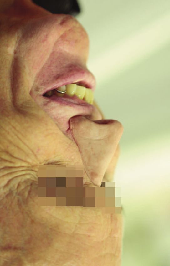
original research article
                                                                                         A lateral view of palatal obturator
                                                                                         Figure 3

                                                                                         prosthesis with extension for ep-
                                                                                         ithesis attachment.

The epithesis allows both prompt inspection of the resection   Frontal view of the patient after superior intraoral prosthesis
Figure 4                                                       Figure 5

site and makes daily care easier.                              and nasal epithesis delivery.

                   ORAL & Implantology - Anno X - N. 3/2017                                                                      279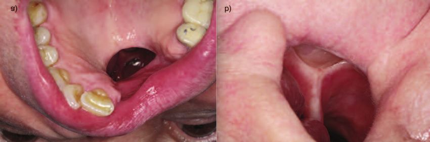
original research article
                                                                                                             Distribution of tumors
                                                                                                             Chart 1.

                                                                                    combination of dentures linked to extraoral fa-
                                                                                    cial prostheses, enabled optimal orofacial reha-
                             Table 1 - The percentage of cancer’s type.             bilitation. Oral functions such as chewing, swal-
                             Squamous Cancer                          36            lowing and speaking were facilitated by the den-
                             Basel Cell Cancer                        20
                                                                                    tures which at the same time worked as a stabil-
                                                                                    isation element for the prostheses.
                             Adenoid Cystic Carcinoma                 16
                                                                                    The facial prostheses were individually adapted
                             Mucoepidermoid Carcinoma                 11            in order to resemble the familiar preoperative
                             Adenocarcinoma                           4             appearance of the patient.
                             Chondrosarcoma                           2             The available ways of prostheses anchorage are
                             Osteosarcoma                             4
                                                                                    four (19, 20): the anatomical (to already existing
                                                                                    structures), the mechanical (to spectacle
                             Giant Cell Tumor                         2
                                                                                    frames), the chemical (using adhesive) and the
                             Other                                    5             surgical anchorage (by osseointegrated titanium
                                                                                    implants with magnets).
                                                                                    The use of fixed facial prostheses is based on a
                            bility (14, 16). This is due to inadequate separa-      close cooperation between prosthodontic and
                            tion of the oral and nasal cavities. After pros-        surgeon to provide the optimal aesthetic and
                            thetic treatment, speech were improved. Howev-          functional outcome.
                            er, maxillary obturators have been cited as being       Maxillofacial prostheses is appropriate (21) in the
                            uncomfortable to wear and some patients find            cases of large midfacial defect after a disfiguring
                            them inconvenient to remove and clean (17, 18).         cancer surgery, since it is very difficult if not im-
                            The success of a prosthesis is related to the ex-       possible to reconstruct these defects by the graft-
                            tent on the size and location of the maxillary de-      ing techniques using autogenous tissue and to
                            fect, as well as the presence of remaining denti-       achieve satisfactory results.
                            tion (10). These factors can affect the stability of    Orofacial rehabilitation of patients with maxillofa-
                            the prosthesis influencing its effectiveness.           cial defects using obturator prostheses is an appro-
                            Maxillary obturators require that a maxillofacial       priate treatment modality. To improve the situation
                            prosthodontist be available for construction and        of patients prior to and after maxillectomy suffi-
                            maintenance of the prosthesis (15, 18).                 cient information about the treatment, adequate
                            With regard to functional and aesthetic aspects,        psychological care and speech therapy should be

                  280                                                         ORAL & Implantology - Anno X - N. 3/2017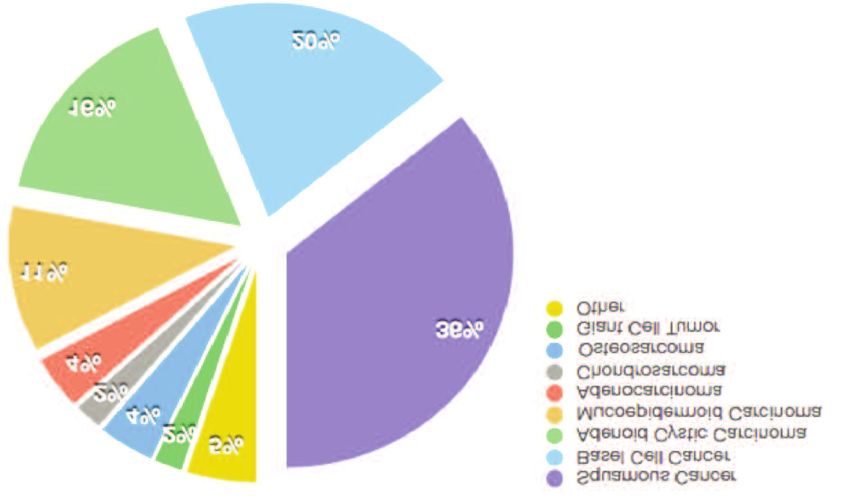
original research article
provided.                                              Authors’ contributions
With the use of obturator protheses and/or epithe-
sis, patients regain self-confidence and assurance.
                                                       Giorgio Gastaldi: clinical procedures, concep-
The patients recounted a high level of satisfaction
                                                       tion and design, drafting the article.
and a positive impact on daily life. According to
                                                       Luca Palumbo: drafting the article, data analy-
Sullivan et al., Wondergem et al. and Rieger et al.,
                                                       sis.
the goal of any intervention, whether surgical or
                                                       Chiara Moreschi: data acquisition and analysis.
prosthetic, is to limit the impact of the oncologic
                                                       Enrico F. Gherlone: critical revision, final ap-
treatment on these aspects of patients’ lives.
                                                       proval.
One of the main pros reported by patients is the
                                                       Paolo Capparé: drafting the article, critical revi-
fact that they are submit to surgery only-once
                                                       sion, clinical procedures.
reaching the same result as the surgery. On the
other hand, disadvantages and limitations of these

                                                       Funding
prostheses include discoloration (as regards ep-
ithesis) and prostheses deterioration and skin reac-
tions (19, 20).
According to our experience, an intra- or extra oral   No funding were requested for this study. The
prostheses rehabilitation should be favored over a     Authors had no conflict of interest in connection
plastic reconstruction in the cases of the previous    to this study.
multiple necessary plastic operations, due to prior
huge surgical resection which makes the surgical

                                                            References
reconstruction technically impossible, or in cases,
the patients prefer the prosthetic solution.
Nevertheless, further studies are needed in order
to draft new guidelines. It is expected that future    1.   Cancer incidence by site. Age-standardized rate per
                                                            100,000. Statistics Canada and the Canadian Council of
improvements in techniques of implantation and              Cancer Registries, Health Protection Branch - Labora-
virtual technique for optical 3d acquisition (22,           tory Centre for Disease Control, 1999.
23) will further improve the overall satisfaction      2.   WHO Mortality Database. Age-standardized rate per
and wellbeing of the patients, but this requires a          100,000. WHO Databank, 1999.
well-considered approach and close collaboration       3.   Murrah VA, Batzakis LG. Proliferative verrucous
                                                            leukoplakia and verrucous hyperplasia. Ann Otol Rhi-
between the surgeon and the prosthodontics (15).            nol Laryngol. 1994;103:660-3.
                                                       4.   Buglione M, Cavagnini R, Di Rosario F, Sottocornola
                                                            L, Maddalo M, Vassalli L, Grisanti S, Salgarello S, Or-
                                                            landi E, Paganelli C, Majorana A, Gastaldi G, Bossi P,
    Conclusion                                              Berruti A, Pavanato G, Nicolai P, Maroldi R, Barasch
                                                            A, Russi EG, Raber-Durlacher J, Murphy B, Magrini
                                                            SM. Oral toxicity management in head and neck can-
The clinical use of fixed facial prostheses is              cer patients treated with chemotherapy and radiation:
based on a close cooperation between surgeon                Dental pathologies and osteoradionecrosis (Part 1) lit-
and prosthodontist to provide the optimal aes-              erature review and consensus statement. Crit Rev On-
                                                            col Hematol. 2016. Jan;97:131-42. doi: 10.1016/j.
thetic and functional outcome.
                                                            critrevonc.2015.08.010.
Within the limitations of this study, after the use    5.   Buglione M, Cavagnini R, Di Rosario F, Maddalo M,
of maxillofacial protheses patients feel more               Vassalli L, Grisanti S, Salgarello S, Orlandi E, Bossi P,
confident and self-assured. Maxillofacial prothe-           Majorana A, Gastaldi G, Berruti A, Trippa F, Nicolai P,
ses are a good solution in order to improve the             Barasch A, Russi EG, Raber-Durlacher J, Murphy B,
                                                            Magrini SM. Oral toxicity management in head and
life’s quality in patients with tumors resections:          neck cancer patients treated with chemotherapy and ra-
prostheses are easy to handle and provide a sat-            diation: Xerostomia and trismus (Part 2). Literature
isfying social interaction for the patients.                review and consensus statement. Crit Rev Oncol Hema-

                 ORAL & Implantology - Anno X - N. 3/2017                                                               281
tol. 2016 Jun;102:47-54. doi: 10.1016/j.critrevonc. j.1365-2842.2001.00754.x.
2016.03.012. 16. Umino S, Masuda G, Ono S, Fujita K. Speech intelligi-
original research article
6. Gherlone EF, Capparé P, Tecco S, Polizzi E, Pantaleo bility following maxillectomy with and without a pros-
G, Gastaldi G, Grusovin MG. Implant Prosthetic Re- thesis: an analysis of 54 cases. J Oral Rehabil. 1998;25:
habilitation in Controlled HIV-Positive Patients: A 153-8. doi:10.1046/j.1365- 2842.1998.00238.x.
Prospective Longitudinal Study with 1-Year Follow- 17. Triana RJ, Uglesic V, Virag M, et al. Microovascular
Up. Clin Implant Dent Relat Res. 2016 Aug;18(4):725- free flap reconstructive options in patients with partial
34. doi: 10.1111/cid.12353. and total maxillectomy defects. Arch Facial Plast Surg.
7. Suarez-Cunqueiro MM, Schramm A, Schoen R, et al. 2000;2:91-101. doi:10.1001/archfaci.2.2.91.
Speech and swallowing impairment after treatment for 18. Browne JD, Burke AJC. Benefits of routine maxillec-
oral and oropharyngeal cancer. Arch Otolaryngol Head tomy and orbital reconstruction with the rectus abdo-
Neck Surg. 2008;134:1299-1304. minis free flap. Otolaryngol Head Neck Surg. 1999;
8. Park SS. Reconstruction of nasal defects larger than 1.5 121:203-9. doi:10.1016/S0194- 5998(99)70172-5.
centimeters in diameter. Laryngoscope. 2000 Aug;110 19. Federspil PA. Auricular prostheses. Adv Otorhino-
(8):1241-50. PubMed PMID: 10942120. laryngol. 2010;68:65-80. doi 10.1159/000314563.
9. Korfage A, Raghoebar GM, Noorda WD, Plaat BE, Epub 2010 May 3. PubMed PMID: 20442562.
Vissink A, Visser A. Recommendations for implant-re- 20. Federspil PA. Implant-retained craniofacial prostheses
tained nasal prostheses after ablative tumor surgery: for facial defects. GMS Curr Top Otorhinolaryngol
Minimal surgical aftercare, high implant survival, and Head Neck Surg. 2009;8:Doc03. doi: 10.3205/cto
satisfied patients. Head Neck. 2016 Apr;38(Suppl 000055. Epub 2011 Mar 10. PubMed PMID:
1):E619-24. doi: 10.1002/hed.24053. Epub 2015 Jul 18. 22073096; PubMed Central PMCID: PMC3199820.
PubMed PMID: 25784187. 21. Scolozzi P, Jaques B. Treatment of midfacial defects
10. Rohrich RJ, Griffin JR, Ansari M, Beran SJ, Potter JK. using prostheses supported by ITI dental implants.
Nasal reconstruction-beyond aesthetic subunits: a 15- Plast Reconstr Surg. 2004 Nov;114(6):1395-404. Re-
year review of 1334 cases. Plast Reconstr Surg. 2004 view. PubMed PMID: 15509925.
Nov;114(6):1405-16; discussion 1417-9. Review. 22. Sansoni G, Trebeschi M, Docchio F, Gastaldi G. State-
PubMed PMID 15509926. of-The-Art and Applications of 3D Imaging Sensors in
11. Kumar TP, Azhagarasan NS, Shankar KC, Rajan M. Industry, Cultural Heritage, Medicine, and Criminal In-
Prosthetic rehabilitation of orofacial donor site fistula vestigation. Sensors (Basel). 2009;9(1):568-601. doi:
following surgical reconstruction: a clinical report. J 10.3390/s90100568. Epub 2009 Jan 20. PubMed
Prosthodont. 2008;17:336-339. PMID: 22389618; PubMed Central PMCID: PMC
12. Sakuraba M, Kimata Y, Ota Y, et al. Simple maxillary 3280764.
reconstruction using free tissue transfer and prostheses. 23. Sansoni G, Cavagnini G, Docchio F, Gastaldi G. Vir-
Plast Reconstr Surg. 2003;111:594-8. doi:10.1097/01. tual and physical prototyping by means of a 3D optical
PRS.0000041941.98504.B6. digitizer: Application to facial prosthetic reconstruction.
13. Davis BK, Roumanas ED, Nishimura RD. Prosthetic- Virtual Physical Prototyping. 2009;4(4):217-226.
surgical collaborations in the rehabilitation of patients
with head and neck defects. Otolaryngol Clin North
Am.1997 Aug;30(4):631-45. Review. PubMed PMID: Correspondence to:
9233862. Dr. Paolo Capparé
14. Arigbede AO, Dosumu OO, Shaba OP, Esan TA. Eval- Department of Dentistry
uation of speech in patients with partial surgically ac- IRCCS San Raffaele Hospital
quired defects: pre and post prosthetic obturation. J Via Olgettina 48
Contemp Dent Pract. 2006;7:89-96. 20132 Milan, Italy
15. Keyf F. Obturator prostheses for hemimaxillectomy Phone: 0039 0226433619; fax 0039 0226432953;
patients. J Oral Rehabil. 2001;28:821-9. doi:10.1046/ E-mail: cappare.paolo@hsr.it
282 ORAL & Implantology - Anno X - N. 3/2017You can also read