Assessment of body balance of patients treated with the Ilizarov method for tibial nonunion
←
→
Page content transcription
If your browser does not render page correctly, please read the page content below
Acta of Bioengineering and Biomechanics                                                                     Original paper
Vol. 22, No. 3, 2020                                                                     DOI: 10.37190/ABB-01633-2020-01

                    Assessment of body balance of patients
              treated with the Ilizarov method for tibial nonunion

        ANDŻELIKA PAJCHERT-KOZŁOWSKA1, ŁUKASZ PAWIK2, ŁUKASZ SZELERSKI3, SŁAWOMIR ŻAREK3,
           RADOSŁAW GÓRSKI3, MALWINA PAWIK4, FELICJA FINK-LWOW4, PIOTR MORASIEWICZ1*
          1
           Wroclaw Medical University, Department and Clinic of Orthopaedic and Traumatologic Surgery, Wrocław, Poland.
                                 2
                                   Department of Physiotherapy in Motor Disorders and Dysfunctions,
                                University School of Physical Education in Wrocław, Wrocław, Poland.
           3
             Medical University of Warsaw, Department of Orthopedics and Musculoskeletal Traumatology, Warsaw, Poland.
         4
           Health Promotion, Faculty of Physiotherapy, University School of Physical Education in Wrocław, Wrocław, Poland.

      Purpose: The biomechanics of the musculoskeletal system in patients after tibial nonunion treatment using the Ilizarov method have
not yet been fully explored. From the orthopaedic and patient point of view, after the treatment, an assessment should be carried out of
the biomechanics of the musculoskeletal system. The aim of this study was to assess the body balance of patients treated with the Ilizarov
method for tibial nonunion. Methods: The research group included 24 individuals with a mean age of 55 years, who were treated for
aseptic tibial nonunion with the Ilizarov method. The control group was matched to the study group in terms of gender and age, and
consisted of 32 subjects with a mean age of 50.5 years and no significant medical history. This study evaluated the balance of patients
with the use of pedobarography. Results: In the control group, a statistically significantly shorter path of centre of gravity was observed.
There were no statistical differences between the study and control groups for the field area of the centre of gravity. There were no sta-
tistical differences between the study and control groups for the minor axis length or major axis length of the centre of gravity. There was
a relationship between the centre of pressure path length and the age of the participants in both the control group and the study group.
Conclusions: Treatment of patients with tibial nonunion with the Ilizarov fixator achieves similar balance to healthy volunteers. In the
pedobarographic evaluation, patients treated for tibial nonunion using the Ilizarov method had similar statics of the musculoskeletal
system to healthy volunteers.

Key words: body balance, pedobarography, nonunion, tibia, Ilizarov method

                   1. Introduction                                     [1], [2], [8]–[10], [12], [15], [20], [21], [28], [29].
                                                                       However, several authors have reported unsatisfactory
                                                                       results of tibial nonunion treatment, where not all pa-
    Tibial nonunion is a significant social problem                    tients achieved bone union [7], [11], [14], [19].
hindering the quality of life of patients [21], [28]. The                  When assessing the outcomes from the treatment
tibia is the most common location of nonunion [21],                    of lower limb diseases, it is important to evaluate the
[28]. The treatment of nonunion is complex and asso-                   limb function in addition to routine clinical and radio-
ciated with numerous complications [1], [8], [9], [11],                logical assessment [3], [5], [6], [23]. From the ortho-
[14], [15], [19], [21], [28]. The Ilizarov method is an                paedic and patient point of view, after the treatment,
internationally recognised and preferred method for                    an assessment should be carried out of the biome-
the treatment of nonunion [1], [2], [7]–[12], [14], [15],              chanics of the musculoskeletal system [5], [6], [17],
[18], [19]–[21], [28], [29]. Most patients treated with                [23]–[25]. Normal joint mobility, muscular strength,
this method have good results and achieve bone union                   proprioception and the absence of pain are all important
______________________________
    * Corresponding author: Piotr Morasiewicz, Wroclaw Medical University, ul. Borowska 213, 50-556 Wrocław, Poland. Phone:
(48)504549666, e-mail: morasp@poczta.onet.pl
    Received: April 20th, 2020
    Accepted for publication: July 6th, 2020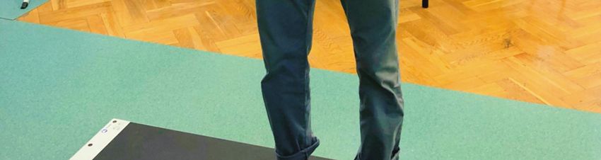
132 A. PAJCHERT-KOZŁOWSKA et al.
factors for restoring the normal biomechanics of the wires. Treatment of tibia nonunion using the Ilizarov
locomotor system [4], [13], [16], [22], [23], [25]–[27]. method was performed through stabilisation and com-
The pedobarographic platform enables reproducible, pression of the nonunion, without bone transport treat-
comparable and objective examination of the statics ment. The distal surface of the proximal tibial frag-
and dynamics of the musculoskeletal system [13], [22], ment and the proximal surface of the distal fragment
[23], [25], [27]. were always drilled with Kirschner wire according to
The biomechanics of the musculoskeletal system Beck.
in patients after tibial nonunion treatment using the Gait with two elbow crutches commenced on the
Ilizarov method have not yet been fully explored. Re- first day following surgery. Clinical and radiological
searchers have focused on the clinical and radiological outpatient follow-up was carried out every 2–6 weeks.
outcomes of patients treated with this method [1], [2], During treatment, the load on the operated limb was
[7]–[12], [14], [15], [19]–[21], [28], [29]; however, no gradually increased up to the point where the use of
studies have assessed the statics of the musculoskele- crutches could be discontinued, allowing patients to
tal system in these patients. walk with full load on the limb.
Previous studies have used pedobarographic plat- The Ilizarov fixtor was removed after observing
forms to evaluate the statics of the musculoskeletal sufficient bone union within the nonunion site con-
system in patients after thigh and lower leg corticot- firmed radiologically and clinically. The radiological
omy using the Iilizarov method [22], [25] and in ankle criterion was the presence of a minimum of three out
arthrodoses stabilised with the Ilizarov fixator [23]. of four cortical layers or the continuous presence of
The aim of this study was to assess the body bal- trabecular transition between the bone fragments, which
ance of patients treated with the Ilizarov method for were visible on X-ray images in the anteroposterior
tibial nonunion. and lateral views. The clinical criteria included a lack
of pain, no pathological movement and no deforma-
tions of the lower limb following the dynamisation of
2. Materials and methods the fixator and a powerful movement attempt in the
area of the nonunion. After removal of the Ilizarov
fixator, patients were recommended to walk with two
The clinical research group included 24 individuals elbow crutches for 4 weeks, with partial relief of the
with a mean age of 55 years (range 26.5 to 82.5 years) operated limb. The load distribution was increased
who were treated for aseptic tibial nonunion with the gradually, taking the progression of nonunion bone
Ilizarov method (Table 1). The control group was remodelling into account, as revealed in the X-ray
matched to the study group in terms of gender and image.
age, and consisted of 32 subjects with a mean age of This study evaluated the balance of patients treated
50.5 years (range 34.0 to 77.7 years) and no signifi- for aseptic tibial nonunion with the Ilizarov method,
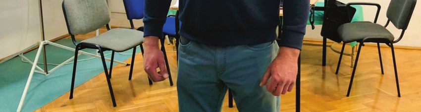
cant medical history (Table 1). using pedobarography. A Zebris Medical GmbH (Fig. 1)
The criteria for inclusion in the study group were: pedobarographic platform was used in the study. The
treatment for aseptic tibia nonunion using the Ilizarov pedobarographic platform has an area of 1580 600 mm
method, minimum follow-up period of 2 years from and includes 11264 sensors, allowing for both static
the completion of treatment, consent to participate in and dynamic tests to be carried out. After connecting
the study, radiological and clinical treatment records, the platform to a computer equipped with the appropriate
data from a pedobarographic examination, and no software (FootPrint), the two- and three-dimensional
other diseases of the lower limbs. The study was ap- distribution of ground reaction forces during gait and
proved by the bioethics committee. All patients were in static conditions were analysed. This platform also
informed about the voluntary nature of participation in allows for the analysis of deviations of the centre of
the study. gravity of the body in both static and dynamic tests.
In the case of nonunion up to a maximum of one- The use of this platform enabled computer registration
third of the distal tibia, the Ilizarov fixator consisted of kinetic gait parameters, which were statistically
of four rings fixed to the tibia and fibula with Kirschner analysed [22], [23].
wires. In the case of nonunion up to a maximum level Balance testing was performed without shoes, first
of the metaphysis and epiphysis of the distal tibia, the with eyes open and then with eyes closed. Before each
Ilizarov stabiliser consisted of three rings fixed to the test, the device was calibrated and the study participant
tibia and fibula with Kirschner wires and one half-ring received detailed instructions and information about the
fixed to the foot bone with three Kirchner “olive” structure of the test. The respondent stood motionless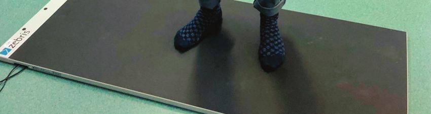
Assessment of body balance of patients treated with the Ilizarov method for tibial nonunion                  133

on the platform in a neutral position with their feet              tion to confirm a normal distribution. All values were
hip-width apart. The 30-s tests were performed with                expressed as the median and the 5th and 95th percen-
both their eyes open and closed. Each study was per-               tiles. An unpaired Student’s t-test was used to exam-
formed three times and the average result was further              ine the differences between the two groups. For data
analysed [22], [23].                                               that was not normally distributed, the significance of
                                                                   differences was analysed using the Mann–Whitney
                                                                   U-test. Linear regression analyses were performed to
                                                                   evaluate the associations between the balance pa-
                                                                   rameters and the age and body mass index (BMI) of
                                                                   the participants. The level of statistical significance
                                                                   was set at p < 0.05.

                                                                                           3. Results

                                                                       The study and control groups did not differ in terms
                                                                   of age, body weight, height or BMI (Table 1).

                                                                               Table 1. Characteristics of the participants

                                                                                     Control group      Patients after
                                                                                                                               P
                                                                                        (n = 32)       surgery (n = 24)
                                                                    Age [years]     50.5 (34.0–77.7)  55.0 (26.5–82.5)        0.758
                                                                    Height [cm]    170 (150.5–191.2) 172.5 (158.3–187.7)      0.297
                                                                    Body mass [kg] 79.5 (56.0–99.8) 79.5 (48.0–105.2)         0.261
                                                                    BMI [kg/m2]     27.2 (21.6–36.4)  27.8 (20.5–36.4)        0.098

                                                                       Data are medians and 5th–95th percentiles, BMI – body mass
                                                                   index.

                                                                       Measurements of the centre of gravity path and
        Fig. 1. Patient on a pedobarographic platform
                                                                   field area for the study and control groups with eyes
                                                                   open and closed are presented in Table 2. In the con-
    Balance was measured as the distance travelled by              trol group, a statistically significantly shorter path of
the centre of gravity of the body during the measure-              centre of gravity was observed (eyes open, p = 0.001;
ment (length of the centre of gravity line created dur-            eyes closed, p < 0.001). There were no statistical dif-
ing the measurement, expressed in centimetres) and                 ferences between the study and control groups for the
using the surface area that the centre of gravity of the           field area of the centre of gravity.
body travelled during the measurement (area of dis-                    The values of minor axis length and major axis
placement of the centre of gravity generated during                length of the centre of gravity for the study and con-
the measurement, expressed in square centimetres),                 trol groups with eyes open and closed are presented in
the minor axis length of the centre of gravity created             Table 3. There were no statistical differences between
during the measurement (expressed in millimetres)                  the study and control groups for the minor axis length
and the major axis length of the centre of gravity cre-            or major axis length of the centre of gravity.
ated during the measurement (expressed in millime-                     To determine whether the centre of pressure (COP)
tres) [22], [23]. All results were compared between                path length is associated with the age of the partici-
the study group and the control group.                             pants, a linear regression analysis was performed. As
                                                                   shown in Fig. 2, there was a relationship between the
   Statistical analysis
                                                                   COP path length and the age of the participants in
   Data were analysed using the SigmaPlot v13 (Systat              both the control group and the study group. The value
Software Inc., San Jose, CA, USA) statistics package.              of this balance parameter increased with the partici-
Continuous variables were firstly analysed using the               pants’ age; this relationship was statistically signifi-
Kolmogorov–Smirnov test with the Lilliefors correc-                cant for all groups examined independently, whether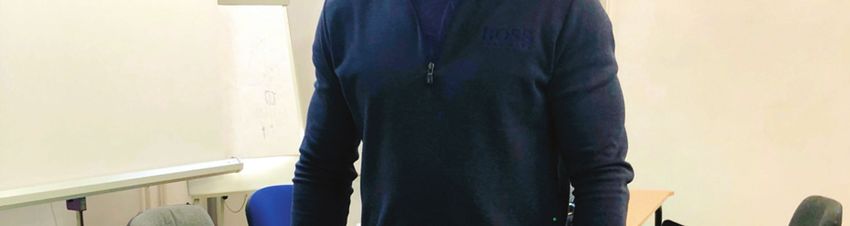
134                                                   A. PAJCHERT-KOZŁOWSKA et al.

                                Table 2. Center of pressure path length and area of the center of presssure

                                                                Control group         Patients after surgery
                             Balance parameter                                                                     P
                                                                  (n = 32)                  (n = 24)
                                                  2
                   95% confidence COP area [cm ]
                                                               1.81 (0.50–4.87)         3.21 (0.48–12.15)        0.130
                   open eyes
                   95% confidence COP area [cm2]
                                                               1.81 (0.52–5.90)         2.13 (0.48–10.15)        0.842
                   closed eyes
                   COP path length [cm]
                                                               30.1 (12.1–73.2)         55.4 (20.3–166.4)        0.001
                   open eyes
                   COP path length [cm]
                                                              50.5 (23.9–114.8)         92.9 (28.1–211.4)
Assessment of body balance of patients treated with the Ilizarov method for tibial nonunion           135

the COP path length was measured with eyes open or                  assessment, balance parameters improved after the
closed. The p-values for all analyses were below 0.05               treatment, but they did not reach the same level as
(Figs. 2A–D); however, the power of tests only ex-                  healthy subjects from the control group [22], [23].
ceeded the desired value of 0.800 in the study group                    The results of the present study are similar to those
(Figs. 2C, D). For the control group, p-values of 0.512             of earlier pedobarographic studies [22], [23], [25].
and 0.605 were obtained for tests performed with eyes               Three out of four balance parameters evaluated in
open and closed, respectively (Figs. 2A, B).                        patients after nonunion treatment did not differ statis-
    The results of the linear regression analyses show              tically from the results of the control group of healthy
that BMI had no effect on the COP path length in both               volunteers. This may indicate a return to almost nor-
the study and control groups.                                       mal static properties of lower limb biomechanics in
                                                                    patients after Ilizarov tibial nonunion treatment.
                                                                        According to the results of pedobarographic as-
                   4. Discussion                                    sessment in the current study, patients who had un-
                                                                    dergone tibial nonunion treatment using the Ilizarov
                                                                    method achieved balance parameters similar to those
    The treatment of tibial nonunion remains a chal-                of healthy individuals. Thus, it can be concluded that in
lenge for orthopaedists [1], [8], [9], [11], [14], [15], [19],      the treatment of tibial nonunion, the Ilizarov method
[21], [28]. The tibia is the most common location of the            allows for pain relief and the restoration of normal
nonunion [21], [28], but the results of treatment are not           muscle strength and joint mobility.
always completely satisfactory [7], [11], [14], [19].                   The results of linear regression analysis lead us to
    Authors of previous studies have focused on pre-                conclude that the COP path length is dependent on the
senting various techniques for the treatment of tibial              age of subjects, as observed in both the study and
nonunion, often only evaluating clinical and radiological           control groups. In the case of the study group, such
results [1], [2], [7]–[12], [14], [15], [19]–[21]. In pre-          a conclusion was expected, as p-values were lower
vious studies, authors focused on forces in the Ilizarov            than 0.05 and the power of analysis was increased to
fixatros [17], [24]. Until now, the biomechanics of the             0.800. This conclusion also seems to be valid for the
musculoskeletal system had not been studied in pa-                  control group ( p < 0.05), but this result should be
tients who had undergone tibial nonunion treatment                  interpreted with caution as the power of analysis was
using the Ilizarov method. Furthermore, no studies have             lower than the desired 0.800, which is probably due to
evaluated the balance of these patients.                            the high variability of data.
    The elimination or reduction of pain and improve-                   The results of the linear regression analyses for both
ment or restoration of normal joint mobility, muscular              the study and control groups suggest that the subject’s
strength and proprioception work together to enhance                BMI had no effect on the COP path length. However,
the biomechanical properties of the musculoskeletal                 this result should be interpreted with caution as the
system [4], [13], [16], [22], [23], [26], [27].                     power of analysis was much lower than the desired
    In previous studies, researchers have observed limi-            0.800, which is probably due to the small group size
tations in the joint mobility of some patients following            and the high variability of data. Thus, it seems possi-
tibial nonunion treatment, which may affect the biome-              ble that a second-order error could have occurred, and,
chanics of the musculoskeletal system [18], [20], [29].             therefore, a firm conclusion about the lack of depend-
    In a study by McHale on 10 patients with tibial non-            ence between COP length and BMI cannot be made.
union who were treated by the Ilizarov method, six pa-                  One of the limitations of our study is the lack of bal-
tients were found to have significantly reduced function            ance assessment prior to treatment. This is due to the
of their lower limbs due to decreased muscle strength               small number of patients with tibial nonunion who were
and limited ankle or knee mobility [18].                            assessed using the pedobarographic platform before and
    Rongies and colleagues evaluated 21 patients with               after surgery. Before treatment, most patients had severe
coxarthrosis using a pedobarographic platform and ob-               pain and significant walking problems due to the tibial
served a correlation between pain relief and enhanced               nonunion, which made it difficult to examine them using
balance [27].                                                       the pedobarographic platform. In the future, we plan to
    Morasiewicz et al. [22], [23], [25] observed an im-             study the statics of the musculoskeletal system before
provement in the statics of the musculoskeletal system              and after tibial nonunion treatment. A strength of our
in patients after thigh and lower leg corticotomy using             work is that the surgery was performed by three sur-
the Ilizarov method and after ankle arthrodesis using               geons, all using the same technique with the same post-
the Ilizarov method. According to the pedobarographic               operative treatment protocol.
136 A. PAJCHERT-KOZŁOWSKA et al.
5. Conclusions [11] KHAN M.S., RASHID H., UMER M., QADIR I., HAFEEZ K.,
IQBAL A., Salvage of infected non-union of the tibia with an
Ilizarov ring fixator, J. Orthop. Surg. (Hong Kong), 2015,
23 (1), 52–55.
In conclusion, it can be stated that treatment of [12] LAURSEN M.B., LASS P., CHRISTENSEN K.S., Ilizarov treat-
patients with tibial nonunion by stabilisation using the ment of tibial nonunions results in 16 cases, Acta Orthop.
Belg., 2000, 66 (3), 279–285.
Ilizarov fixator achieves similar balance to healthy vol- [13] LORKOWSKI J., TRYBUS M., HŁADKI W., BRONGEL L., Underfoot
unteers. Furthermore, in the pedobarographic evaluation, pressure distribution of a patient with unilateral ankylosis of
patients treated for tibial nonunion using the Ilizarov talonavicular joint during rheumatoid arthritis – case report,
method had similar statics of the musculoskeletal system Przegl. Lek., 2008, 65, 54–56.
to healthy volunteers. [14] MADHUSUDHAN T.R., RAMESH B., MANJUNATH K., SHAH H.M.,
SUNDARESH D.C., KRISHNAPPA N., Outcomes of Ilizarov ring
fixation in recalcitrant infected tibial non-unions – a pro-
spective study, J. Trauma Manag. Outcomes, 2008, 23, 2 (1), 6,
Conflict of interests DOI: 10.1186/1752-2897-2-6.
[15] MAGADUM M.P., BASAVARAJ YADAV C.M., PHANEESHA M.S.,
There was no conflict of interest for all authors. RAMESH L.J., Acute compression and lengthening by the Ilizarov
technique for infected nonunion of the tibia with large bone de-
fects, J. Orthop. Surg. (Hong Kong), 2006, 14 (3), 273–279.
References [16] MAJEWSKI M., BISCHOFF-FERRARI H., GRUNEBERG C., DICK W.,
ALLUM J.H., Improvements in balance after total hip re-
placement, J. Bone Joint Surg. Br., 2005, 87, 1337–1343
[1] ABUOMIRA I.E., SALA F., ELBATRAWY Y., LOVISETTI G., [17] MARTYNIUK B., MORASIEWICZ P., WUDARCZYK S., DRAGAN S.F.,
ALATI S., CAPITANI D., Distraction osteogenesis for tibial FILIPIAK J., The impact of configuration of the Ilizarov fixa-
nonunion with bone loss using combined Ilizarov and Taylor tor on its stiffness and the degree of loading of distraction
spatial frames versus a conventional circular frame, Strate- rods, Clin. Biomech. (Bristol, Avon), 2019, 63, 79–84, DOI:
gies Trauma Limb Reconstr., 2016, 11 (3), 153–159. 10.1016/j.clinbiomech.2019.02.020. Epub. 2019, Feb. 27.
[2] BARUAH R.K., Ilizarov methodology for infected non union of [18] MCHALE K.A., ROSS A.E., Treatment of infected tibial non-
the Tibia: Classic circular transfixion wire assembly vs. hybrid unions with debridement, antibiotic beads, and the Ilizarov
assembly, Indian J. Orthop. [serial online], 2007, 41, 198–203. method, Mil. Med., 2004, 169 (9), 728–734.
Available from: http://www.ijoonline.com/text.asp?2007/41/ [19] MCNALLY M., FERGUSON J., KUGAN R., STUBBS D., Ilizarov
3/198/33682 [accessed: January 14, 2019], Treatment Protocols in the Management of Infected Nonunion
[3] BEDNARZ P., BEAL’S T., MANILA A., Subtalar distraction of the Tibia, J. Orthop. Trauma, 2017, 31 Suppl 5, S47–S54.
bone blok fusion: an assessment of outcome, Foot Ankle Int., [20] MEGAS P., SARIDIS A., KOUZELIS A., KALLIVOKAS A.,
1997, 18, 785–791. MYLONAS S., TYLLIANAKIS M., The treatment of infected
[4] BHAVE A., PALEY D., HERZENBERG J., Improvement in Gait nonunion of the tibia following intramedullary nailing by the
Parameters After Lengthening for the Treatment of Limb-Length Ilizarov method, Injury, 2010, 41 (3), 294–299, DOI: 10.1016/
Discrepancy, J. Bone Joint Surg. Am., 1999, 81, 529–534. j.injury.2009.09.013.
[5] CHAHAL J., STEPHEN D.J., BULMER B., DANIELS T., KREDER H.J., [21] MELEPPURAM J.J., IBRAHIM S., Experience in fixation of in-
Factors associated with outcome after subtalar arthrodesis, fected non-union tibia by Ilizarov technique – a retrospective
J. Orthop. Trauma, 2006, 20 (8), 555–561. study of 42 cases, Rev. Bras. Ortop., 2016, 30, 52 (6), 670–675,
[6] DALAT F., TROUILLET F., FESSY M.H., BOURDIN M., BESSE J.L., DOI: 10.1016/j.rboe.2016.11.008.
Comparison of quality of life following total ankle arthro- [22] MORASIEWICZ P., DRAGAN S., DRAGAN S.Ł., WRZOSEK Z.,
plasty and ankle arthrodesis: Retrospective study of 54 cases, PAWIK Ł., Pedobarographic analysis of body weight distri-
Orthop. Traumatol. Surg. Res., 2014, 100 (7), 761–766. bution on the lower limbs and balance after Ilizarov corti-
[7] DRÓŻDŻ M., RAK S., BARTOSZ P., BIAŁECKI J., MARCZYŃSKI W., cotomies, Clin. Biomech., 2016, 31, 2–6.
Results of the Treatment of Infected Nonunions of the Lower [23] MORASIEWICZ P., KONIECZNY G., DEJNEK M., URBAŃSKI W.,
Limbs Using the Ilizarov Method, Ortop. Traumatol. Rehabil., DRAGAN S.Ł., KULEJ M., DRAGAN S.F., PAWIK Ł., Assess-
2017, 12, 19 (2), 111–125. ment of the distribution of load on the lower limbs and bal-
[8] ERALP İ.L., KOCAOĞLU M., DIKMEN G., AZAM M.E., BALCI H.İ., ance before and after ankle arthrodesis with the Ilizarov
BILEN F.E., Treatment of infected nonunion of the juxta-articular method, Sci. Rep., 2018, 24, 8(1), 15693, DOI: 10.1038/
region of the distal tibia, Acta Orthop. Traumatol. Turc., 2016, s41598-018-34016-3.
50 (2), 139–146, DOI: 10.3944/AOTT.2015.15.0147. [24] MORASIEWICZ P., FILIPIAK J., KONIETZKO M., DRAGAN S.,
[9] HOSNY G., SHAWKY M.S., The treatment of infected non- The impact of the type of derotation mechanism on the stiffness
union of the tibia by compression-distraction techniques of the Ilizarov fixator, Acta Bioeng. Biomech., 2012, Vol. 14,
using the Ilizarov external fixator, Int. Orthop., 1998, 22 (5), No. 1, 67–73.
298–302. [25] MORASIEWICZ P., DRAGAN S., Pedobarographic evaluation
[10] HOSNY G.A., AHMED A.A., HUSSEIN M.A., Clinical outcomes of body weight distribution on the lower limbs and balance
with the corticotomy-first technique associated with the after derotation corticotomies using the Ilizarov method,
Ilizarov method for the management of the septic long bones Acta Bioeng. Biomech., 2013, Vol. 15, No. 2, 91–96.
non-union, Int. Orthop., 2018, 42(12), 2933–2939, DOI: [26] RADLER C., KRANZL A., MANNER H.M., HÖGLINGER M.,
10.1007/s00264-018-3924-9. Epub. 2018, Apr. 7. GANGER R., GRILL F., Torsional profile versus gait analysis:Assessment of body balance of patients treated with the Ilizarov method for tibial nonunion 137
consistency between the anatomic torsion and the resulting [28] WANI N.B., SYED B., Ilizarov ring fixator in the management
gait pattern in patients with rotational malignment of the of infected non-unions of tibia, SICOT J., 2015, 29, 1, 22,
lower extremity, Gait Posture, 2010, 32, 405–410. DOI: 10.1051/sicotj/2015022.
[27] RONGIES W., BAK A., LAZAR A., A trial of the use of pedo- [29] XU J., JIA Y.C., KANG Q.L., CHAI Y.M., Management of
barography in the assessment of the effectiveness of rehabilita- hypertrophic nonunion with failure of internal fixation by
tion in patients with coxarthrosis, Ortop. Traumatol. Rehabil., distraction osteogenesis, Injury, 2015, 46 (10), 2030–2035,
2009, 11, 242–252. DOI: 10.1016/j.injury.2015.06.020. Epub 2015 Jun 17.You can also read