Duplex-Ultrasound Assisted Mynx Closure of Superficial Femoral Artery Antegrade Access Following Lower-Extremity Endovascular Intervention
←
→
Page content transcription
If your browser does not render page correctly, please read the page content below
Original Contribution

Duplex-Ultrasound Assisted Mynx Closure
of Superficial Femoral Artery Antegrade
Access Following Lower-Extremity
Endovascular Intervention
  Jorge A. Miranda, MD; Zachary Pallister, MD; Natasha Hansraj, MD;

                                                                                                               ns
  Miguel Montero-Baker, MD

                                                                                                             io
                                              y at
                                            nl ic
Abstract

                                           O un
Purpose. This analysis seeks to describe the technique of ultrasound-assisted percutaneous superficial femoral arterial

                                         se m
access closure with a Mynx device (Cordis Corporation). No comparable analysis has been reported utilizing this method
for closure. The study aim was to demonstrate the technical considerations, benefits, efficacy, and safety of this technique.
                                      l U om
Methods. A retrospective review was performed in 100 patients who underwent ipsilateral antegrade superficial femoral
artery access for angiography and subsequent ultrasound-assisted Mynx closure of the access arteriotomy. Patients were
                                    na C
followed for 6 months to evaluate potential long-term complications. Duplex ultrasound was used to visualize device po-
sitioning, deployment, and probe-guided pressure application. Demographics, complications, and procedural success were
                                  so P

evaluated and descriptive statistics were used to report data. Results. One hundred patients were followed for 6 months
                                er HM

following the index procedure. At 1 month, 2 complications occurred, ie, an immediate occlusion of the superficial femoral
artery causing acute limb ischemia requiring reintervention and a large hematoma that did not require further intervention.
Fifty-seven patients were followed for 6 months, with no additional procedure-related complications. At 1 month, 5 patients
                            r P 21

experienced a major adverse limb event (MALE); at 6 months, 10 patients had a MALE. None of the MALEs were directly
related to complications from the closure or index procedure. Conclusion. The utilization of duplex ultrasound-assisted
                         Fo 20

Mynx closure of superficial femoral artery antegrade percutaneous access arteriotomies led to excellent results with
minimal morbidity. This technique is safe and effective to use as an adjunct to ambulatory lower-extremity endovascular
interventions for peripheral arterial disease.
                             ht

J CRIT LIMB ISCHEM 2021;1(2):E56-E61. Epub 2021 March 26.
                          ig

Key words: antegrade approach, deployment technique, superficial femoral artery, ultrasound, vascular closure devices
                   yr
           op
      C

Endovascular interventions have become the standard treat-                       When percutaneous access is approached in an ipsilateral
ment approach for chronic limb-threatening ischemia (CLTI),                  antegrade manner, new challenges arise in performing the
with balloon angioplasty, stenting, and atherectomy largely                  arteriotomy closure. In many instances, the superficial femo-
replacing open surgical procedures.1,2 To date, percutaneous                 ral artery is accessed over the common femoral artery when
arterial access closure can be performed with direct manual                  performing an ipsilateral antegrade approach. However, the
pressure of the arteriotomy or use of percutaneous vascular                  anatomic location of the superficial femoral artery makes
closure devices (VCDs). VCDs have been found to improve pa-                  manual pressure ineffective for closure. Furthermore, severe
tient and physician comfort by avoiding prolonged pressure                   calcification, obesity, uncontrolled hypertension, and female
and allowing earlier patient ambulation.3 A Cochrane review                  gender can lead to VCD failure and potential complications.5,6
demonstrated reduced time for hemostasis compared with                       To mitigate these risks, we routinely implement MynxGrip and
manual compression and no relative risk increase for death or                MynxControl (Cordis Corporation) ultrasound-guided deploy-
infection with closure device usage.4                                        ment for small sheath size (5-7 Fr) procedures. We report our

E56                                                                                                    Journal of Critical Limb Ischemia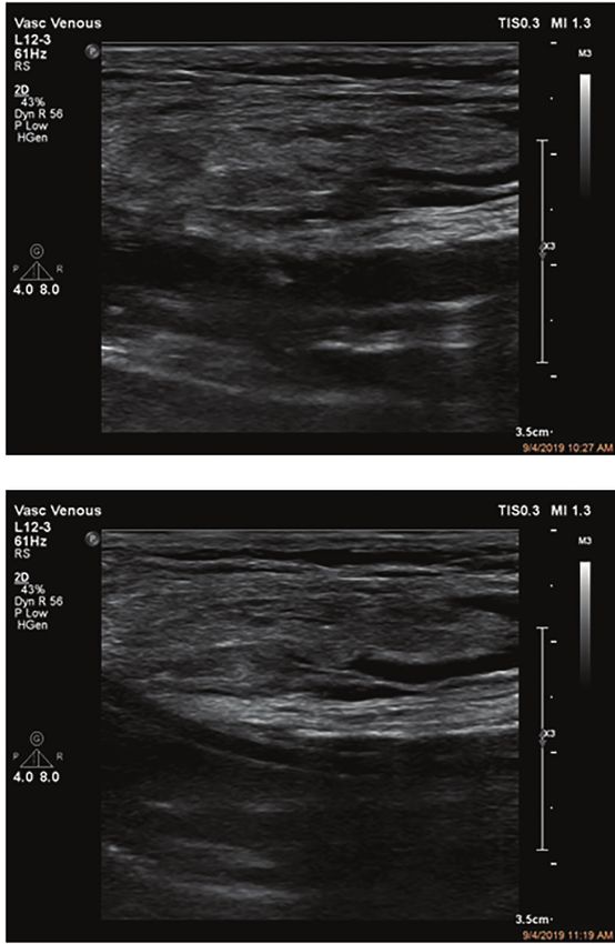
SFA Access Closure With a Mynx Device Post Endovascular Intervention                                                               MIRANDA, et al.

                                                                                                               ns
                                                                                                             io
 A                                                                            B

                                             y at
                                           nl ic
                                          O un
                                        se m
                                     l U om
                                   na C
                                 so P
                               er HM
                           r P 21

C                                                                            D
                        Fo 20

Figure 1. (A) Longitudinal view of proximal superficial femoral artery. (B) Needle access within lumen of superficial femoral artery. (C) Wire passage
within lumen of superficial femoral artery. (D) Sheath introduced to superficial femoral artery.
                            ht
                         ig
                   yr

experience utilizing ultrasound-guided Mynx closure systems                  with ischemic rest pain, gangrene, or lower-limb ulceration of
           op

to assist with closure of the superficial femoral artery percuta-            >2 weeks with combined hemodynamic evidence of impaired
neous access with a focus on safety and success of the technique.            perfusion (ankle brachial index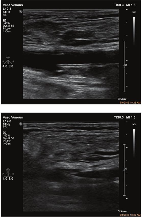
SFA Access Closure With a Mynx Device Post Endovascular Intervention MIRANDA, et al.
ns
io
A B
y at
nl ic
O un
se m
l U om
na C
so P
er HM
r P 21
C D
Fo 20
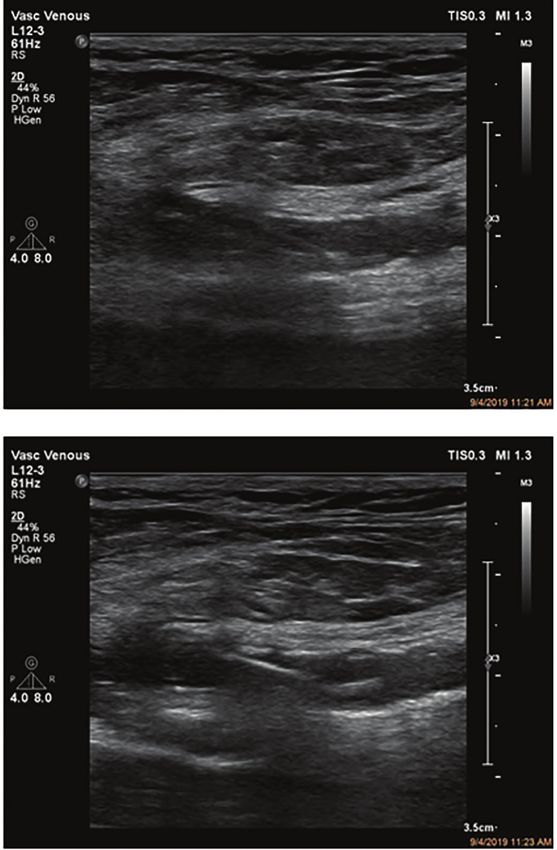
Figure 2. (A) Sheath retracted under ultrasound guidance to just distal to arteriotomy. (B) Active visualization of Mynx device passing intraluminally. (C)
Mynx balloon retracted under ultrasound guidance to completely oppose the arteriotomy. (D) Post-closure color Doppler examination without evidence
ht
of extravasation and triphasic Doppler signal.
ig
yr
intervention was performed if indicated. All patients were re- a 45° angle using ultrasound guidance in both the transverse
op
viewed at 1-month follow-up for major adverse limb event (MALE), and longitudinal projections. In the longitudinal projection,
major amputation, or death. Patients who were >6 months from the proximal superficial femoral artery was visualized just
C
the procedure at the time of data collection were also reviewed after the common femoral bifurcation and the soft tissue was
at 6-month follow-up for MALE, major amputation, or death. inspected. After the needle was introduced intra-arterially,
MALE was defined as need for an above-ankle amputation of the wire was passed under ultrasound guidance to ensure
the index limb or major reintervention (new bypass graft, jump/ a luminal course and then under fluoroscopic guidance to
interposition graft revision, or thrombectomy/thrombolysis).8 ensure an antegrade path without resistance or deviation
Major amputations included through-tibial and through-femur (Figure 1). Diagnostic angiography was obtained through the
amputations. Descriptive statistics were used for demographic 5 Fr sheath and then upsized to either a 6 Fr or 7 Fr sheath to
data and for 1-month and 6-month outcomes. accommodate the intervention planned.
MynxGrip- or MynxControl-assisted closure of the percu-
Technique. All patients underwent ultrasound-guided ante- taneous access arteriotomy was performed upon conclusion of
grade percutaneous access of the proximal superficial femoral the intervention. Heparin was reversed to ensure an activated
artery utilizing a micropuncture access needle and wire. Me- clotting time (ACT) of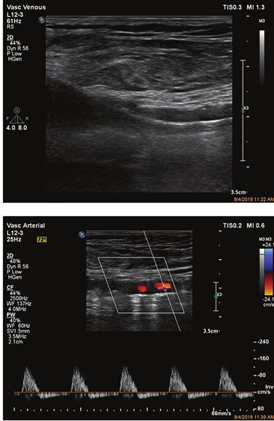
SFA Access Closure With a Mynx Device Post Endovascular Intervention                                                 MIRANDA, et al.

 Table 1. Rutherford chronic limb ischemia stage.
                                                                    Results
                    Rutherford Class                 Patients (n)       We identified 100 consecutive patients who underwent
 3                                                       12         ipsilateral antegrade percutaneous access of the superficial
                                                                    femoral artery for angiography and subsequent endovascular
 4                                                       22
                                                                    revascularization for lower-extremity claudication or CLTI. All
 5                                                       42         patients underwent attempted closure with a Mynx VCD. The
 6                                                       24         median age of intervention was 71 ± 12.63 years (range, 39-95
 Data presented as counts.
                                                                    years). The study population comprised 66 men (66%) and 34
                                                                    women (34%). Rutherford stages of chronic lower-extremity

                                                                                                   ns
                                                                    ischemia were evaluated (Table 1); 66 patients were in stages
 Table 2. Arterial segment of intervention.                         4 and 5. All but 5 patients underwent intervention in addition

                                                                                                 io
                    Arterial Segment                 Patients (n)   to diagnostic angiography (levels of intervention are further

                                                  y at
                                                                    detailed in Table 2). Various sheath sizes were utilized; 62 pro-
 Superficial femoral artery                              58
                                                                    cedures were completed through a 5 Fr sheath, 36 through a 6 Fr

                                                nl ic
 Popliteal artery                                        52         sheath, and 2 through a 7 Fr sheath. All 100 patients received full

                                               O un
 Anterior tibial artery                                  23         anticoagulation with heparin to achieve an ACT >250 seconds.
                                                                    Eight patients received pharmacothrombolytic treatment with

                                             se m
 Tibioperoneal trunk                                     16
                                                                    intra-arterial catheter-directed recombinant tissue plasminogen
 Peroneal artery                          l U om         15
                                                                    activator infusion during their interventions.
 Posterior tibial artery                                 22             Immediate access-site complications were encountered
                                                                    in 2 patients (2%). The first patient was a 94-year-old woman
                                        na C
 Data presented as counts.
                                                                    who underwent successful percutaneous atherectomy and
                                                                    angioplasty of the femoropopliteal short segment chronic total
                                      so P

 Table 3. Follow-up outcomes.                                       occlusion (CTO) through a 6 Fr sheath to treat ischemic rest
                                    er HM

               Outcome                 1 Month (n)   6 Months (n)   pain. The MynxGrip-assisted closure failed to obtain hemostasis,
                                                                    but subsequent manual pressure was successful. However, the
 Major adverse limb event                     5          10
                                                                    patient developed acute limb ischemia from prolonged manual
                                r P 21

 Major amputation                             1           2         compression (>30 minutes) requiring immediate reintervention,
 Death                                        0           2         at which time the access site and superficial femoral artery
                             Fo 20

                                                                    were found to be occluded. This was successfully revascularized
 Data presented as counts.
                                                                    using thrombolytic infusion and suction thrombectomy from a
                                                                    contralateral percutaneous access. At 1-month follow-up, the
                                 ht

                                                                    intervention segment was still patent and no amputation or
                              ig

of access arteriotomy, with either a 5 Fr or 6-7 Fr Mynx device.    further intervention were required. The second patient was a
The Mynx device balloon was prepped before use with a 50:50         63-year-old man who underwent percutaneous atherectomy and
                    yr

contrast-to-saline ratio. Next, the Mynx device was introduced      angioplasty of multisegment, high-grade (80%) superficial femoral
            op

and the balloon was inflated under ultrasound guidance. Then,       artery stenoses and peroneal artery CTO to treat Rutherford 4
the device and the sheath were slowly retracted to the arteri-      chronic ischemia with tissue loss. The Mynx Grip closure failed
      C

otomy site under constant ultrasound visualization. Excellent       and required manual compression. A large hematoma developed
resolution of the balloon location within the vessel can be         in the area tracking along the sartorius muscle. Ultrasound
obtained, as shown in Figure 2. When intraluminal anterior          demonstrated no further hemorrhage from the access site fol-
wall apposition of the balloon was seen on ultrasound, the          lowing prolonged ultrasound-guided compression. The patient
polyethylene glycol plug was deployed. This wall apposition of      remained hemodynamically stable and the limb was successfully
the balloon prevents inadvertent intra-arterial polyethylene        revascularized. Duplex ultrasonography on postoperative day 1
glycol introduction. Tension was held and then the plug was         revealed no evidence of pseudoaneurysm and the patient had a
released per the device instructions for use (IFU). Following       stable hemoglobin and hematocrit compared with his preoper-
the requisite deployment steps, additional ultrasound-guided        ative levels. No further intervention was required. At 1-month
pressure over the arteriotomy site and sheath tract was per-        follow-up, the patient was without further MALE or major
formed for 5 minutes. At that time, duplex ultrasonography was      amputation. None of the patients who received intra-arterial
performed to evaluate for evidence of hematoma formation,           thrombolytic therapy experienced an access-site complication.
active bleeding, or pseudoaneurysm formation.                           At 1-month follow-up, the cohort in total had 5 patients

Vol. 1 · no. 2 June 2021                                                                                                          E59
SFA Access Closure With a Mynx Device Post Endovascular Intervention MIRANDA, et al.
(5%) with a MALE. Four patients required reintervention for also observed a high rate of psuedoaneurysms.12 The etiology
continued ischemia with reocclusion of the treated segment on for these complications can have several explanations. First,
duplex or angiography. One patient had severe persistent pain the balloon of the device can get entrapped in atherosclerotic
and progressed to below-knee amputation. She was a 77-year- and severely calcific lesions upon retraction. This can lead to
old woman who underwent only diagnostic angiography as her an intraluminal deployment of the plug if the operator does
index procedure for this study. She was not revascularized and not confirm appropriate balloon apposition to the anterior wall
did not have evidence of access-site complication. There were of the vessel at the arteriotomy. Second, the balloon can also
no deaths during the 1-month follow-up period. rupture on intraluminal plaque, which would lead to sheath
At 6-month follow-up, 57 patients were available for review. removal along with device removal if the operator is not utiliz-
Ten patients in this cohort (17.5%) had experienced a MALE ing imaging. Adjunct techniques, including opening the sheath
ns
during the follow-up period. Two patients progressed to major side arm and fluoroscopic guidance of a contrast-filled balloon,
amputation due to severity of peripheral arterial disease and 8 can assist with safe retraction, but they do not allow for luminal
io
patients required reintervention on the ipsilateral limb for ongoing identification of the balloon and avoidance of plaques. Finally,
y at
ischemia. There were no access-site complications appreciated confirming complete balloon apposition to the luminal wall
during the follow-up period. Two of the 57 patients reviewed at without ultrasound is not feasible.
nl ic
6 months expired during the follow-up period. Neither death The final step in our technique is ultrasound-guided probe
O un
was associated with access-site complications. Our follow-up compression on the arteriotomy. This allows for precision with
data are summarized in Table 3. pressure application and confirms that no residual arterial jet
se m
or pseudoaneurysm has formed when interrogated with color
Discussion l U om Doppler. If there is evidence of this occurring, we can increase the
period of pressure or decide to intervene prior to the development
Antegrade superficial femoral artery access is not a novel of a large hematoma. Additionally, given the large compliance of
na C
technique; however, it is avoided by many practitioners due to the thigh, clinical findings of a hematoma often only occur with
difficulty with closure. When performing distal lower-extremity large hematomas and would otherwise be neglected.
so P
interventions, ipsilateral antegrade superficial femoral artery Currently, VCD use in the superficial femoral artery is con-
er HM
access provides many advantages, such as improved “pushabil- sidered off-label from the IFU for all devices; however, a previous
ity” and “maneuverability” of the catheters and wires due to retrospective study demonstrated safe usage of early-generation
decreased length. It also avoids difficulty in subselecting the VCDs in the superficial femoral artery.13 Using a VCD decreases
r P 21
superficial femoral artery if ipsilateral antegrade or contralat- patient and physician discomfort, and shortens time to ambulation
eral retrograde common femoral artery access is obtained. With and time to hemostasis. Furthermore, real-time imaging with
Fo 20
ultrasound guidance, it has been demonstrated that access of the ultrasound of the balloon retraction under direct observation
superficial femoral artery is feasible and safe.9 Ultrasound guid- allows operators to avoid balloon rupture, arterial wall damage,
ance is paramount, ensuring 12 o’clock access to the vessel and and intraluminal deployment of the hemostatic plug. In our study,
ht
confirming wire position within the lumen prior to exchanging only 1 intervention required immediate attention to treat an ac-
ig
the needle to a sheath.10 One of the challenges one must overcome cess-site complication. Ninety-eight percent of the study population
with superficial femoral artery antegrade access is to exclude underwent safe and successful percutaneous ultrasound-guided
yr
significant inflow disease; however, all of our patients undergo access closure utilizing the Mynx system. Additionally, the lack
op
preoperative imaging. of permanent sealant or intraluminal component allows for
The superficial femoral artery is prone to significant calcifi- reintervention on that same vessel segment if progression or
C
cation and hemodynamically significant lesions.11 This vessel can recurrence of disease occurs. This was demonstrated with our
have a greater risk of suboptimal percutaneous access, but we data in which 8 patients required reintervention at 6 months.
mitigate this risk with the use of ultrasound. Ultrasound-guided
access allows the user to avoid heavily calcific areas that can lead Study limitations. Our study is limited by its retrospective
to access failure. Additionally, our described approach does not design. Due to the lack of a comparison group, we are unable to
limit the treatment of the superficial femoral artery unless the ascertain whether this technique is an improvement over the
lesion is located proximal to the access. In this study, 58 patients standard VCD technique. Furthermore, data were not collected on
underwent treatment of the SFA. This demonstrates the versatility the number of punctures necessary to obtain luminal superficial
of the antegrade approach while still allowing many patients to femoral artery access, potentially contributing to complication
undergo below-knee interventions at much shorter treatment rate; albeit, we strive for a single puncture to minimize potential
lengths for wires, catheters, and devices. complications. Lastly, operator experience with ultrasound-guided
Fields et al demonstrated that the Mynx device can lead to closure requires time to develop, and this technique could prove
intravascular sealant (potentially causing distal ischemia) and difficult for those less adept with ultrasound. Nonetheless, the
E60 Journal of Critical Limb IschemiaSFA Access Closure With a Mynx Device Post Endovascular Intervention MIRANDA, et al.
ability to observe the device in real time allows for more accu- 6. Waksman R, King SB III, Douglas JS. Predictors of groin complications after balloon
and new-device coronary intervention. Am J Cardiol. 1995;75:886-889.
rate deployment. With this study, we have added a large cohort
of patients to previous data and demonstrate the safety and 7. Conte MS, Bradbury AW, Kolh P, et al; GVG Writing Group. Global vascular guide-
lines on the management of chronic limb-threatening ischemia. J Vasc Surg.
benefit of utilizing ultrasound-guided closure with the Mynx
2019;69:3S-125S.e40.
closure system.
8. Conte MS, Geraghty PJ, Bradbury AW, et al. Suggested objective performance goals
Conclusion and clinical trial design for evaluating catheter-based treatment of critical limb
ischemia. J Vasc Surg. 2009;50:1462-1473.e1-e3.
9. Gutzeit A, Schoch E, Sautter T, et al. Antegrade access to the superficial fem-
Antegrade superficial femoral artery access for ipsilateral oral artery with ultrasound guidance: feasibility and safety. J Vasc Interv Radiol.
revascularization offers many potential advantages when treating
ns
2010;21:1495-1500.
lower-extremity peripheral arterial disease. However, anatomic 10. Lo RC, Fokkema MT, Curran, T. Routine use of ultrasound-guided access reduces
challenges and the potential for immediate access-site complica-
io
access site-related complications after lower extremity percutaneous revascular-
tions have limited the widespread use of this approach. We have ization. J Vasc Surg. 2015;61:405-412.
y at
demonstrated that utilizing the Mynx closure devices under direct 11. Okuno S, Iida O, Shiraki T, et al. Impact of calcification on clinical outcomes after
ultrasound guidance for antegrade superficial femoral artery
nl ic
endovascular therapy for superficial femoral artery disease: assessment using the
peripheral artery calcification scoring system. J Endovasc Ther. 2016;23:731-737.
access closure can be safe and effective to facilitate successful
O un
antegrade percutaneous access when treating patients with 12. Fields JD, Liu DS, Lee SJ, et al. Femoral artery complications associated with the
Mynx closure device. Am J Neuroradiol. 2010;31:1737-1740.
endovascular therapy for peripheral arterial disease.
se m
13. Gutzeit A, van Schie B, Schoch E, et al. Feasibility and safety of vascular closure
l U om devices in an antegrade approach to either the common femoral artery or the
References superficial femoral artery. Cardiovasc Intervent Radiol. 2012;35:1036-1040.
1. Goodney PP, Tarulli M, Faerber AE, et al. Fifteen-year trends in lower limb amputa-
na C
tion, revascularization, and preventive measures among medicare patients. JAMA
Surg. 2015;150:84-86.
so P
2. Gallagher KA, Meltzer AJ, Ravin RA. Endovascular management as first therapy
for chronic total occlusion of the lower extremity arteries: comparison of balloon
er HM
angioplasty, stenting, and directional atherectomy. J Endovasc Ther. 2011;18:624-
637.
From the Division of Vascular Surgery and Endovascular Therapy, Michael E. DeBakey Depart-
3. Noori VJ, Eldrup-Jørgensen J. A systematic review of vascular closure devices for ment of Surgery, Baylor College of Medicine, Houston, Texas.
r P 21
femoral artery puncture sites. J Vasc Surg. 2018;68:887-899.
Disclosure: The authors have completed and returned the ICMJE Form for Disclosure of Potential
4. Robertson L, Andras A, Colgan F. et al. Vascular closure devices for femoral arterial Conflicts of Interest. The authors report no conflicts of interest regarding the content herein.
Fo 20
puncture site haemostasis. Cochrane Database Syst Rev. 2016;3:CD009541.
Manuscript accepted March 15, 2021.
5. Hackl G, Gary T, Belaj K, et al. Risk factors for puncture site complications after
endovascular procedures in patients with peripheral arterial disease. Vasc Endovasc Address for correspondence: Jorge A. Miranda, MD, 7200 Cambridge Street, Suite 6B, Houston,
ht
TX 77030. Email: Jorge.miranda@bcm.edu
Surg. 2015;49:160-165.
ig
yr
op
C
Vol. 1 · no. 2 June 2021 E61You can also read