The use of a score-based protocol in pediatric appendicitis decreases CT scan utilization when evaluating children in a community hospital
←
→
Page content transcription
If your browser does not render page correctly, please read the page content below
Hulka et al. Annals of Pediatric Surgery (2021) 17:44 Annals of Pediatric Surgery
https://doi.org/10.1186/s43159-021-00109-4
ORIGINAL RESEARCH Open Access
The use of a score-based protocol in
pediatric appendicitis decreases CT scan
utilization when evaluating children in a
community hospital
Frieda Hulka1,2* , Bryn Morris3, Paige Elliott2 and Bogna Targonska4
Abstract
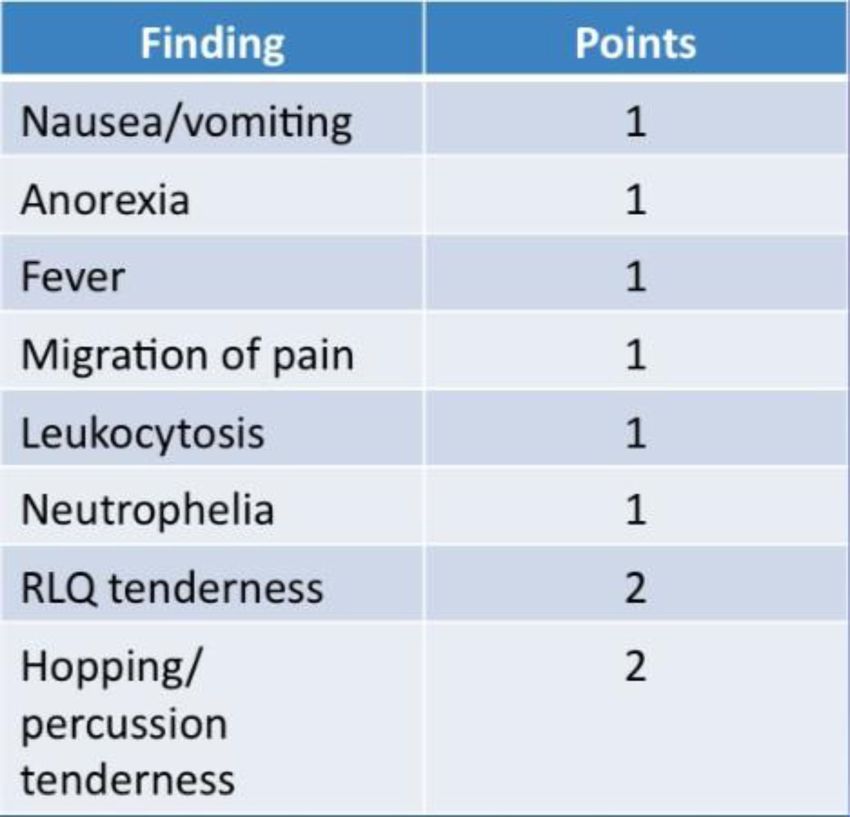
Background: The Pediatric Appendicitis Score (PAS) is a validated scoring system assessing children with
abdominal pain. Prior to 2016, children with abdominal pain in our community hospital were evaluated primarily
using CT scans. A protocol using PAS and ultrasound (US) as the primary radiologic modality was adopted in 2016
for evaluating children with abdominal pain. The protocol consisted of three tiers with low PAS requiring no
radiologic evaluation; moderate PAS requiring US and high PAS requiring initial surgical consultation. Retrospective
chart review of children presenting with clinically suspected appendicitis was performed from January 2015
through December 2017, representing 1 year before and 2 years after implementation of PAS protocol. PAS scoring
was assigned retrospectively to patients not scored in the emergency physician’s note, and statistical analysis of the
patient cohorts was performed using SPSS, version 17. This study was approved by the University of Nevada
Institutional Review Board.
Results: Application of PAS scoring system increased use of US as the primary radiologic test from 59% pre-
protocol to 91% post protocol and decreased use of CT scans from 41 to 8% (p < .05). Physician adherence to
protocol improved from 59 to 71%, increasing further to 81% in the 2nd year post-protocol (p < .05). The highest
rate of non-compliance was noted when providers ordered an US in patients with a low PAS, followed by ordering
any radiologic tests in patients with a high PAS.
Conclusion: Implementation of PAS-based protocol altered clinician behavior in a community hospital when
evaluating children with clinically suspected appendicitis. Improved adherence to the protocol over time with
significant decrease of CT scans ordered thereby reducing radiation exposure in the pediatric population. Future
improvements will be aimed at decreasing radiologic testing in patients with a low PAS and involving surgeons
earlier with patients who have a high PAS as clinical acceptance to the protocol matures.
Keywords: Pediatric Appendicitis Score, CT scan, Ultrasound, Evaluation, Community Hospital
Background This has prompted an increased use of CT scans over
Appendicitis is the most common surgical disease in the years to aid in the diagnosis. An abdominal CT de-
children and can be clinically challenging to diagnose. livers approximately 8 millisieverts (mSV) of radiation
[1]. The average person in the USA receives more than 3
* Correspondence: fhulka@westernsurgical.com mSV of background radiation (excluding medical
1
Department of Surgery, University of Nevada, Reno School of Medicine, sources) per year, rendering an abdominal CT almost
Reno, USA three times the average person’s annual exposure [2].
2
Western Surgical Group, 75 Pringle Way Suite 1002, Reno, NV 89502, USA
Full list of author information is available at the end of the article Studies have shown exposure to CT can increase the risk
© The Author(s). 2021 Open Access This article is licensed under a Creative Commons Attribution 4.0 International License,
which permits use, sharing, adaptation, distribution and reproduction in any medium or format, as long as you give
appropriate credit to the original author(s) and the source, provide a link to the Creative Commons licence, and indicate if
changes were made. The images or other third party material in this article are included in the article's Creative Commons
licence, unless indicated otherwise in a credit line to the material. If material is not included in the article's Creative Commons
licence and your intended use is not permitted by statutory regulation or exceeds the permitted use, you will need to obtain
permission directly from the copyright holder. To view a copy of this licence, visit http://creativecommons.org/licenses/by/4.0/.Hulka et al. Annals of Pediatric Surgery (2021) 17:44 Page 2 of 6 of developing leukemia and brain tumors in pediatric pa- US as the primary radiologic evaluation and encourage tients [3]. Therefore, the benefit of CT scan utilization earlier surgical involvement. for diagnostic purposes must be weighed against the cost of radiation exposure and potential for future Methods oncogenesis. A retrospective chart review of pediatric patients (under Validated scoring systems, such as the Pediatric Ap- 18 years of age) presenting with clinically suspected ap- pendicitis Score (PAS) and the Alvarado Score, have pendicitis was conducted from January 2015 through been shown to help physicians accurately predict appen- December 2017. This review covered the 1 year before dicitis in pediatric patients presenting with abdominal and 2 years after implementation of the PAS protocol. pain [4, 5] (Table 1). The use of clinical practice guide- The protocol divided patients into three groups based lines applied in the emergency department (ED) to po- on their PAS. If the PAS was 0 to 2, the patient was to tential pediatric appendicitis patients has been shown to be discharged with a follow-up call or instructions to re- decrease the use of CT scans [6]. turn to ER within 8 to 12 h for re-evaluation. If the PAS Prior to 2016, children with clinically suspected appen- was 3 to 6, an abdominal ultrasound (US) was to be ob- dicitis in our institution were evaluated primarily using tained. If the US was positive, a surgical consultation CT scans. At the start of 2016, our ED adopted a proto- was obtained. If US was negative or inconclusive, the ER col utilizing PAS as the principal method for evaluation physician was given the option to obtain an abdominal of possible pediatric appendicitis to try and decrease the CT scan or call the surgeon to discuss management. If use of CT scans and thereby radiation exposure. The the PAS was 7 to 10, the ER physician was to obtain a protocol consisted of three tiers with low PAS requiring surgical consultation without initial imaging. no radiologic evaluation, moderate PAS requiring ultra- Charts were then abstracted for demographic data, sound (US), and high PAS requiring initial surgical con- PAS, any imaging obtained, any surgical intervention, sultation. The primary goal was to increase the use of and length of stay. The PAS was assigned retrospectively Table 1 Pediatric Appendicitis Score
Hulka et al. Annals of Pediatric Surgery   (2021) 17:44                                                                     Page 3 of 6

to patients not scored in the emergency physician’s note.              first year and 20% in the second year which was statisti-
There were three time periods reviewed: (1) 1 year prior               cally significant (Table 4). The reasons for protocol vio-
to protocol implementation, (2) first year of protocol,                lations were most frequently obtaining imaging in either
and (3) second year of protocol. Patients in these groups              the low PAS group (29 to 27%) and the high PAS group
were compared for protocol adherence and use of im-                    (29 to 17%). The second most common reason for
aging studies as well as surgical intervention and patho-              protocol violations was CT scans being ordered before
logic findings. Statistical analysis of the patient cohorts            US (58 to 16%) but this became significantly less fre-
was performed using SPSS, version 17 and R statistics.                 quent in the second post protocol year (8%).
Statistical significance was assigned to p values < .05.                  Subset analysis of the negative appendectomy patients
                                                                       demonstrated that all but one in each time frame re-
Results                                                                ceived an imaging study (Table 5). One third to one half
Over the three time periods reviewed, 1758 pediatric pa-               of those studies were interpreted as positive for appendi-
tients were evaluated in our ER with symptoms concern-                 citis. Indications for surgery in the normal or equivocal
ing for appendicitis. The pre protocol group was                       studies were based on clinical appearance. The PAS
compared to the first and second year post protocol                    score in this subset averaged 5.3 to 6.
groups with no significant differences in demographics                    US sensitivity improved over the study period (Table
(Table 2). After application of the PAS protocol, there                6). Specificity remained high throughout the study
was a significant decrease in the usage of CT as the pri-              period, greater than 96%. US reading of the “appendix
mary radiologic test in the first post protocol year com-              not seen” ranged from 59 to 66% of the readings. Missed
pared to the pre protocol year (42 to 16%). There was a                appendicitis on US dropped from 31% in the pre proto-
further decrease in the second post protocol year (16 to               col time frame to 11% and 14% respectively.
8%), demonstrating increasing compliance with the                         Review of patients with low PAS found that 2.5 to 8%
protocol. The use of US as the primary radiologic mo-                  of them returned for re-evaluation (Table 7). There were
dality concomitantly increased significantly in the first              two patients in the pre protocol year and two patients in
and second year post protocol implementation.                          the first post protocol year who were discharged and
   Approximately one-third of the patients who pre-                    later returned and underwent surgery at their second
sented in each time frame underwent appendectomy.                      visit. This number increased to 10 patients (26%) in the
There was a minimal increase in the negative appendec-                 second post protocol year. Appendicitis was found in 50
tomy rate over time (12.5% pre protocol; 18% in second                 to 100% of these re-evaluated patients who underwent
year post protocol) (Table 3). The vast majority of pro-               surgery.
cedures performed were laparoscopic appendectomies at
greater than 90%. The diagnosis of appendicitis was                    Discussion
based on pathologic findings. The length of stay for non-              The use of clinical scoring systems to evaluate children
perforated appendectomies was 25 h in the pre protocol                 with possible appendicitis has been used for over 20
time frame. There was no significant difference in the                 years [7]. Our findings concur that a protocol based sys-
first and second post protocol groups (28 h and 30 h).                 tem can decrease the use of CT and increase the use of
   Analysis found protocol violations present in 42% of                US to help diagnose pediatric appendicitis. If a patient
patients in the pre protocol group, which was applied                  presented to our ER pre protocol, 42% would undergo
retrospectively for comparison. After implementation of                CT scan. By the end of the study, CT usage as the first
the PAS protocol, the violations dropped to 27% in the                 modality dropped to 8%. Parenthetically, US usage

Table 2 Demographics and radiologic tests
Demographic                                          Pre-protocol                Post protocol year 1          Post protocol year 2
Total patients                                       400                         927                           431
Gender—M/F                                           190/210 (49% M)             441/486 (48% M)               134/237 (47% M)
Age                                                  9.67 years                  9.46 years                    9.77 years
Length of symptoms                                   3.4 days                    3.5 days                      2.0 days
PAS Score                                            3.6                         3.0                           4.3
Number of patients with radiologic tests             220 (55%)                   455 (50%)                     380 (88%)
US obtained first                                    129 (58%)                   381 (84%)                     345 (91%), p < .001
CT obtained first                                    88 (42%)                    71 (16%)                      33 (8%), p < .001
Total CTs completed (% of all patients)              133 (37%)                   151 (16%)                     112 (25%)
Hulka et al. Annals of Pediatric Surgery (2021) 17:44 Page 4 of 6 Table 3 Procedures and findings Procedures/findings Pre-protocol (400) Post protocol year 1 (927) Post protocol year 2 (431) Operative procedure 127 (32%) 143 (15%) 125 (29%) Laparoscopic appendectomy 120 (95%) 132 (92%) 117 (94%) Open appendectomy 7 ( 5%) 11 ( 8%) 8 ( 6%) Acute appendicitis 100 (79%) 107 (75%) 88 (70%) Perforated appendicitis 11 ( 9%) 15 (10%) 14 (11%) Normal appendix 16 (12%) 15 (15%) 14 (18%) Length of stay—non perforated 25 h (4–168) 28 h (4–500) 30 h (4–166) Length of stay—perforated 108 h (32–208) 92 h (23–216) 135 h (65–280) increased from 58 to 92% in those needing radiologic ordered on pediatric patients with abdominal pain, usu- evaluation. ally before consulting with them [15]. This concern led Both ultrasound (US) and computed tomography (CT) to the implementation of the Pediatric Appendicitis have been shown to improve diagnostic accuracy in ap- Score in our institution and this retrospective study was pendicitis [8]. There is more evidence over the past dec- used to evaluate our own outcomes with this change in ade that implementation of diagnostic algorithms can practice. Increased focus on US usage as the primary decrease CT utilization thereby decreasing radiation ex- modality improved our sensitivity from 42 to 60–80%, posure in the pediatric population [9–11]. Ultrasound comparable to other studies [16]. has been an important tool for the diagnosis of appendi- Our findings after initiating the PAS protocol concur citis since the 1990s [12]. There have been substantial with other recent reviews and supports the conclusion advances in ultrasound technology and the graded com- that protocols alter clinician behavior [11]. The number pression technique that have allowed for improved of CT scans ordered decreased over time with improving visualization of the appendix [13]. US presents an advan- compliance with the protocol. This was evidenced by an tage over other imaging modalities as it is noninvasive, increase in ultrasounds ordered and fewer violations in can be rapidly performed, and is relatively inexpensive the post protocol group compared to the pre protocol [14]. group. Protocol violations were most common in pa- Ultrasound, however, is highly operator dependent. tients with a low PAS, where our study revealed that While technical expertise and diagnostic accuracy is im- many clinicians opted to order an imaging study. In proving in high volume centers, for smaller community those with a high PAS, we found that many clinicians hospitals, this can present some challenges and doubts ordered imaging before notifying the general surgeon on as to accuracy. False-negative ultrasound results may call. Time of presentation and clinical presentation may lead to delayed diagnosis, increased risk of perforation, have altered the clinical decision making in these cases. and increased sepsis-related morbidity. False-positive re- By replacing CT with ultrasound as first-line imaging sults may lead to unnecessary surgery and risk of com- for clinically suspected appendicitis, there was a modest plications. CT scans provide more detailed images than increase in negative appendectomy rate, which did not US, but they carry an increased risk of exposure to ioniz- prove statistically significant. Mandatory imaging in clin- ing radiation. Over the past decade, there has been more ically suspected appendicitis can decrease the negative attention given to the risks of radiation exposure in the appendectomy rate, but this also requires subjecting pediatric population. Despite publication of evidence- pediatric patients to ionizing radiation, in the case of CT based reviews and Cochrane recommendations for in- use, or relying on operator-dependent ultrasounds [17]. creased use of US, the surgeons in our community hos- The protocol deemed imaging unnecessary in patients pital were continuing to see a large number of CT scans with a high PAS, based on the idea that surgeon Table 4 Protocol violations Protocol violations Pre-protocol (400) Post protocol year 1 (927) Post protocol year 2 (431) Number of protocol violations 167 (42%) 272 (27%) 89 (20%), p < .001 Imaging in patients with PAS < 3 49 (29%) 95 (34%) 24 (29%) Imaging in patients with PAS > 7 49 (29%) 23 ( 8%) 15 (17%) CT done without US 86 (58%) 71 (16%) 33 ( 8%)
Hulka et al. Annals of Pediatric Surgery               (2021) 17:44                                                                                 Page 5 of 6

Table 5 Negative appendectomy                                                     Table 7 Re-evaluated patients with low PAS
Negative                   Pre-             Post protocol         Post protocol   Re-evaluation with low         Pre-           Post              Post
appendectomies             protocol         year 1 (18)           year 2 (23)     PAS                            protocol       protocol          protocol
                           (16)                                                                                  (14)           year 1 (24)       year 2 (38)
Imaging testing            15 (93%)         17 (93%)              22 (93%)        Number of patients             2 (14%)        2 (8%)            10 (26%)
performed                                                                         undergoing
                                                                                  appendectomy
Positive imaging for       8 (53%)          6 (33%)               9 (40%)
appendicitis                                                                      Pathology findings             1—             1—positive        5—positive
                                                                                                                 positive       appy              appy
Averaging PAS              6                5.3                   5.3
                                                                                                                 appy           1—normal          2—
                                                                                                                 1—             appy              perforated
                                                                                                                 perforated                       appy
prediction for appendicitis is more accurate than im-                                                            appy                             3—normal
aging or clinical findings, alone and in these cases, the                                                                                         appy
benefits of definitive surgical treatment outweighed the
risks of imaging or missing appendicitis [18].                                       Based on the positive impact of implementation of use
                                                                                  of PAS and guidelines for evaluation of abdominal pain
Conclusions                                                                       in our pediatric patients, we continue to see an increase
Implementation of a PAS protocol altered clinician be-                            in compliance with these guidelines. Future improve-
havior in a community hospital when evaluating children                           ments will be aimed at decreasing radiologic testing in
with clinically suspected appendicitis, demonstrated by                           patients with a low PAS and involving surgeons in pa-
increased adherence to the protocol over time and a sig-                          tient care sooner in those with a high PAS. The use of
nificantly lower number of CT scans ordered thereby re-                           longer observation prior to surgical intervention may
ducing radiation exposure in the pediatric population.                            also improve our negative appendectomy rate.
Future improvements will be aimed at decreasing radio-
                                                                                  Abbreviations
logic testing in patients with a low PAS and involving                            PAS: Pediatric Appendicitis Score
surgeons earlier with patients who have a high PAS.
                                                                                  Acknowledgements
                                                                                  We would like to acknowledge Yan Liu, PhD, Assistant Professor of
Limitations                                                                       Biostatistics, School of Community Health Sciences at the University of
Limitations of our study are comparable to other retro-                           Nevada, Reno, for his assistance with analysis of the data.
spective reviews. It is possible that patient charts did not                      We also would like to acknowledge Kristina Deeter, MD, for her editorial
                                                                                  assistance of this manuscript.
provide a complete clinical picture of the patient. It is
difficult to assess in a chart review whether a physician                         Authors’ contributions
had a specific reason for ordering an imaging study in                            FH designed the study and aided in the collection of data, analysis and
patients with a higher or lower score. There often is not                         interpretation of the data, and writing of the manuscript. BM aided in the
                                                                                  collection of data and the writing of the manuscript. PE aided in the
documentation of timing of calls to a surgeon or even                             collection of the data. BT aided in the writing of the manuscript. All authors
hallway discussions in the ED which may have led to im-                           have read and approved the final manuscript.
aging documented prior to official consultation.
                                                                                  Funding
                                                                                  There was no funding required in the in the designing, collection, analysis,
Table 6 Evaluation of ultrasound                                                  and interpretation of data or the writing of the manuscript.
Ultrasound        Pre-protocol Post protocol                  Post protocol
findings          (129)        year 1 (382)                   year 2 (345)        Availability of data and materials
                                                                                  All data generated or analyzed during this study are included in this
Appendix not      74 (57%)            232 (60%)               229 (60%)           published article.
seen
Positive          23 (53%)            26 (11%)                33 (14%)            Declarations
appendicitis
                                                                                  Ethics approval and consent to participate
Normal            30 (23%)            88 (23%)                53 (23%)            This study was approved by the ethics committee of the University of
appendix                                                                          Nevada Institutional Review Board. No reference number was provided.
Positive          1 (1%)              0                       0                   Consent to participate was not applicable.
appendicitis
                                                                                  Consent for publication
Positive          21 (16%)            53 (14%)                63 (28%)            Not applicable
appendicitis
Negative          3 (2%)              6 (3%)                  10 (4%)             Competing interests
appendix                                                                          The authors declare that they have no competing interests.

Sensitivity       42%                 81%                     62%                 Author details
                                                                                  1
                                                                                   Department of Surgery, University of Nevada, Reno School of Medicine,
Specificity       96%                 97%                     86%
                                                                                  Reno, USA. 2Western Surgical Group, 75 Pringle Way Suite 1002, Reno, NV
Hulka et al. Annals of Pediatric Surgery (2021) 17:44 Page 6 of 6
89502, USA. 3University of Nevada, Reno School of Medicine, Reno, USA.
4
Reno Radiological Associates, Reno, NV, USA.
Received: 24 January 2021 Accepted: 8 June 2021
References
1. Power SP, Moloney F, Twomey M, James K, O'Connor OJ, Maher MM.
Computed tomography and patient risk: Facts, perceptions and
uncertainties. World J Radiol. 2016;8(12):902–15. https://doi.org/10.4329/wjr.
v8.i12.902.
2. Environmental Protection Agency: https://www.epa.gov/radiation/radiation-
sources-and-doses
3. Pearce MS, Salotti JA, Little MP, McHugh K, Lee C, Kim KP, et al. Radiation
exposure from CT scans in childhood and subsequent risk of leukaemia and
brain tumours: a retrospective cohort study. Lancet. 2012;380(9840):499–505.
https://doi.org/10.1016/S0140-6736(12)60815-0.
4. Goldman RD, et al. Prospective validation of the Pediatric Appendicitis
Score. J Pediatr. 2013;153(2):278–82.
5. Macklin CP, Radliffe GS, Merei JM, Stringer MD. A prospective evaluation of
the modified Alvarado score for acute appendicitis is children. Ann R Coll
Surg Engl. 1997;79(3):203–5.
6. Russell WS, Schuh AM, Hill JG, Hebra A, Cina RA, Smith CD, et al. Clinical
practice guidelines for Pediatric Appendicitis evaluation can decrease
computed tomography utilization while maintaining diagnostic accuracy.
Pediatr Emerg Care. 2013;29:568–73.
7. Samuel M. Pediatric appendicitis score. J Pediatr Surg. 2002;37(6):877–81.
https://doi.org/10.1053/jpsu.2002.32893.
8. Bachur RG, Hennelly K, Callahan MJ, Chen C. Monutreaux. Diagnostic
imaging and negative appendectomy rates in children; effects of age and
gender. Pediatrics. 2012;129(5):877–84. https://doi.org/10.1542/peds.2011-33
75.
9. Shah SR, Sinclair KA, Theut SB, Johnson KM, Holcomb GW, Peter SDS.
Computed tomography utilization for the diagnosis of acute appendicitis in
children decreases with a diagnostic algorithm. Ann Surg. 2016;264(3):474–
81. https://doi.org/10.1097/sla.0000000000001867.
10. Kim DY, Shim DH, Cho KY. Use of the pediatric appendicitis score in a
community hospital. Indian Pediatr. 2016;53(3):217–20. https://doi.org/10.1
007/s13312-016-0823-2.
11. Nielsen JW, Boomer L, Kurtovic K, Lee E, Kupzyk K, Mallory R, et al. Reducing
computed tomography scans for appendicitis by introduction of a
standardized and validated ultrasonography report template. J Pediatr Surg.
2015;50(1):144–8. https://doi.org/10.1016/j.jpedsurg.2014.10.033.
12. Sivit CJ, Newman KD, Boenning DA, Nussbaum-Blask AR, Bulas DI, Bond SJ,
et al. Appendicitis: usefulness of US in a pediatric population. Radiology.
1992;185(2):549–52. https://doi.org/10.1148/radiology.185.2.1410371.
13. Davidson PM, Douglas CD, Hosking CS. Graded compression
ultrasonography in the assessment of the “tough decision” acute abdomen
in childhood. Pediatric Surgery. 1999;15(1):32–5. https://doi.org/10.1007/s003
830050506.
14. Wan MJ, Krahn M, Ungar WJ, Sung L, Medina LS, Doria AS. Acute
appendicitis in young children: cost-effectiveness of US versus CT in
diagnosis – a Markov decision analytic model. Radiology. 2009;250(2):378–
86. https://doi.org/10.1148/radiol.2502080100.
15. Wild JRL, Abdul N, Ritchie JE, Rud B, Freels S, Nelson RL. Ultrasonography for
diagnosis of acute appendicitis. Cochrane Database Syst Rev. 2013;2:1465–858.
16. Mittal MK, Dayan PS, Macias CG, Bachur RG, Bennett J, Dudley NC, et al.
Performance of ultrasound in the diagnosis of appendicitis in children in a
multicenter cohort. Acad Emerg Med. 2013;20(7):697–702. https://doi.org/1
0.1111/acem.12161.
17. Castro SMD, Geerdink TH, Macco S, Veen RNV, Jensch S, Vrouenraets BC.
Mandatory imaging in the work-up of children suspected of having
appendicitis reduces the rate of unnecessary surgeries. J Pediatr Surg. 2018;
53(10):2028–31. https://doi.org/10.1016/j.jpedsurg.2018.02.050.
18. Yangyang RY, Rosenfeld EH, Dadjoo S, et al. Accuracy of surgeon prediction
of appendicitis severity in pediatric patients. J Pediatr Surg. 2019;54(11):
2274–8. https://doi.org/10.1016/j.jpedsurg.2019.04.007.
Publisher’s Note
Springer Nature remains neutral with regard to jurisdictional claims in
published maps and institutional affiliations.You can also read