Short- and long-term outcomes of endoscopic submucosal dissection for non-ampullary duodenal neuroendocrine tumors
←
→
Page content transcription
If your browser does not render page correctly, please read the page content below
ORIGINAL ARTICLE                                                                                  Annals of Gastroenterology (2020) 33, 1-7

Short- and long-term outcomes of endoscopic submucosal
dissection for non-ampullary duodenal neuroendocrine tumors

Masafumi Nishioa, Kingo Hirasawaa, Yuichiro Ozekia, Atsushi Sawadaa, Ryosuke Ikedaa,
Takehide Fukuchia, Ryosuke Kobayashia, Makomo Makazua, Chiko Satoa, Shin Maedab
Yokohama City University Medical Center; Yokohama City University Graduate School of Medicine, Japan

Abstract                            Background Endoscopic resection is recommended for non-ampullary duodenal neuroendocrine
                                    tumors (NAD-NETs) ≤10 mm in diameter and confined to the submucosal layer, without lymph
                                    node or distant metastasis. However, the efficacy and safety of endoscopic submucosal dissection
                                    (ESD) for NAD-NET remains unclear. The aim of this study was to assess the short-term efficacy
                                    and safety and the long-term outcomes of ESD for NAD-NET.
                                    Methods Eight patients with 8 NAD-NETs who underwent ESD between 2015 and 2018 were
                                    included. The indications for ESD were: i) tumor ≤10 mm in diameter; ii) NET G1; iii) confined
                                    to the submucosal layer; and iv) without lymph node or distant metastasis. We retrospectively
                                    assessed the short- and long-term outcomes and safety.
                                    Results The median patient age was 69 (48-76) years. All tumors were located in the duodenal
                                    bulb and showed 0-Is morphology. The median size was 6.4 (3-9.3) mm. The rates of en bloc
                                    resection, histologically free horizontal and vertical margins, and curative resection were 100%,
                                    88%, and 88%, respectively. Intraoperative and postoperative perforation each occurred in 13% of
                                    patients, all of whom were treated conservatively and avoided emergent surgery. Delayed bleeding
                                    was not observed. No local, lymph node or distant recurrence was observed during a median
                                    follow-up period of 34 (18.5-62.5) months.
                                    Conclusions The rates of en bloc and curative resection, and histologically free margins were sufficiently
                                    high. Although intraoperative and postoperative perforations occurred, emergency surgery was not
                                    needed. The results show that ESD is an efficacious and safe treatment for NAD-NET.
                                    Keywords Duodenum, endoscopic resection, endoscopic submucosal dissection, neuroendocrine
                                    tumor, treatment outcome
                                    Ann Gastroenterol 2020; 33 (3): 1-7

Introduction                                                             of the total number [1,2]. However, these tumors are being
                                                                         recognized at an increasing frequency with the widespread use
   The gastrointestinal tract is the most frequent site of               of gastrointestinal screening endoscopy [3].
neuroendocrine tumors (NETs) [1]. Duodenal NETs occur less                   According to the European Neuroendocrine Tumor
frequently than gastric and rectal NETs, accounting for
2 M. Nishio et al
however, it achieves better en bloc and curative resection rates exclusive pump (Telfusion pump; TERUMO, Tokyo, Japan) and
than EMR [13-18]. Since there are only a few published studies pentazocine (15 mg). A single-channel upper gastrointestinal
with a very small number of patients, we aimed to assess the endoscope with a water-jet system (GIF-Q260J; Olympus)
efficacy and safety of ESD for NAD-NET over a longer period was used. Several spots were marked at least 5 mm outside
and with a slightly higher number of cases. the border of the lesion with the Dual knife (Olympus). After
injection of 0.4% hyaluronic acid solution (MucoUp; Johnson
& Johnson Medical Co., Tokyo, Japan) into the submucosa, the
mucosal incision was performed outside of the markings using
Patients and methods the Dual knife to achieve negative horizontal margins. After
mucosal incision, submucosal dissection was also performed
Enrolled patients and tumors using the Dual knife (1.5 mm). To achieve negative vertical
margins, submucosal dissection was performed as close to the
Between January 2015 and September 2018, 8 muscle layer as possible. A high-frequency generator (VIO
consecutive patients with 8 NAD-NETs underwent ESD 300D; ERBE, Tübingen, Germany) was used during mucosal
at Yokohama City University Medical Center. In all cases, incision and submucosal dissection: mucosal incision was
esophagogastroduodenoscopy (EGD), endoscopic ultrasound performed using ENDO CUT I mode (Effect 2), and submucosal
(EUS; high-frequency miniprobe, UM-2R, 20MHz; Olympus, dissection was performed using SWIFT COAG mode (Effect
Tokyo, Japan), and computed tomography (CT) were 3, 40W). Carbon dioxide insufflation was used during all ESD
performed before ESD. We confirmed that all patients met cases. In 7 cases, the artificial ulcer that developed after ESD
the following criteria before ESD: i) histological diagnosis of was covered with a polyglycolic acid (PGA) sheet (Neoveil;
NET G1 via endoscopic biopsy; ii) tumor ≤10 mm in diameter Gunze Co., Kyoto, Japan) and fixed in place with fibrin glue
on EUS; iii) confined to submucosal layer on EUS; and iv) no (Beriplast P Combi-Set; CSL Behring Pharma, Tokyo, Japan)
regional lymph node enlargement or distant metastasis on to prevent delayed perforation. All procedures were performed
CT. The procedures were performed in accordance with the by an experienced endoscopist who had previously performed
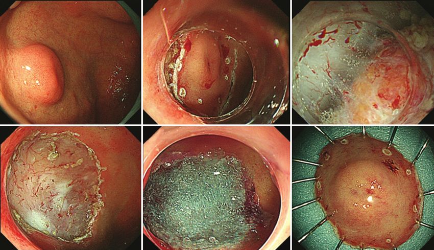
Helsinki Declaration of the World Medical Association. more than 20 duodenal ESDs for epithelial tumors (Fig. 1).
ESD procedures Histological evaluation
All patients underwent ESD under sedation with After fixing in 10% formalin and serial sectioning at
intravenous propofol (0.8-2.0 mg/kg/h) administered using an 2-mm intervals, the resected specimens were assessed
A B C
D E F
Figure 1 Endoscopic submucosal dissection technique. (A) A non-ampullary duodenal neuroendocrine tumor is observed in the anterior wall of
the duodenal bulb. (B) Mucosal incision is performed using the Dual knife after marking with dots around the tumor. (C) Submucosal dissection is
performed using the Dual knife (1.5 mm) as close to the muscle layer as possible. Since few Brunner’s glands exist just below the tumor, we should
be careful not to injure the tumor when submucosal dissection is at that site. In contrast, when the lifting of the submucosal injection around the
tumor is insufficient because of abundant Brunner’s glands, we should be careful not to injure the muscle layer to avoid intraoperative perforation.
(D) The tumor is completely removed with no intraoperative perforation. (E) The artificial ulcer after endoscopic submucosal dissection is covered
with a polyglycolic acid sheet, fixed in place with fibrin. (F) The tumor is resected en bloc
Annals of Gastroenterology 33Efficacy of ESD for duodenal NETs 3
histologically. Experienced gastrointestinal pathologists alternative versions were obtained from all patients included
assessed the histological type, macroscopic appearance, tumor in the study.
size, depth of invasion, lymphatic and vascular involvement,
and horizontal and vertical margins. For classification of
histological type, the World Health Organization (WHO)
2010 classification of tumors of digestive system was used [22]. Result
Immunohistochemical analysis with 2 well-characterized
neuroendocrine markers, chromogranin A and synaptophysin, Clinicopathological characteristics
was performed to reach an accurate diagnosis. In addition, the
Ki-67 index was assessed in all cases to classify tumors as G1, The clinicopathological characteristics of the study cohort
G2, or G3 according to the WHO classification. are summarized in Table 1. Five (63%) of the 8 patients were
men. The median age at the time of diagnosis was 69 (range
48-78) years. All patients were asymptomatic, and the tumors
Definition were detected incidentally during a screening EGD. All tumors
were located in the duodenal bulb and showed 0-Is endoscopic
En bloc resection was defined as resection of the lesion in a morphology. The median tumor diameter on EUS was 6.4
single piece with no endoscopically visible residual tumor. R0 (range 3-9.3) mm. During the study period, all NAD-NETs
resection was defined as en bloc resection with histologically free were included in the indication of endoscopic resection (ESD
horizontal and vertical margins. Curative resection was defined or EMR) and none was treated by surgery.
as en bloc resection of tumor ≤10 mm in diameter confined to
the submucosal layer, and without lymphovascular invasion.
According to the time of onset, bleeding was subdivided into Short-term outcome and pathological findings
intraoperative and delayed bleeding [23]. Delayed bleeding was
defined as hematemesis or melena that required an endoscopic The short-term outcome and pathological findings of all
hemostatic procedure using hemostatic forceps or clips [24]. cases is summarized in Table 2. The median ESD procedural
Intraoperative perforation was defined as perforation occurring time was 43 (range 25-98) min. En bloc, R0 and curative
during the procedure. Delayed perforation was diagnosed when resection were achieved in 100% (8/8), 88% (7/8), and 88%
sudden high fever with peritoneal or retroperitoneal free air on (7/8) of tumors, respectively. In the single case in which
CT occurred postoperatively, in the absence of intraoperative an R0 resection was not achieved, the horizontal margin
perforation and signs of free air on CT immediately after tumor was negative but the vertical margin of the specimen was
removal [25]. positive. All tumors were confined to the submucosal layer
and were positive for chromogranin A and/or synaptophysin
on immunohistochemical staining. The Ki-67 index was less
Follow up than 2% in all tumors; therefore, all tumors were diagnosed
as NET G1 based on the WHO classification. None of the
All patients received a standard intravenous dose of proton tumors exhibited lymphovascular invasion. Additional
pump inhibitors (PPIs) for 3 days and were switched to oral PPIs surgical resection was offered to the patient in whom a curative
for 4 weeks after the ESD procedure. All patients were followed- resection was not achieved because of positive vertical margins.
up by an annual EGD to check for local recurrence and an However, she rejected the additional treatment and underwent
annual CT to identify lymph node and distant metastases. careful observation using EGD and CT after the ESD.
Table 1 Characteristics of patients and tumors in the present study
Statistical analysis Characteristics Value
Age in years, median (range) 69 (48-78)
Statistical analysis was performed using JMP Pro 12
software (SAS Institute, Cary, NC). Continuous variables are Sex, n (%)
presented as median and range, and categorical variables are Male 5 (63)
presented as percentage. The Kaplan-Meier method was used Female 3 (37)
for the analysis of long-term outcomes.
Tumor location, n (%)
Bulb 8 (100)
Ethical approval Descending part 0 (0)
Tumor morphology, n (%)
This study was approved by the Institutional Review Board
0-Is 8 (100)
of Yokohama City University Medical Center (D1602024). All
patients were informed of the risks and benefits of treatment Tumor size on EUS, median, mm (range) 6.4 (3-9.3)
before they underwent the procedure. Informed consent or EUS, endoscopic ultrasound
Annals of Gastroenterology 334 M. Nishio et al
Long-term outcome Complications
The details of all patients in the present study are shown in Intraoperative perforation occurred in 1 patient (13%).
Table 3. Patients were followed up for a median of 34.0 (range Stitching using an endoscopic clip was performed immediately
18.5-62.5) months after ESD. None of the patients showed after removing the tumor and emergent surgery was avoided.
evidence of local recurrence, metachronous lymph metastasis, Delayed perforation occurred in 1 patient (13%) on the day
or distant metastasis during the follow-up period. Based following ESD. In this case, free air around the duodenum
on the Kaplan-Meier survival analysis, both the estimated was found on emergency CT. Since there was no obvious
overall survival and progression-free survival rates were 100% perforation site and the artificial ulcer was fully covered
(Fig. not shown). by the PGA sheet during emergency EGD, she was treated
conservatively and avoided emergent surgery. Notably, this
patient showed positive vertical margins. None of the patients
Table 2 Short and long-term outcome of patients with non-ampullary
duodenal NETs who underwent ESD developed belayed bleeding.
Outcome Value
Procedural time, median, min (range) 43 (25-98)
Discussion
Pathological type, n (%)
NET G1 8 (100) In the present study, we assessed the short- and long-term
Tumor size on pathology, median, mm (range) 6.5 (4-9.5) treatment outcomes of ESD for NAD-NET in 8 consecutive
Invasive depth, n (%)
patients; to the best of our knowledge, this is the largest number
reported to date. Soga et al reported that metastasis was found
submucosal layer 8 (100)
in 9.8% of patients with duodenal NET larger than 10 mm
En bloc resection, n (%) 8 (100) in diameter [26]. Therefore, several guidelines recommend
R0 resection, n (%) 7 (88) endoscopic resection, rather than significantly more invasive
open surgery, for duodenal NETs ≤10 mm in diameter
Curative resection, n (%) 7 (88)
and confined to the submucosal layer, with no evidence of
Complication, n (%) lymph node or distant metastasis on CT [4,5,27,28]. Various
delayed bleeding 0 (0) endoscopic resection methods, such as EMR, EMR-L, EMR-P,
intraoperative perforation 1 (13) EMR-C, and ESD, have been reported in the treatment of
duodenal NETs [7,11-14,16-18,29,30]. However, there is no
delayed perforation 1 (13)
consensus regarding the preferred method of endoscopic
Follow-up time, median, months, (range) 34.0 (18.5-62.5) resection. We have employed ESD for NAD-NET since 2015 for
Local recurrence, n (%) 0 (0) the following reasons: i) intraoperative perforation requiring
Lymph metastasis, n (%) 0 (0) emergent surgery occurred in one patient who underwent
EMR-L for NAD-NET in 2015; and ii) as we had previously
Distant metastasis, n (%) 0 (0)
reported, ESD was significantly superior to EMR-L for rectal
ESD, endoscopic submucosal dissection; NET, neuroendocrine tumor
Table 3 Clinicopathological characteristics of patients with NAD-NET underwent ESD
No. Age, Sex Location Size, Depth Pathological Procedural En bloc R0 Curative Complication
year mm Morphology time, min resection resection resection
1 70 M Bulb, AW 5 SM NET G1 76 Yes Yes Yes No
2 55 F Bulb, AW 5 SM NET G1 98 Yes Yes Yes No
3 48 M Bulb, AW 6 SM NET G1 35 Yes Yes Yes No
4 77 M Bulb, SW 7 SM NET G1 30 Yes Yes Yes No
5 67 F Bulb, AW 4 SM NET G1 57 Yes No No Delayed perforation
(pVM1) (pVM1)
6 76 M Bulb, AW 8 SM NET G1 31 Yes Yes Yes No
7 78 M Bulb, AW 7 SM NET G1 25 Yes Yes Yes Intraoperative
perforation
8 66 F Bulb, AW 9.5 SM NET G1 50 Yes Yes Yes No
AW, anterior wall; ESD, endoscopic submucosal dissection; NAD-NET, non-ampullary duodenal neuroendocrine tumor; NET, neuroendocrine tumor; SM,
submucosal; SW, superior wall; VM, vertical margin
Annals of Gastroenterology 33Efficacy of ESD for duodenal NETs 5
NETs in terms of the en bloc, R0 and curative resection is very thin and the narrow lumen leads to poor endoscopic
rates [19]. maneuverability [13,35,36]. This technical difficulty is
In the present study, we observed en bloc, R0 and curative considered to be associated with the high frequency of
resection rates of 100%, 88%, and 88%, respectively, after ESD intraoperative perforations. Two small case series of ESD
for NAD-NET; these were similarly favorable compared to the for NAD-NET reported a high frequency of intraoperative
previously reported results for rectal NETs (100%, 100%, and perforation: 67% (2/3) [18] and 29% (2/5) [17]. Therefore,
83%, respectively). Several previous studies have shown that ESD for duodenal NETs should be performed by experienced
ESD achieves better en bloc resection, R0 resection and curative endoscopists. Although ESD is useful for en bloc resection and
resection rates than EMR [19-21]. We had reported that in R0 resection, duodenal ESD is technically difficult and is only
rectal NETs, the en bloc, R0 and curative resection rates were performed in a few countries. Therefore, duodenal ESD might
73%, 63% and 50%, respectively, in an EMR-L group (n=22), be unacceptable in western countries. On the other hand,
and 100%, 100% and 83% in an ESD group (n=24) [19]. the efficacy of EFTR using an FTRD for duodenal superficial
In NAD-NETs, several previous studies have reported that tumors has been reported [31,32]. Since the evidence of EFTR
the en bloc, R0 and curative resection rates of ESD were higher for duodenal NETs is still unclear, comparative studies of the
than those of EMR, EMR-L and EMR-C [13-18]. Kim et al efficacy and safety of ESD and EFTR are needed. In addition,
reported 38 patients with 41 duodenal NETs treated by EMR exposure of the artificial ulcer after ESD to bile and pancreatic
(n=18), EMR-L (n=16), EMR-P (n=3), or ESD (n=4). In their juice can also lead to delayed perforation [35]. Closure with an
study, the en bloc resection rates in the EMR, EMR-L, EMR-P endoscopic clip is commonly used for the mucosal defect after
and ESD groups were 89%, 100%, 100% and 100%, respectively, ESD. However, in ESD for duodenal bulbus, closure with the
while curative resection was achieved in 56%, 25%, 33% and endoscopic clip is technically difficult because of the narrow
100% [13]. To avoid an additional, invasive surgical resection, working space and proximity to the pylorus. Therefore, we
accurate pathological evaluation of horizontal and vertical selected a PGA sheet with fibrin glue for covering the artificial
margins is important. EMR is associated with positive vertical ulcer after ESD in most cases of the present study. Coverage by
a PGA sheet with fibrin glue has been shown to be helpful for
margins, or a crush and burn effect on the resected specimen,
prevention of delayed perforation after duodenal ESD [37,38].
which leads to difficulties in performing a precise pathological
However, delayed perforation did occur in 1 patient who
evaluation. Recently, endoscopic full-thickness resection
received PGA sheet coverage with fibrin glue in the present
(EFTR) with the full-thickness resection devise (FTRD;
study. Therefore, careful observation of the clinical course
Ovesco Endoscopy, Tübingen, Germany) for duodenal tumors
after ESD and rapid assessment with blood tests and CT scan
had been reported [31,32]. Bauder et al reported that complete
are needed if delayed perforation is suspected.
resection rates were 80% in five subepithelial tumors treated
The patients in the present study were followed-up by CT
by FTRD. Their results suggested that EFTR is effective
scan for detecting metastasis. The European Neuroendocrine
for NAD-NET, but the evidence is insufficient because of
Tumors Society (ENETS) consensus guideline suggests that
the small sample size [32]. Therefore, at the moment, ESD
somatostatin receptor scintigraphy and 68Ga-DOTA-NOC
is the preferred method for NAD-NET to ensure accurate
positron emission tomography/CT are useful for diagnosis;
pathological diagnosis and avoid additional surgical resection however, this modality can lead to follow-up effectiveness. The
for residual tumor. evidence for these modalities is still insufficient and a study of
We also assessed the long-term outcomes of patients with a larger cohort over a longer term is needed.
NAD-NET who underwent ESD. In the present study, none The present study has several limitations. First, it was a
of the patients showed any evidence of local recurrence, or single-center, retrospective study that assessed the outcome
lymph node or distant metastasis, during a median follow-up of ESD for NAD-NET. Second, the present study had a small
period of 34.0 (range 18.5-62.5) months. In previous studies, number of patients. However, to the best of our knowledge,
local recurrence occurred in 0-18% of patients with NAD-NET this is the largest number of patients undergoing ESD for
who underwent EMR [7,13,14,29,30,33,34], whereas no local NAD-NET reported to date. Third, the follow-up period in the
recurrences were observed in patients after ESD [13-15,18]. present study is somewhat insufficient for detecting metastasis.
These studies indicated that en bloc resection and R0 resection Considering the rarity of NAD-NET in the general population,
is important for avoiding local recurrence. Similarly to our a multicenter study involving a larger number of patients with
own, the results from previous studies suggest that ESD is more a long follow-up period is needed.
effective than EMR for maintaining recurrence-free survival In conclusion, the present study showed that if there is
after NAD-NET treatment. neither lymph node nor distant metastasis evident on CT, ESD
We next assessed the complications occurring during is effective and safe for NAD-NETs measuring ≤10 mm in size
ESD for NAD-NET. ESD for duodenal tumors was associated and confined to the submucosal layer. En bloc resection was
with a higher risk of complications, including bleeding and achieved in all cases and R0 resection in most cases, and there
perforation, than EMR [13,35,36]. In the present study, were no recurrences during the follow-up period. However,
no delayed bleeding occurred; however, intraoperative further multicenter, prospective studies involving larger
perforation and delayed perforation each occurred in 1 patient numbers of patients are needed to assess the efficacy and safety
(13%). ESD for duodenal tumors is more technically difficult of ESD and to determine the preferred endoscopic resection
than for stomach or colon lesions, because the duodenal wall method in patients with NAD-NETs.
Annals of Gastroenterology 336 M. Nishio et al
carcinoids in the duodenal bulb using the band ligation technique
Summary Box with the Duette mucosectomy device. Endoscopy 2013;45(Suppl 2
UCTN):E365-E366.
11. Otaki Y, Homma K, Nawata Y, Imaizumi K, Arai S. Endoscopic
What is already known: mucosal resection with circumferential mucosal incision
of duodenal carcinoid tumors. World J Gastrointest Endosc
• Endoscopic resection is recommended for non- 2013;5:197-200.
ampullary duodenal neuroendocrine tumors 12. Karagiannis S, Eshagzaiy K, Duecker C, Feyerabend B,
(NAD-NETs) ≤10 mm in diameter without Mozdzanowski E, Faiss S. Endoscopic resection with the cap
technique of a carcinoid tumor in the duodenal bulb. Endoscopy
lymphovascular invasion
2009;41(Suppl 2):E288-E289.
• Previous studies have reported that endoscopic 13. Kim GH, Kim JI, Jeon SW, et al; Korean College of Helicobacter
submucosal dissection (ESD) was superior to and Upper Gastrointestinal Research. Endoscopic resection for
endoscopic mucosal resection for achieving en duodenal carcinoid tumors: a multicenter, retrospective study.
bloc, R0 and curative resection of rectal NETs J Gastroenterol Hepatol 2014;29:318-324.
• However, the efficacy and safety of ESD for NAD- 14. Kim SH, Park CH, Ki HS, et al. Endoscopic treatment of duodenal
NETs remains unclear neuroendocrine tumors. Clin Endosc 2013;46:656-661.
15. Li QL, Zhang YQ, Chen WF, et al. Endoscopic submucosal
dissection for foregut neuroendocrine tumors: an initial study.
What the new findings are: World J Gastroenterol 2012;18:5799-5806.
16. Matsumoto S, Miyatani H, Yoshida Y. Endoscopic submucosal
• In NAD-NETs treated by ESD, the rates of en bloc dissection for duodenal tumors: a single-center experience.
Endoscopy 2013;45:136-137.
and curative resection, and histologically free
17. Matsumoto S, Miyatani H, Yoshida Y, Nokubi M. Duodenal
margins were sufficiently high carcinoid tumors: 5 cases treated by endoscopic submucosal
• Intraoperative and delayed perforation occurred in dissection. Gastrointest Endosc 2011;74:1152-1156.
a few cases, but no emergency surgery was needed 18. Suzuki S, Ishii N, Uemura M, et al. Endoscopic submucosal
• ESD is an efficacious and safe treatment for NAD- dissection (ESD) for gastrointestinal carcinoid tumors. Surg Endosc
NET 2012;26:759-763.
19. Kaneko H, Hirasawa K, Koh R, et al. Treatment outcomes of
endoscopic resection for rectal carcinoid tumors: an analysis of
the resectability and long-term results from 46 consecutive cases.
References Scand J Gastroenterol 2016;51:1489-1494.
20. Lee DS, Jeon SW, Park SY, et al. The feasibility of endoscopic
1. Modlin IM, Sandor A. An analysis of 8305 cases of carcinoid submucosal dissection for rectal carcinoid tumors: comparison
tumors. Cancer 1997;79:813-829. with endoscopic mucosal resection. Endoscopy 2010;42:647-651.
2. Modlin IM, Lye KD, Kidd M. A 5-decade analysis of 13,715 21. Park HW, Byeon JS, Park YS, et al. Endoscopic submucosal
carcinoid tumors. Cancer 2003;97:934-959. dissection for treatment of rectal carcinoid tumors. Gastrointest
3. Yao JC, Hassan M, Phan A, et al. One hundred years after Endosc 2010;72:143-149.
“carcinoid”: epidemiology of and prognostic factors for 22. Bosman F CF. World Health Organization Classification of
neuroendocrine tumors in 35,825 cases in the United States. J Clin Tumours, Pathology and Genetics of Tumours of the Digestive
Oncol 2008;26:3063-3072. System. Lyon: IARC Press 2010.
4. Delle Fave G, O’Toole D, Sundin A, et al; Vienna Consensus 23. Oda I, Suzuki H, Nonaka S, Yoshinaga S. Complications of gastric
Conference participants. ENETS Consensus Guidelines Update for endoscopic submucosal dissection. Dig Endosc 2013;25(Suppl 1):71-78.
Gastroduodenal Neuroendocrine Neoplasms. Neuroendocrinology 24. Hoteya S, Kaise M, Iizuka T, et al. Delayed bleeding after endoscopic
2016;103:119-124. submucosal dissection for non-ampullary superficial duodenal
5. Lipiński M, Rydzewska G, Foltyn W, et al. Gastroduodenal neoplasias might be prevented by prophylactic endoscopic closure:
neuroendocrine neoplasms, including gastrinoma - management analysis of risk factors. Dig Endosc 2015;27:323-330.
guidelines (recommended by the Polish Network of 25. Inoue T, Uedo N, Yamashina T, et al. Delayed perforation: a
Neuroendocrine Tumours). Endokrynol Pol 2017;68:138-153. hazardous complication of endoscopic resection for non-ampullary
6. Dasari BVM, Al-Shakhshir S, Pawlik TM, et al. Outcomes of duodenal neoplasm. Dig Endosc 2014;26:220-227.
surgical and endoscopic resection of duodenal neuroendocrine 26. Soga J. Early-stage carcinoids of the gastrointestinal tract: an
tumours (NETs): a Systematic review of the literature. J Gastrointest analysis of 1914 reported cases. Cancer 2005;103:1587-1595.
Surg 2018;22:1652-1658. 27. Dalenbäck J, Havel G. Local endoscopic removal of duodenal
7. Gincul R, Ponchon T, Napoleon B, et al. Endoscopic treatment of carcinoid tumors. Endoscopy 2004;36:651-655.
sporadic small duodenal and ampullary neuroendocrine tumors. 28. Delle Fave G, Kwekkeboom DJ, Van Cutsem E, et al; Barcelona
Endoscopy 2016;48:979-986. Consensus Conference participants. ENETS Consensus Guidelines
8. Kumta NA, Desai A, Doshi R, Kahaleh M, Sharaiha RZ. for the management of patients with gastroduodenal neoplasms.
Endoscopic mucosal resection of duodenal carcinoid. Endoscopy Neuroendocrinology 2012;95:74-87.
2016;48(Suppl 1):E158-E159. 29. Oono Y, Shinmura K, Hori K, et al. Endoscopic submucosal
9. Harada H, Suehiro S, Shimizu T, Katsuyama Y, Hayasaka K, resection using a ligation device without injection for duodenal
Ito H. Ligation-assisted endoscopic submucosal resection with neuroendocrine tumors. Surg Endosc 2019;33:2008-2014.
circumferential mucosal incision for duodenal carcinoid tumor. 30. Mahmud N, Tomizawa Y, Stashek K, Katona BW, Ginsberg GG,
World J Gastroenterol 2015;21:10041-10044. Metz DC. Endoscopic resection of duodenal carcinoid tumors:
10. Neumann H, Ramesh J, Wilcox CM, Mönkemüller K. Resection of a single-center comparison between simple polypectomy and
Annals of Gastroenterology 33Efficacy of ESD for duodenal NETs 7
endoscopic mucosal resection. Pancreas 2019;48:60-65. 35. Honda T, Yamamoto H, Osawa H, et al. Endoscopic submucosal
31. Schmidt A, Meier B, Cahyadi O, Caca K. Duodenal endoscopic dissection for superficial duodenal neoplasms. Dig Endosc
full-thickness resection (with video). Gastrointest Endosc 2015; 2009;21:270-274.
82:728-733. 36. Hoteya S, Furuhata T, Takahito T, et al. Endoscopic submucosal
32. Bauder M, Schmidt A, Caca K. Endoscopic full-thickness resection dissection and endoscopic mucosal resection for non-ampullary
of duodenal lesions-a retrospective analysis of 20 FTRD cases. superficial duodenal tumor. Digestion 2017;95:36-42.
United European Gastroenterol J 2018;6:1015-1021. 37. Takimoto K, Imai Y, Matsuyama K. Endoscopic tissue shielding
33. Min BH, Kim ER, Lee JH, et al. Management strategy for small method with polyglycolic acid sheets and fibrin glue to prevent
duodenal carcinoid tumors: does conservative management with delayed perforation after duodenal endoscopic submucosal
close follow-up represent an alternative to endoscopic treatment? dissection. Dig Endosc 2014;26(Suppl 2):46-49.
Digestion 2013;87:247-253. 38. Takimoto K, Toyonaga T, Matsuyama K. Endoscopic tissue
34. Park SB, Kang DH, Choi CW, Kim HW, Kim SJ. Clinical shielding to prevent delayed perforation associated with
outcomes of ligation-assisted endoscopic resection for duodenal endoscopic submucosal dissection for duodenal neoplasms.
neuroendocrine tumors. Medicine (Baltimore) 2018;97:e0533. Endoscopy 2012;44(Suppl 2 UCTN):E414-E415.
Annals of Gastroenterology 33You can also read