Prenatal Presentation of Medulloepithelioma: Case and Literature Review - Cureus
←
→
Page content transcription
If your browser does not render page correctly, please read the page content below
Open Access Case
Report DOI: 10.7759/cureus.5018
Prenatal Presentation of
Medulloepithelioma: Case and Literature
Review
Nidhi Arora 1 , Chanchal Ahmad 2 , Arpit Gupta 3 , Nitin Ghonge 4 , Anita Kaul 1
1. Fetal Medicine, Indraprastha Apollo Hospitals, Delhi, IND 2. Fetal Medicine, Madhukar Rainbow
Children's Hospital, Delhi, IND 3. Neonatology, Metropolitan Hospital Center, New York, USA 4.
Radiology, Indraprastha Apollo Hospitals, Delhi, IND
Corresponding author: Nidhi Arora, drnidhiarora84@gmail.com
Disclosures can be found in Additional Information at the end of the article
Abstract
Congenital brain tumors (CBTs) are extremely rare and account for only 0.5%-1.9% of all
pediatric brain tumors. Medulloepithelioma is one of the rare tumors with an incidence of
about 1% among all CBTs with a very dismal prognosis and typically diagnosed at the median
age of 24 months. The objective is reporting medulloepithelioma presenting in the intrauterine
period with very few prior cases being reported in the prenatal period, and to add to the limited
existing literature on medulloepithelioma. We present a rare case of medulloepithelioma
referred to us in the antenatal period at 27 weeks and subsequently causing intrauterine fetal
demise. Prenatal MRI of the fetal brain and postnatal histopathological findings on autopsy
were suggestive of intracranial medulloepithelioma.
Categories: Obstetrics/Gynecology, Pathology, Radiology
Keywords: ultrasound, fetal mri, intracranial tumor, prenatal diagnosis, medulloepithelioma
Introduction
Congenital brain tumors (CBTs), defined as tumors presenting within 60 days after birth, are
extremely rare and account for only 0.5-1.9% of all pediatric brain tumors [1-3]. The most
frequently reported tumors are teratomas (63%) and gliomas (30%) [4, 5]. Embryonal tumors
(7%) account for rest of the CBTs. Most of the time intracranial tumors (ICTs) are detected
incidentally on routine fetal imaging as a mass with or without hydrocephalus with
macrocephaly as a late feature. Prognosis is usually guarded with an overall neonatal survival
rate of 28% [6]. Medulloepithelioma is one of the rare embryonal tumors with an incidence of
about 1% among all CBTs with a very dismal prognosis [6]. Making a definitive diagnosis
Received 06/03/2019 requires histopathology after tissue biopsy which is very difficult in the fetal period. Therefore,
Review began 06/05/2019
the diagnosis is usually made on the basis of fetal sonogram and fetal MRI findings [7, 8].
Review ended 06/17/2019
Published 06/27/2019
We here present a case of medulloepithelioma which was initially diagnosed with glioma based
© Copyright 2019
Arora et al. This is an open access
on fetal imaging findings but later on diagnosed as medulloepithelioma based on tissue
article distributed under the terms of histopathology. Medulloepithelioma tumors are rare with an average age of two years at
the Creative Commons Attribution presentation with very few cases diagnosed in the neonatal period [6, 9]. Most of the
License CC-BY 3.0., which permits information on medulloepithelioma is based on the prior reported cases. The objective of
unrestricted use, distribution, and
reporting this case is to add to the very limited information on the medulloepithelioma
reproduction in any medium, provided
the original author and source are presentation especially in the fetal period and its findings in the fetal imaging (ultrasound and
credited. MRI). We will also do a brief review on medulloepithelioma based on previously reported
cases [9-14].
How to cite this article
Arora N, Ahmad C, Gupta A, et al. (June 27, 2019) Prenatal Presentation of Medulloepithelioma: Case and
Literature Review. Cureus 11(6): e5018. DOI 10.7759/cureus.5018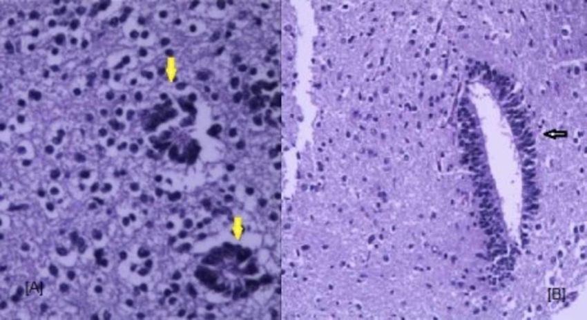
Case Presentation
                                   A 35-year-old gravida three and para one female was referred to the fetal medicine department
                                   in view of an intracranial mass detected on routine growth scan at 27 weeks. Anomaly scan at
                                   19 weeks was normal. The mother gave no history of fever with rash, bleeding disorders,
                                   radiation exposure, drug intake or substance abuse. She was not hypertensive or diabetic and
                                   was not on any medication apart from iron and calcium supplementation. There was no
                                   personal or family history of malignancy in either partner.

                                   Ultrasound was done using Voluson E-Radiance (GE Healthcare, Milwaukee, WI) equipped with
                                   a convex 4-8 MHz abdominal probe, and 6-12 MHz endovaginal probe. Two-dimensional
                                   ultrasound (Figure 1A-1C) showed an intracranial mass in the fetal right frontal lobe measuring
                                   4.5 x 3.8 x 3 cm with echogenicity similar to the adjacent normal brain. The mass was crossing
                                   the midline. A detailed neurosonogram was done. There was no associated ventriculomegaly.
                                   The posterior fossa structures were normal. Transvaginal ultrasound was done to confirm the
                                   findings and to determine the spread of the lesion. On color Doppler, feeding vessels were
                                   identifiable (Figure 1 D). There was no other structural abnormality. Fetal echocardiography
                                   was normal. Fetal growth was within the normal range for gestation. There was
                                   polyhydramnios (amniotic fluid volume above the 95th centile). Diagnosis of an isolated
                                   intracranial mass was made.

                                      FIGURE 1: Ultrasound images of the intracranial mass.
                                      (A) 2D image showing homogenous mass in right frontal lobe in transthalamic plane (white arrow).
                                      (B) Coronal plane (thick white arrow). (C) Normal posterior fossa. (D) Color Doppler showing

2019 Arora et al. Cureus 11(6): e5018. DOI 10.7759/cureus.5018                                                                           2 of 7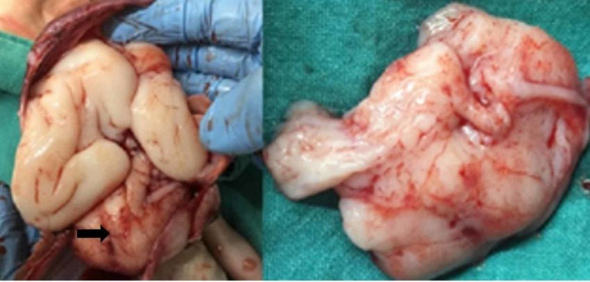
feeding vessels into the mass.
Fetal MRI was performed on a 3 Tesla mode, Philips 3T scanner. T2-weighted axial, coronal and
sagittal images were acquired, along the fetal planes using half Fourier acquired single shot
turbo spin echo (HASTE) sequences for fetal central nervous system (CNS). Fetal MRI showed a
focal intra-axial mass lesion in the right frontal location. Posterosuperiorly the extent was up to
right basal ganglia and thalamic region with indentation over the third ventricle. The lesion
was not seen separate from the crus cerebri. There was compression over the septum
pellucidum, which was displaced to the left by 2-3 mm. The lesion measured 4.8 cm x 4.0 cm x
2.8 cm in antero-posterior, transverse, and craniocaudal dimension, respectively. It was
hypointense on T2W imaging as compared to white matter and showed hypointense to
isointense signal on T1W images. The fetal ventricular system showed extrinsic compression,
mainly over the right lateral ventricle and the third ventricle. The fourth ventricle was not
dilated. The posterior fossa structures were normal. No definite signs of proptosis or
intraorbital extension were seen (Figure 2). A provisional diagnosis of a glioma, possibly of
hypothalamic/thalamic origin was made.
FIGURE 2: Fetal MRI brain.
(A) Axial T2WI HASTE image showing ill-defined round mass (arrow) in left frontal lobe. (B) Coronal
image showing inferior extent of the lesion into the thalami (star).
HASTE: Half-Fourier acquired single shot turbo spin echo
A joint consultation with the neonatologist, pediatric neurosurgeon, and pediatric neurologist
was done. The couple was counseled regarding the expected poor prognosis of antenatally
diagnosed intracranial tumors in view of its imaging findings. The timing of delivery and the
need for close follow-up was also discussed. Follow-up ultrasound one week later at 28 weeks
showed no fetal cardiac activity. Induction of labor was done and a stillborn male fetus
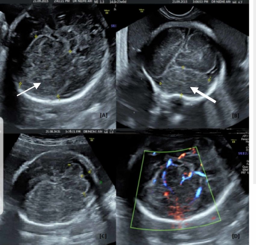
weighing 1300 grams was delivered vaginally. The couple consented for fetal autopsy. The
external examination was normal. There were no dysmorphic features. On autopsy, there was a
large, homogenous, right-sided frontal tumor extending to the base of the skull. There were no
necrotic areas and no hemorrhages (Figure 3). The cerebellum was normal. Histopathology gave
the unexpected diagnosis of medulloepithelioma which is a rare tumor of embryonal origin
2019 Arora et al. Cureus 11(6): e5018. DOI 10.7759/cureus.5018 3 of 7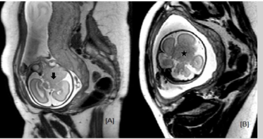
(Figure 4). The histopathology showed a tumor, composed of nests, tubules, and trabecular
                                   arrangement of malignant cells, lined by pseudostratified epithelia, resembling primitive neural
                                   tube, sheets of poorly differentiated cells with hyperchromatic nuclei. Immunohistochemistry
                                   was positive for synaptophysin, and vimentin suggestive of medulloepithelioma.

                                      FIGURE 3: Gross examination of the tumor.
                                      (A) Solid homogenous mass arising from right frontal lobe (arrow). (B) Resected tumor mass.

                                      FIGURE 4: Histopathology of the resected mass.
                                      (A) 20X magnification - Dyscohesive sheets of cells forming rosettes (yellow arrows) at focal places.
                                      The rosettes show well-defined empty small lumen bordered by a thick membrane. (B) 10X
                                      magnification - Scattered few elongated canals (black arrow) lined by stratified columnar type
                                      epithelium at the interface with adjacent brain tissue.

                                   Discussion
                                   Medulloepitheliomas are very rare embryonal tumors with an incidence of 1% to 1.5% among

2019 Arora et al. Cureus 11(6): e5018. DOI 10.7759/cureus.5018                                                                           4 of 7
all CBTs [6]. They were earlier described under the umbrella of primitive neuroendocrine
tumors (PNETs), but as per the latest 2016 WHO classification of brain tumors, they fall under
the category of embryonal tumors with multilayered rosettes (ETMR) or embryonal tumors with
abundant neuropil and true rosettes (ETANTR) [15].
Many of these rare tumors display amplification of the C19MC region on chromosome 19
(19q13.42) and hence described as ETMR C19MC-altered. In the absence of C-19 amplification,
they can be described as ETMR NOS, and if the histopathology is suggestive of
medulloepithelioma, then should be named as medulloepithelioma. However, significant
proportions of medulloepithelioma have not shown C19MC alterations [15]. The main
histopathology description for this tumor is the papillary, tubular, or trabecular arrangement of
pseudostratified neuroepithelium resembling an embryonic neural tube. Others are multi-
layered rosettes and evidence of multiple lines of differentiation including neuronal, glial and
mesenchymal elements [1,6,14]. Majority of these tumors are immunoreactive for vimentin,
and sometimes are immunoreactive for glial and neuronal antigens. Regardless of the change in
the classification, medulloepitheliomas are still considered as highly malignant grade IV
tumor [15].
It was first described by Bailey and Cushing in 1926 [11]. It is a rare malignant tumor of
childhood with an average age at diagnosis is two years. Only very few congenital patients have
been reported [6,14]. There is a very limited data on medulloepithelioma identified as early as
27 weeks post-conceptual age, and causing fetal demise. Cassart et al. in an extensive review
reported 27 cases of fetal intracranial tumors over a period of 14 years [16]. They
retrospectively analyzed imaging and clinical findings in 27 cases of fetal intracranial tumors
assessed by ultrasound and MR imaging followed with histologic confirmation. They diagnosed
15 germinal tumors, four glial tumors, two craniopharyngiomas, and three hamartomas. No
PNET was diagnosed. Isaacs reported only three cases of medulloepithelioma out of 250 cases
(1.3%) in one of the extensive literature review of brain tumors diagnosed in a fetal and
neonatal period [6]. All of the three cases presented with macrocephaly with some neurological
signs upon presentation with no cases of stillbirth.
Majority of medulloepithelioma carries a bad prognosis with a dismal survival of patients.
Molloy et al. reported a case series of eight cases of CNS medulloepithelioma diagnosed in their
center over a period of 14 years [9]. The reported incidence of medulloepithelioma was eight
out of 800 cases (1%, similar to what reported by Isaacs) of primary brain tumors diagnosed by
histopathology. The mean age of diagnosis ranged from six to 52 months with a median age of
24.5 months. Six out of eight cases underwent surgery with extensive resection of brain tissue
followed with adjuvant therapy postoperatively (radiation therapy or chemotherapy). Six
patients died within three days to 20 months post-diagnosis. Two patients survived but with
significant neurological impairment.
In the same case series by Molloy et al., the majority of medulloepithelioma were either
hypointense (four of five cases) or isointense (one of five cases) on T1-weighted MR imaging.
The tumors were well circumscribed, mildly heterogeneous mass with no evidence of
hemorrhage at presentation. The intracranial tumor mass, in this case, was also mildly
hypointense on T1-weighted imaging with no evidence of hemorrhage upon presentation (at 27
weeks). On histopathology, the mass had a characteristic appearance of medulloepithelioma as
described above. The C19MC amplification test was not performed in this case. However, that
does not rule out the diagnosis of medulloepithelioma, since not all medulloepithelioma
tumors manifest C19MC amplification (WHO new classification) [15].
A rare tumor, intraorbital medulloepithelioma primarily in the ciliary body is histologically
similar to an intracranial tumor. However, it carries a better prognosis and exhibits less
2019 Arora et al. Cureus 11(6): e5018. DOI 10.7759/cureus.5018 5 of 7malignant course. Patients with intraorbital medulloepithelioma, treated only with enucleation
achieve excellent long-term survival compared with the dismal survival of patients with
intracranial medulloepitheliomas. The exact reason for better prognosis in the intraorbital
location is still unexplained [17].
The fetus, in this case, had normal fetal anatomy sonogram at 19 weeks but noted to have a
large intracranial mass at 27 weeks measuring 4.8 cm x 4.0 cm x 2.8 cm. Subsequent follow-up
one week later revealed stillbirth. The small time period from undetected mass at 19 weeks to a
relatively huge mass at 27 weeks and subsequent unexplained intrauterine fetal demise may
indicate an aggressive nature and the lethality of intracranial medulloepithelioma in the fetal
period. On average, the gestational age at diagnosis is 27 weeks for teratomas, 21 weeks for
hamartomas, and 34 weeks for gliomas [16, 18]. No information is available on the relevant
gestational age for the other types of CBTs including medulloepithelioma.
Conclusions
Medulloepithelioma is a rare tumor with a general perception of it being an early childhood
tumor. The objective of reporting this case is to add to the limited literature about the
medulloepithelioma especially its fetal presentation and fetal imaging findings. It may guide
the clinicians to consider medulloepithelioma as a differential while approaching the family.
However, the chances of intracranial fetal mass being a medulloepithelioma will be still very
small looking at the overall incidence of only 1% among all ICTs. Looking at the overall grim
prognosis of medulloepithelioma and similar embryonal tumors, it is important to provide
detailed information to family and involve them in the decision-making process along with
providing support to the family to determine the course of treatment after the diagnosis.
Additional Information
Disclosures
Human subjects: Consent was obtained by all participants in this study. Conflicts of interest:
In compliance with the ICMJE uniform disclosure form, all authors declare the following:
Payment/services info: All authors have declared that no financial support was received from
any organization for the submitted work. Financial relationships: All authors have declared
that they have no financial relationships at present or within the previous three years with any
organizations that might have an interest in the submitted work. Other relationships: All
authors have declared that there are no other relationships or activities that could appear to
have influenced the submitted work.
References
1. Buetow PC, Smirniotopoulos JG, Done S: Congenital brain tumors: a review of 45 cases . AJR
Am J Roentgenol. 1990, 155:587-593. 10.2214/ajr.155.3.2167004
2. Shekdar KV, Schwartz ES: Brain tumors in the neonate . Neuroimag Clin N Am. 2017, 27:69-
83. 10.1016/j.nic.2016.09.001
3. Mazewski CM, Hudgins RJ, Reisner A, Geyer R: Neonatal brain tumors: a review . Semin
Perinatol. 1999, 23:286-298. 10.1016/S0146-0005(99)80037-8
4. Rivera-Luna R, Medina-Sanson A, Leal-Leal C, et al.: Brain tumors in children under 1 year of
age: emphasis on the relationship of prognostic factors. Childs Nerv Syst. 2003, 19:311-314.
10.1007/s00381-003-0738-9
5. Jaing TH, Wu CT, Chen SH, Hung PC, Lin KL, Jung SM, Tseng CK: Intracranial tumors in
infants: a single institution experience of 22 patients. Childs Nerv Syst. 2011, 27:415-419.
10.1007/s00381-010-1298-4
6. Isaacs H Jr: Perinatal brain tumors: a review of 250 cases . Pediatr Neurol. 2002, 27:333-342.
10.1016/S0887-8994(02)00459-9
7. Parmar HA, Pruthi S, Ibrahim M, Gandhi D: Imaging of congenital brain tumors . Semin
2019 Arora et al. Cureus 11(6): e5018. DOI 10.7759/cureus.5018 6 of 7Ultrasound CT MR. 2011, 32:578-589. 10.1053/j.sult.2011.07.001
8. Milani HJ, Araujo Júnior E, Cavalheiro S, et al.: Fetal brain tumors: prenatal diagnosis by
ultrasound and magnetic resonance imaging. World J Radiol. 2015, 7:17-21.
10.4329/wjr.v7.i1.17
9. Molloy PT, Yachnis AT, Rorke LB, et al.: Central nervous system medulloepithelioma: a series
of eight cases including two arising in the pons. J Neurosurg. 1996, 84:430-436.
10.3171/jns.1996.84.3.0430
10. Auer RN, Becker LE: Cerebral medulloepithelioma with bone, cartilage, and striated muscle:
light microscopic and immunohistochemical study. J Neuropathol Exp Neurol. 1983, 42:256-
267. 10.1097/00005072-198305000-00004
11. Bailey P, Cushing H: A Classification of Tumors of the Glioma Group on a Histogenetic Basis
with a Correlated Study of Progress. J. B. Lippincott, Philadelphia, PA; 1926.
12. Best PV: Posterior fossa medulloepithelioma. Report of a case . J Neurol Sci. 1974, 22:511-518.
10.1016/0022-510X(74)90084-7
13. Sato T, Shimoda A, Takahashi T, Daita G, Goto S, Takamura H, Hirama M: Congenital
cerebellar neuroepithelial tumor with multiple divergent differentiations. Acta Neuropathol.
1980, 50:143-146. 10.1007/BF00692865
14. Khoddami M, Becker LE: Immunohistochemistry of medulloepithelioma and neural tube .
Pediat Pathol Lab Med. 1997, 17:913-925. 10.1080/15513819709168755
15. Louis DN, Perry A, Reifenberger G, et al.: The 2016 World Health Organization classification
of tumors of the central nervous system: a summary. Acta Neuropathol. 2016, 131:803-820.
10.1007/s00401-016-1545-1
16. Cassart M, Bosson N, Garel C, Eurin D, Avni F: Fetal intracranial tumors: a review of 27 cases .
Eur Radiol. 2008, 18:2060-2066. 10.1007/s00330-008-0999-5
17. Chidambaram B, Santosh V, Balasubramaniam V: Medulloepithelioma of the optic nerve with
intradural extension -- report of two cases and a review of the literature. Childs Nerv Syst.
2000, 16:329-333. 10.1007/s003810050527
18. Cavalheiro S, Moron AF, Hisaba W, Dastoli P, Silva NS: Fetal brain tumors. Childs Nerv Syst.
2003, 19:529-536. 10.1007/s00381-003-0770-9
2019 Arora et al. Cureus 11(6): e5018. DOI 10.7759/cureus.5018 7 of 7You can also read