The EM Educator Series - emDocs
←
→
Page content transcription
If your browser does not render page correctly, please read the page content below
The EM Educator Series
The EM Educator Series: Ankle and foot can’t-miss diagnoses
Author: Alex Koyfman, MD (@EMHighAK) // Edited by: Brit Long, MD (@long_brit) and Manpreet Singh,
MD (@MprizzleER)

Case 1:
A 19-year-old male presents with left ankle pain after an ankle inversion injury sustained while playing
soccer. His ankle is swollen and tender along the lateral malleolus.

Case 2:
A 44-year-old female presents with severe mid-foot and plantar surface pain after stepping down from a
horse she was riding. You find significant plantar surface bruising and pain to palpation.

Questions for Learners:
   1. What are important features of the history and exam for patients with foot or ankle injury?
   2. What comprise the Ottawa ankle/foot rules?
   3. How can patient with a ruptured Achilles tendon present, and what are important exam and
       imaging findings?
   4. What are important considerations in patients with ankle fractures or suspected dislocation?
   5. How do you interpret foot and ankle x-rays?
   6. What are features of a syndesmosis injury? Why are these important to diagnose?
   7. What comprises a Maisonneuve fracture?
   8. How can a calcaneus fracture present? What other injuries should you consider? What imaging
       should you obtain?
   9. Why are talus fractures often missed? When should they be considered?
   10. What is a Lisfranc fracture? What are the exam features, and what should you look for on
       radiograph?
   11. What is the difference between a pseudo-Jones and Jones fracture?
   12. What are several types of foot infections found in diabetic patients? How should these be
       managed?
   13. What is a Charcot joint?

Suggested Resources:
• Articles:
     o ALiEM – Ankle exam
     o MD Calc – Ottawa ankle rule
     o CORE EM – Achilles Tendon Rupture
     o CORE EM – Ankle Fractures
     o Ortho Bullets – Ankle Fractures
     o ALiEM – Ankle Dislocation
     o Emergency Medicine Cases – Commonly Missed Ankle Injuries
     o Don’t Forget the Bubbles – Ankle X-rays
     o ALiEM – Traumatic Foot X-ray
     o ALiEM – Traumatic Ankle X-ray
     o CORE EM – Calcaneus fractures
     o Ortho Bullets – Calcaneus fractures
     o Ortho Bullets – Talus fractures

1|Page From emDOCs.net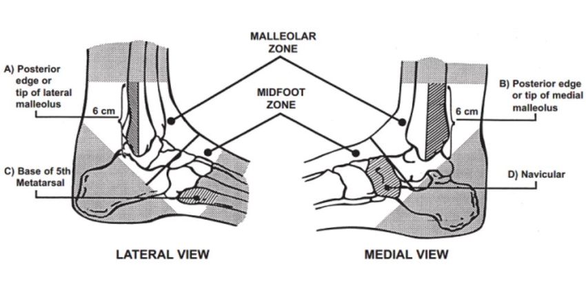
oCORE EM – Lisfranc Injuries
        oCORE EM – Jones Fractures
        oEM@3AM – 5th metatarsal fractures
        oemDocs – Diabetic Foot Infections
        oemDocs – Osteomyelitis
        oemDocs – Charcot Joint
•   Journal Articles
      o Emergency Medicine Clinics of North America – Foot and Ankle Pain

Answers for Learners:

1. What are important features of the history and exam for patients with foot or ankle injury?

Key Questions for Your History
   1. What was the mechanism of injury and what symptoms occurred afterwards? Subsequent
       symptoms include swelling, inability to bear weight, fevers/chills, and erythema
   2. What is the location of pain?
   3. Was there a previous injury or surgery to the affected ankle?
   4. Has the patient experienced similar pain previously?

Key Points for the Ankle Exam
Develop a structured approach to your ankle exam and you won’t miss an injury. Here’s our suggestion:
   1. Visually inspect the ankle and ask the patient to take at least 4 steps.
   2. Evaluate the medial and lateral malleolus, lateral 5th toe, and 1st dorsal web space for sensation.
   3. Palpate for a posterior tibial (PT) and dorsalis pedis (DP) pulse.
   4. Evaluate for effusion and reduced range of motion.
   5. Evaluate for focal tenderness especially at the proximal tibia/fibula, posterior edge of
       lateral/medial malleolus, base of 5th metatarsal (MT), navicular.
   6. Test ankle dorsi/plantar-flexion, eversion, and inversion strength.
   7. Test ankle stability by performing the anterior drawer and talar tilt.
   8. Test syndesmotic stability by performing the squeeze test.
   9. Test achilles tendon injury by performing the Thompson test.

We recommend performing these steps in the sequence described as it allows quick triage and
prioritizes the neurovascular exam. The 9 step exam can be broken down into 3 critical questions:
    1. Is the patient neurovascularly intact?
    2. Is the ankle stable?
    3. Is there a fracture?

2. What comprise the Ottawa ankle/foot rules?

    •       Ottawa Ankle Rule
                o Should be applied to determine which patients with traumatic ankle pain need imaging
                o Test Characteristics: Sensitivty 100%, Specificity 40.1% (Stiell 1992)

2|Page From emDOCs.net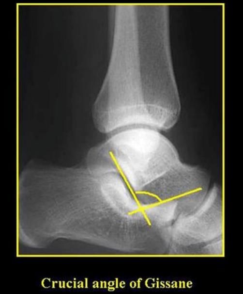
3. How can patient with a ruptured Achilles tendon present, and what are important exam and
imaging findings?
➔ Achilles tendon rupture is a clinical diagnosis. The Thompson Test should be applied in all suspected
cases. Imaging in form of plain films is limited, but POCUS can be very helpful.
Physical Exam
• Weakness with plantar flexion
• Increased resting ankle dorsiflexion on affected side in prone position with knees bent
• Palpable gap in dorsal portion of heel
• Calf atrophy in chronic cases
• Usually in absence of bony tenderness unless accompanied by other injury
• Thompson Test (video)
o Place the patient in the prone position, with feet hanging over the end of a stretcher or
table. If patient is not able to lay down/there are no stretchers, the patient can kneel on
a stool or chair
o Squeeze the calf of the normal limb. You will notice the squeeze will cause the ankle to
plantar flex appropriately
o Squeeze the calf of the limb with the suspected Achilles tendon rupture. You will notice
the squeeze will cause no motion if there is a full rupture/tear, and diminished motion if
there is a partial tear
o Performance Characteristics (Garras 2012)
Sensitivity Specificity (+) LR (-) LR
96-100% 93-100% 13.7 0.04
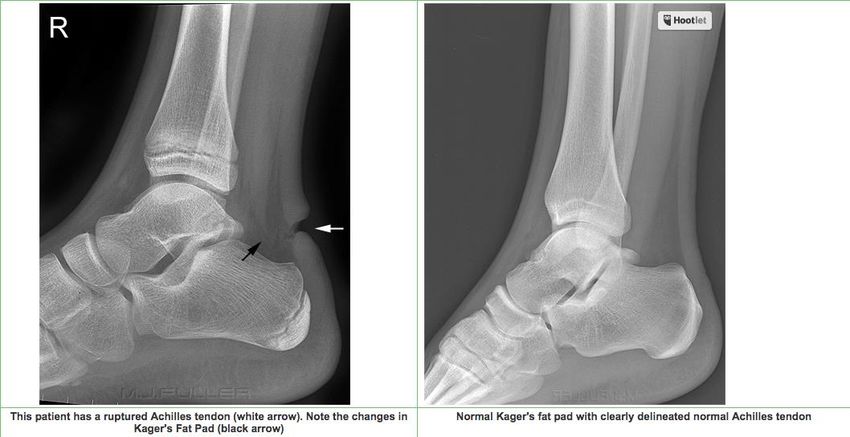
Imaging
• X-Rays
o Used to rule out other or concurrent pathology
o May show soft tissue swelling and destruction of pre-Achilles fat pad (Kager’s Fat Pad)
o Findings are non-specific as tear of tendon unable to be visualized
3|Page From emDOCs.net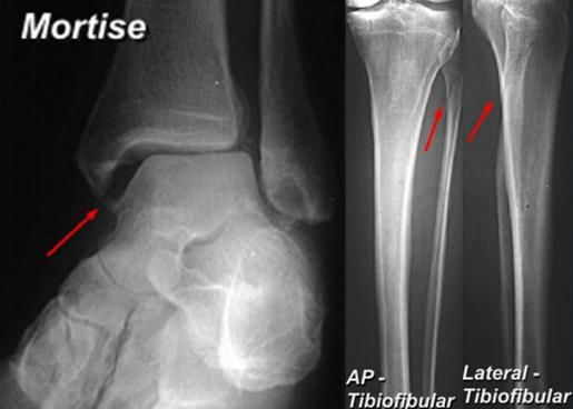
Kager’s Fat Pad (wikiradiography.net)

  •   Ultrasound
          o Ultrasound is helpful if obvious findings present and to distinguish between partial vs
              complete tears, however only around 50% sensitive for detecting only partial tears
              (Kayser 2005)
          o Normal Achilles tendon . In the image below, you can visualize the intact tendon in
              between the two white crosses. You will notice there is no tear or surrounding
              edema/fluid

             Case courtesy of Dr Maulik S Patel, Radiopaedia.org. From the case rID: 9759
  •   Full thickness rupture of Achilles Tendon: The retracted tendon appears to the left, with an
      anechoic area representing a defect full of fluid adjacent to ruptured tendon (to the right, as
      labeled)

           Case courtesy of Dr Maulik S Patel, Radiopaedia.org. From the case rID: 13548

4|Page From emDOCs.net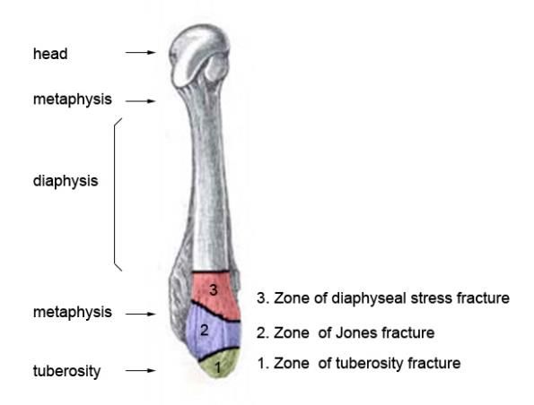
•   Partial thickness rupture: In the image below there is a partial thickness tear indicated in
       between the white arrows. Partial tears show a more hypoechoic and thickened portion of the
       tendon as you near the tear, as seen to the right of the midline of the image

                               Partial Thickness Tear (orthobullets.com)

   •   MRI
           o   Gold-standard imaging modality
           o   Rarely, if ever, necessary in the ED
           o   Used for equivocal physical exam/alternate imaging findings or for assessing the severity
               of the tear for possible operative management
           o   Findings
                   ▪ A full-thickness tear often shows a tendinous gap filled with edema or blood
                   ▪ Complete rupture shows retraction of tendon ends

4. What are important considerations in patients with ankle fractures or suspected dislocation?

   •   Closed ankle dislocations without any other fracture are rare.
   •   Posterior ankle dislocations are the most common type of ankle dislocation.
   •   If patients have signs of neurovascular compromise or skin tenting from a posterior dislocation,
       they should be reduced immediately without waiting for radiographs. With an assistant, bend
       the knee, plantar-flex the ankle, and apply traction with an anterior force to the heel.
   •   If high energy / complex fracture-dislocation, consider a post-reduction CT to ensure there are
       no additional injuries.
   •   Place patients in a short posterior leg splint ± stirrup after reduction.
   •   Bosworth fracture-dislocations, which will require an open reduction internal fixation, may be
       missed on plain film with CT imaging providing better visualization.
   •   Emergent orthopedics consultation should be obtained for an ankle dislocation if it is
       irreducible, associated with a fracture, demonstrates neurovascular compromise, or is open.

5. How do you interpret foot and ankle x-rays?

➔ Check out these resources for an in-depth explanation of interpretation.
  • https://dontforgetthebubbles.com/ankle-x-ray-interpretation/
  • https://www.aliem.com/emrad-foot-x-ray/
  • https://www.aliem.com/emrad-ankle/

5|Page From emDOCs.net
6. What are features of a syndesmosis injury? Why are these important to diagnose?
The joint between the tibia and fibula are held together by ligaments. If this ligament is sprained then
this is a syndesmotic injury → important to diagnosis as these are usually unstable injuries and require
operative repair.
There can be widening of the clear space between the medial border of the fibula and the lateral border
of the posterior tibia (>5mm). You can also get overlap of the fibula and the anterior tibial tubercle
(>6mm on the AP views, >1mm on the mortise view).
7. What comprises a Maisonneuve fracture?
• Spiral fracture of the proximal third of the fibula with an associated fracture of the medial
malleolus or rupture of the deep deltoid ligament (PER4 fracture)
• Palpation along the entire length of the fibular is important in order to pick up signs of this injury
as it can be seen in patients with relatively unremarkable ankle X-rays
• If the patient is discovered to have either a medial malleolus fracture or high fibular fracture on
X-ray, tibia-fibula films are indicated to rule out this type of fracture.
8. How can a calcaneus fracture present? What other injuries should you consider? What imaging
should you obtain?
• Always suspect calcaneus fractures in patients with axial loading injuries to the lower
extremities. If a calcaneus injury is found, look for concomitant fractures of the ankle and
vertebrae.
o Vertebral injuries secondary to axial loading injury (10%)
o Contralateral calcaneal injury (10%)
• Watch out for compartment syndrome of the foot which occurs in 10% of calcaneal fractures
and results in significant morbidity
Diagnostic Imaging
• Standard X-rays: AP, lateral, oblique foot
• Optional X-rays (See Image Gallery below)
o Broden views: allows visualization of posterior facet
o Harris View: visualizes tuberosity fragment widening, shortening, and varus positioning
o AP ankle: demonstrates fibular impingement if lateral wall extrusion is present
• Findings in calcaneal fractures:
• Reduced Bohler angle
6|Page From emDOCs.neto Created with lines drawn tangiental to anterior and posterior aspects of superior
calcaneus
o Normal is 20-40 degrees
• Increased angle of Gissane
o Formed by downward and upward slopes of calcaneus
o Normal is 95-105 degrees
• Non-contrast CT
o Obtain when clinical suspicion of a fracture is high despite negative x-rays
o Gold standard imaging test for any calcaneal fracture (except for stress fractures)
o Will almost always be obtained for pre-operative planning but does not necessarily need
to be obtained in the ED
9. Why are talus fractures often missed? When should they be considered?
A snowboarder’s fracture is a lateral process of talus fracture that is commonly misdiagnosed as a
simple ankle sprain. Yet again, the mechanism of injury is external rotation of the ankle usually
associated with dorsiflexion and an axial load upon landing. The talus is an often overlooked bone on
physical exam. Just as we are trained to assess the scaphoid bone for occult fracture for all wrist injuries
we should assess the talus bone for occult fracture for all ankle injuries. Get a good mortise view of your
ankle to pick up this fracture, which can be very subtle or occult on x-ray. If in doubt on your x-ray, place
the patient in a posterior slab, non-weight-bearing and have them follow in up fracture clinic. If
available, get a CT ankle.
The talus is the “scaphoid bone of the ankle”. Make sure to always assess the talus on physical exam
of the ankle.
Common situations in which talus fractures are missed:
• Patients who injured their ankle in a boot/skate
• Patients in an MVC with distracting injuries
• Older patients with osteoporosis
• Patients diagnosed with ankle sprain returning weeks later with ongoing pain
7|Page From emDOCs.net10. What is a Lisfranc fracture? What are the exam features, and what should you look for on
radiograph?
• The Lisfranc joint complex is a tarso-metatarsal articulation named for Jacques Lisfranc (1790-
1847), one of Napoleon’s battlefield surgeons.
o Specifically, it is the articulation of the 1st, 2nd, and 3rd metatarsals with the medial,
intermediate, and lateral cuneiforms, respectively, as well as the articulation of the 4th
and 5th metatarsals with the cuboid. (Englanoff 1995)
o The Lisfranc (or Oblique) ligament secures the second metatarsal to the medial
cuneiform, serving as a mortise joint anchoring the entire complex and preventing
medio-lateral or plantar displacement.
o Fractures and concomitant disarticulations of this joint are termed Lisfranc fracture-
dislocations
▪ A Lisfranc injury must be part of the differential for any midfoot trauma because
of the significant morbidity associated with missed diagnosis
• Physical exam findings, including deformity, swelling and ecchymosis, may be subtle or absent
• Normal foot x-rays do not rule out a Lisfranc injury, weight-bearing views or CT are essential
o Diastasis (separation beyond normal) of the space between the bases of the 1st and 2nd
metatarsals
▪ Diastasis is a measurement >2mm in a normal foot, or >1mm relative to the
contralateral foot in people with widened joint spaces at baseline
▪ Bilateral films are thus necessary when obtaining weight-bearing views.
11. What is the difference between a pseudo-Jones and Jones fracture?
• Zone 1: Avulsion or “Pseudo-Jones” fracture
o Proximal tubercle (rarely enters 5th tarsometatarsal joint)
o Caused when bony fragment is detached by ligament or other connective tissue
▪ Typically long plantar ligament, lateral band of plantar fascia, or contraction of
peroneus brevis
• Zone 2: Jones fracture
o Metaphyseal-diaphyseal junction (4th-5th metatarsal articulation)
o Vascular watershed area
▪ Proximal 5th metatarsal has unique blood supply with a watershed area at the
metaphyseal-diaphyseal junction
▪ Fractures in this area are at a high risk for non-union due to the fracture
interrupting the already limited blood supply in this area
8|Page From emDOCs.netVascular Watershed Zone (orthobullets.com)
• Zone 3: stress fracture
o Proximal diaphyseal fracture (distal to 4th-5th metatarsal articulation)
o Associated with cavovarus foot deformities or sensory neuropathies
12. What are several types of foot infections found in diabetic patients? How should these be
managed?
Most DFI are polymicrobial. The most common causative source is staphylococcus (S. aureus), though
streptococci and enterobacteriaceae are also likely pathogens. In ischemic and necrotic wounds, aerobic
gram-negative rods and anaerobes are often also present. It is important to note that not all ulcers are
infectious. Therefore, careful examination and classification of the ulcer should guide treatment.
The IDSA guidelines propose empiric antibiotic treatment recommendations for DFI based on infection
severity and causative pathogen. The IDSA recommends 1-2 weeks of antibiotics for mild infections, 1-3
weeks for moderate, and 2-4 weeks for severe infections. Remember to check with your pharmacists
and institutional policies before following these guidelines.
13. What is a Charcot joint?
Charcot Joint is progressive deterioration of a joint seen in patients with peripheral neuropathy. Jean-
Martin Charcot and colleagues first described the disease, as it was caused by tertiary syphilis in the late
1800s. However in the modern age, diabetes mellitus is the most common cause of this neuro-
arthropathy, and emergency physicians must have a high index of suspicion to be able to differentiate it
from other disease entities such as osteomyelitis, arthritis, or gout.
The pathophysiology of Charcot Joint is multifactorial, but ultimately it is a combination of mechanical
and vascular complications from diabetes. Loss of pain sensation and proprioception may lead to
repeated trauma, which results in joint laxity, joint instability, and the release of pro-inflammatory
cytokines. This is further complicated by autonomic vascular reflexes that cause hyperemia,
arteriovenous shunts, and periarticular osteopenia. This eventually progresses to bony foot deformities,
particular in the mid-foot.
It is important to recognize Charcot Joint early to prevent morbidity, as the management greatly differs
from other disease entities. Patients should be referred to diabetic foot specialists where conservative
management with pressure-relieving methods can be initiated. Total contact casting, which requires
precise molding and frequent reapplication, is considered gold standard to offload weight. Crutches and
wheelchairs are also options. Gradually the patient is transitioned back to weight-bearing as erythema
and edema resolve and radiographic findings are stabilized.
9|Page From emDOCs.netYou can also read